0022-538X/06/$08.00
⫹0 doi:10.1128/JVI.01557-06
Copyright © 2006, American Society for Microbiology. All Rights Reserved.
Increased Frequency of Circulating CCR5
⫹
CD4
⫹
T Cells in Human
Immunodeficiency Virus Type 2 Infection
䌤
Rui Soares,
1Russell Foxall,
1Adriana Albuquerque,
1Catarina Cortesa
˜o,
1Miguel Garcia,
1,2Rui M. M. Victorino,
1,3and Ana E. Sousa
1*
Unidade de Imunologia Clı´nica, Instituto de Medicina Molecular, Faculdade de Medicina de Lisboa, Av. Prof. Egas Moniz,
1649-028 Lisbon, Portugal
1; Instituto Superior de Cieˆncias da Sau
´de Egas Moniz, Quinta da Granja,
2829-511 Monte da Caparica, Portugal
2; and Medicina 2, Hospital de Santa Maria,
Av. Prof. Egas Moniz, 1649-035 Lisbon, Portugal
3Received 20 July 2006/Accepted 28 September 2006
CCR5 expression determines susceptibility to infection, cell tropism, and the rate of human
immunodefi-ciency virus type 1 (HIV-1) disease progression. CCR5 is also considered the major HIV-2 coreceptor in vivo,
in spite of broad coreceptor use in vitro. Here we report a significantly increased proportion of memory-effector
CD4 T cells expressing CCR5 in HIV-2-infected patients correlating with CD4 depletion. Moreover, HIV-2
proviral DNA was essentially restricted to memory-effector CD4, suggesting that this is the main target for
HIV-2. Similar levels of proviral DNA were found in the two infection categories. Thus, the reduced viremia and
slow rate of CD4 decline that characterize HIV-2 infection seem to be unrelated to coreceptor availability.
Human immunodeficiency virus type 2 (HIV-2)
immunode-ficiency is characterized by slow disease progression with
lim-ited impact on the survival of the majority of infected adults
(20, 33, 45). The rate of CD4
⫹T-cell decline is much slower in
HIV-2 than in HIV-1 disease, and there is a low plasma viral
load irrespective of disease stage (2, 4, 9, 17, 20, 32, 41, 43).
The factors contributing to the suggested decreased rate of
virus production in HIV-2 infection remain largely unknown.
In spite of the promiscuity of coreceptor usage exhibited by
HIV-2 in in vitro experimental settings (8, 13, 16, 22, 29, 39,
40), several lines of evidence show that CCR5 and CXCR4 are
the major coreceptors for HIV-2 infection in vivo (6, 24, 25).
* Corresponding author. Mailing address: Unidade de Imunologia
Clı´nica, Instituto de Medicina Molecular, Faculdade de Medicina de
Lisboa, Av. Prof. Egas Moniz, 1649-028 Lisboa, Portugal. Phone: 351
21 799 95 25. Fax: 351 21 799 95 27. E-mail: [email protected].
䌤
Published ahead of print on 11 October 2006.
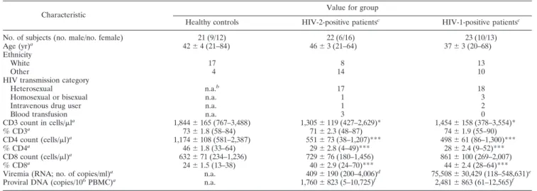
TABLE 1. Cohort characterization
Characteristic
Value for group
Healthy controls HIV-2-positive patientsc HIV-1-positive patientsc
No. of subjects (no. male/no. female) 21 (9/12) 22 (6/16) 23 (10/13)
Age (yr)a
42⫾ 4 (21–84) 46⫾ 3 (21–64) 37⫾ 3 (20–68)
Ethnicity
White 17 8 13
Other 4 14 10
HIV transmission category
Heterosexual n.a.b
17 18
Homosexual or bisexual n.a. 1 3
Intravenous drug user n.a. 1 2
Blood transfusion n.a. 3 0
CD3 count in cells/la 1,844⫾ 165 (767–3,488) 1,305⫾ 119 (427–2,629)* 1,454⫾ 158 (378–3,554)* % CD3a 73⫾ 1.8 (58–84) 71⫾ 2.3 (48–87) 74⫾ 1.9 (55–90) CD4 count (cells/l)a 1,174⫾ 108 (581–2,387) 551⫾ 73 (38–1,207)*** 498⫾ 61 (86–1,300)*** % CD4a 46⫾ 1.8 (33–64) 29⫾ 2.8 (4–49)*** 28⫾ 2.4 (9–52)*** CD8 count (cells/l)a 632⫾ 71 (234–1,236) 729⫾ 76 (180–1,456) 861⫾ 100 (269–2,007) % CD8a 24⫾ 1.5 (13–38) 40⫾ 2.9 (24–70)*** 44⫾ 2.4 (28–64)*** Viremia (RNA; no. of copies/ml)a
n.a. 409⫾ 190 (200–4,006)d
75,508⫾ 30,429 (118–548,631)e
Proviral DNA (copies/106
PBMC)a
n.a. 1,760⫾ 823 (5–10,725)f
2,481⫾ 863 (61–12,565)f aData are means⫾ standard errors of the means with limits in brackets.
bn.a., not applicable.
cSignificance in comparison with healthy control results is represented as follows: *, P⬍ 0.05; ***, P ⬍ 0.0001. There were no significant differences between the
HIV-2 and HIV-1 cohorts except for the viremia results (P⬍ 0.0001).
dHIV-2 viremia was quantified by a reverse transcriptase PCR-based test (41); the results were below 200 RNA copies/ml (cutoff) for 19 out of the 21 patients studied.
In these cases the cutoff value was used.
eHIV-1 viremia was quantified by reverse transcriptase PCR (Ultrasensitive Test; Roche Molecular Systems, Branchburg, NJ). The cutoff was 50 RNA copies/ml. fProviral DNA was quantified by real-time PCR and was detected in all 17 HIV-2-infected patients and all 16 HIV-1-infected patients investigated.
In HIV-1-infected patients, CCR5 expression determines
susceptibility to infection, cell tropism, and the rate of disease
progression and is currently an important target of new
anti-retroviral drugs (10, 11, 21, 27, 34, 35).
There are scanty data available on CCR5 and CXCR4
ex-pression in HIV-2-infected patients. A previous study of a
Senegalese cohort reported lower CCR5 expression in HIV-2
than in HIV-1 infection, but as these patients, unlike our
co-horts, were not stratified according to CD4 depletion or
vire-mia, direct comparison is problematic (38).
We analyzed here CCR5 and CXCR4 expression in freshly
isolated peripheral blood mononuclear cells (PBMC) from
untreated HIV-2- and HIV-1-infected subjects who were
cur-rently living in Portugal and attending outpatient clinics in
Lisbon and who exhibited no known ongoing opportunistic
infections or tumors. The epidemiological and clinical features
FIG. 1. CCR5 and CXCR4 expression in circulating CD4
⫹T cells from HIV-2- and HIV-1-infected patients as well as healthy controls. Freshly
isolated PBMC were surface stained with anti-CCR5 (clone 2D7) or anti-CXCR4 (clone 12G5) monoclonal antibodies (phycoerythrin conjugated;
BD Biosciences, San Jose, CA), acquired, and analyzed in a FACSCalibur flow cytometer using CellQuest software (BD Biosciences). Percentages
of CCR5- and CXCR4-expressing cells within total CD4 T cells (CD4
⫹), a naı¨ve CD4 subset (CD4
⫹CD45RA
⫹), and a memory CD4 subset (CD4
⫹CD45RA
neg) are shown in panels A and C, respectively. Bars represent means
⫾ standard errors of the means. Representative contour plots of
the flow cytometric analysis of CD4
⫹gated T cells of an HIV-2-infected patient with 291 CD4
⫹T cells/
l are illustrated in panel B. (D) Correlation
between the percentage of CCR5
⫹cells within the CD4 subset and the frequency of circulating CD4
⫹T cells. Each dot represents one individual.
The Pearson correlation coefficients for the different cohorts are shown. The median fluorescence intensities (MedianFI) of the whole population
of CCR5- and CXCR4-expressing CD4
⫹T cells are shown in panels E and F, respectively. Each dot represents one individual; bars represent
means. Statistical analysis was performed using GraphPad Prism version 4.00 (GraphPad Software, San Diego, CA). The data were compared using
an unpaired t test. Spearman’s correlation coefficients were used to determine the correlations between two variables. P values
⬍ 0.05 were
considered significant and are depicted in the figure.
of these cohorts, as well as of healthy controls, are summarized
in Table 1. Of note, HIV-2 and HIV-1 cohorts exhibited
sim-ilar levels of CD4 depletion but striking differences in viremia.
CCR5 and CXCR4 expression were assessed by flow
cytom-etry in freshly isolated PBMC as previously described (42).
HIV-2-positive patients exhibited a higher frequency of
CCR5
⫹cells within the CD4
⫹subset that reaches statistical
significance than that seen with healthy subjects (Fig. 1A). In
HIV-2 patients CCR5 expression was also largely confined to
the memory CD4
⫹CD45RA
⫺population, as illustrated in the
representative contour plot of Fig. 1B.
The frequencies of CXCR4
⫹cells within CD4 T cells
showed no significant differences among the three cohorts,
although there was a trend to lower frequencies in the HIV-2
cohort in both the naı¨ve and memory subsets (Fig. 1C).
A significant correlation between the frequency of CCR5
⫹cells within the CD4 subset and the degree of CD4 depletion
was observed with the HIV-2 cohort that was not observed with
HIV-1-positive patients (Fig. 1D).
We have previously shown that CD4 depletion is directly
linked to immune activation in both HIV-2 and HIV-1
infec-tions in spite of the striking differences in viremia (15, 42).
Since CCR5 is up-regulated upon T-cell activation, we looked
for a possible correlation between the frequency of CCR5
⫹cells and the expression of HLA-DR, a marker widely used to
quantify immune activation in HIV disease (15). A significant
correlation was observed with the HIV-2 cohort (r
⫽ 0.68; P ⫽
0.0009) that was not found with HIV-1-positive patients (r
⫽
0.25; P
⫽ 0.2814).
These data illustrated the link between the expansion of
CCR5
⫹cells and immune activation in HIV-2 infection. The
lower frequency of circulating CCR5
⫹cells in HIV-1 infection
compared to HIV-2 results despite the similarities in
height-ened immune activation may be related to a continuous
deple-tion of the CCR5 pool in associadeple-tion with the high level of
viremia (19, 23).
The frequency of CCR5
⫹cells may be underestimated
due to binding-induced receptor internalization (26, 30). In
fact, the assessment of the median fluorescence intensity
(MedianFI) of CCR5
⫹CD4
⫹T cells revealed similar and
significant down-regulation results for both 2- and
HIV-1-infected cohorts in comparison with the results seen with
healthy subjects (Fig. 1E). This contrasts with the absence of
differences in MedianFI of CXCR4
⫹CD4
⫹T cells for the
three cohorts (Fig. 1F). It is noteworthy that HIV-2 infection
has been associated with high levels of production of RANTES,
MIP-1
␣, and MIP-1 (1, 18, 28), possibly contributing to the
CCR5 down-regulation.
In order to exclude the possibility that CCR5
⌬32 mutations
contribute to the low MedianFI of CCR5 (44), we screened the
cohorts for the presence of this allele using the primers
de-scribed in Table 2. None of the HIV-2- or HIV-1-infected
patients exhibited the CCR5
⌬32 allele. There were five healthy
subjects heterozygous for CCR5
⌬32. The exclusion of these
individuals from the analysis resulted in an even more
signifi-cant difference in the results of down-regulation of CCR5
MedianFI between HIV cohorts and healthy subjects (P
⫽
0.0019 for HIV-2 and P
⬍ 0.0001 for HIV-1).
On the other hand, despite differing levels of viremia, we did
not find significant differences between HIV-2 and HIV-1
pro-viral DNA levels, suggesting the presence of similar numbers
of infected cells in the two infection categories (Table 1), in
agreement with previous reports (3–5, 14, 31). Proviral DNA
was assessed by absolute quantitative real-time PCR using an
ABI PRISM 7000 sequence detection system (Applied
Biosys-tems) with a detection range of 7 orders of magnitude and a
sensitivity of five copies. Reactions containing 150 ng of
genomic DNA extracted from 10
6PBMC by use of an ABI
PRISM 6100 nucleic acid extractor (Applied Biosystems), 25
l of Platinum quantitative PCR SuperMix-UDG, 1 l ROX
reference dye (Invitrogen) (50
⫻), 5 mM MgCl
2, 300 nM
primer (each), and 200 nM probe (Table 2) were run in
du-plicate. Albumin was used to standardize DNA input.
It is worth noting that no correlation was found between the
TABLE 2. Primer and probe sequences
aPrimer or probe Sequenceb
HIV-2
Forward primer ...5
⬘-CGC GAG AAA CTC CGT CTT G-3⬘
Reverse primer...5
⬘-CAC ACA ATA TGT TTT AGC CTG
TAC TTT TT-3
⬘
Probe ...5
⬘-FAM-CCG GGC CGT AAC CT-MGB-3⬘
HIV-1
Forward primer ...5
⬘-GGG AGA ATT AGA TCG ATG GGA
AA-3
⬘
Reverse primer...5
⬘-CTG CTT GCC CAT ACT ATA TGT
TTT AAT TTA-3
⬘
Probe ...5
⬘-FAM-CCC TGG CCT TAA CCG AAT
T-MGB-3
⬘
Albumin
Forward primer ...5
⬘-TGC ATG AGA AAA CGC CAG TAA-3⬘
Reverse primer...5
⬘-ATG GTC GCC TGT TCA CCA A-3⬘
Probe ...5
⬘-FAM-TCA CCA AAT GCT GCA CAG
A-MGB-3
⬘
CCR5
cForward primer ...5
⬘-TTC ATT ACA CCT GCA GCT CT-3⬘
Reverse primer...5
⬘-CAC AGC CCT GTG CCT CTT CTT
CTC ATT TCG-3
⬘
aPrimers and probes were designed using Primer Express 2.0 software
(Ap-plied Biosystems, Foster City, CA) and checked against the Los Alamos HIV database.
bFAM, 6-carboxyfluorescein; MGB, minor groove binding.
cFifty nanograms of genomic DNA was amplified by PCR using 25l of
Platinum PCR SuperMix (Invitrogen, Carlsbad, CA), 300 nM primer (each), and 5 mM MgCl2and was run on a 2% agarose gel with DNA bands stained with
ethidium bromide.
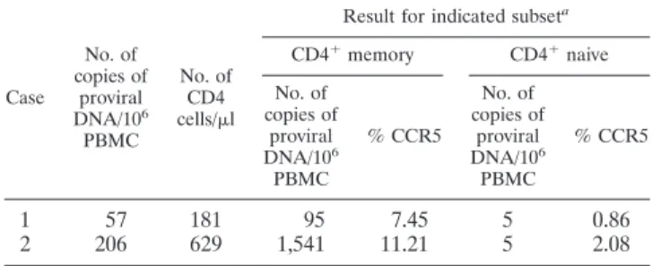
TABLE 3. HIV-2 proviral DNA in CD4 naı¨ve and memory subsets
Case No. of copies of proviral DNA/106 PBMC No. of CD4 cells/l
Result for indicated subseta
CD4⫹memory CD4⫹naive No. of copies of proviral DNA/106 PBMC % CCR5 No. of copies of proviral DNA/106 PBMC % CCR5
1
57
181
95
7.45
5
0.86
2
206
629
1,541
11.21
5
2.08
aFreshly isolated PBMC were successively gated using CD4⫹and CD45RA⫹
(naı¨ve) or CD45RA⫺(memory); the subsets were purified using FACSAria (BD Biosciences), with purity higher than 98%.
frequency of CCR5
⫹cells within the CD4 subset and the levels
of HIV-2 proviral DNA (r
⫽ 0.08; P ⫽ 0.7483).
In order to evaluate the possibility that the similar levels of
proviral DNA in the presence of the dissimilar HIV-1 and
HIV-2 viremia results might be due to differences in cell
tar-gets, we purified the naı¨ve and the memory CD4 T cells from
PBMC of two HIV-2 patients with different levels of CD4
depletion by high-speed cell sorting using FACSAria (BD
Bio-sciences).
As depicted in Table 3, the levels of HIV-2 proviral DNA
documented in the naı¨ve subset were minimal. Therefore,
these data suggest that memory CD4 T cells are the main
targets for HIV-2 infection in vivo, reinforcing the idea of a
major role of CCR5 coreceptor in HIV-2 infection. This was in
agreement with data on HIV-1 infection in which integrated
proviruses are preferentially detected within the memory
sub-set (7, 12, 36).
In summary, HIV-2-infected patients showed an increase in
the proportion of CCR5
⫹cells within the memory-effector
CD4
⫹T cells in correlation with the degree of CD4 depletion
and immune activation. In contrast, in HIV-1 infection there
was dissociation between CCR5 and other markers of immune
activation which could be interpreted as an indirect evidence of
depletion of the CCR5
⫹cells by HIV-1. However, the HIV-2
proviral load was also mainly restricted to memory-effector
CD4 T cells, suggesting these are the major HIV-2 targets,
which is consistent with CCR5 being the main HIV-2
corecep-tor in vivo. Moreover, the levels of HIV-2 proviral load were
similar to those observed in untreated HIV-1-infected
individ-uals, suggesting equivalent numbers of infected cells resulting
from the two diseases in spite of viremia being undetectable in
the majority of the HIV-2 patients.
The presence of reduced HIV-2 viremia seems to be
unre-lated to coreceptor availability. Since HIV-2 is no less
cyto-pathic per se than HIV-1 (37), other host factors must be
implicated in the control of viral replication in spite of
signif-icant proviral DNA levels in HIV-2-positive patients. The
fur-ther investigation of the mechanisms contributing to this
con-trol of HIV-2 viremia in the absence of antiretroviral therapy
may prove to be useful in defining complementary therapeutic
strategies to control viral reservoirs in HIV-1.
This work was supported by grants from Fundac¸a
˜o para a Cie
ˆncia e
a Tecnologia (FCT) and Comissa
˜o Nacional de Luta Contra a SIDA to
A.E.S. R.S., R.F., and C.C. received scholarships from FCT.
We gratefully acknowledge Perpe
´tua Gomes for the quantification
of HIV-2 viremia, Ana Caetano for cell sorting technical assistance,
and the clinical collaboration of the following colleagues: E. Valadas,
F. Antunes, L. Pinheiro, M. Doroana, M. Lucas, and R. Marc¸al.
REFERENCES
1. Ahmed, R. K., H. Norrgren, Z. da Silva, A. Blaxhult, E. L. Fredriksson, G. Biberfeld, S. Andersson, and R. Thorstensson.2005. Antigen-specific beta-chemokine production and CD8 T-cell noncytotoxic antiviral activity in HIV-2-infected individuals. Scand. J. Immunol. 61:63–71.
2. Andersson, S., H. Norrgren, Z. da Silva, A. Biague, S. Bamba, S. Kwok, C. Christopherson, G. Biberfeld, and J. Albert. 2000. Plasma viral load in HIV-1 and HIV-2 singly and dually infected individuals in Guinea-Bissau, West Africa: significantly lower plasma virus set point in HIV-2 infection than in HIV-1 infection. Arch. Intern. Med. 160:3286–3293.
3. Ariyoshi, K., N. Berry, A. Wilkins, D. Ricard, P. Aaby, A. Naucler, P. T. Ngom, O. Jobe, S. Jaffar, F. Dias, R. S. Tedder, and H. Whittle.1996. A community-based study of human immunodeficiency virus type 2 provirus load in rural village in West Africa. J. Infect. Dis. 173:245–248.
4. Berry, N., K. Ariyoshi, S. Jaffar, S. Sabally, T. Corrah, R. Tedder, and H.
Whittle.1998. Low peripheral blood viral HIV-2 RNA in individuals with high CD4 percentage differentiates HIV-2 from HIV-1 infection. J. Hum. Virol. 1:457–468.
5. Berry, N., K. Ariyoshi, O. Jobe, P. T. Ngum, T. Corrah, A. Wilkins, H. Whittle, and R. Tedder.1994. HIV type 2 proviral load measured by quan-titative polymerase chain reaction correlates with CD4⫹lymphopenia in HIV type 2-infected individuals. AIDS Res. Hum. Retrovir. 10:1031–1037. 6. Blaak, H., P. H. Boers, R. A. Gruters, H. Schuitemaker, M. E. van der Ende,
and A. D. Osterhaus.2005. CCR5, GPR15, and CXCR6 are major corecep-tors of human immunodeficiency virus type 2 variants isolated from individ-uals with and without plasma viremia. J. Virol. 79:1686–1700.
7. Brenchley, J. M., B. J. Hill, D. R. Ambrozak, D. A. Price, F. J. Guenaga, J. P. Casazza, J. Kuruppu, J. Yazdani, S. A. Migueles, M. Connors, M. Roederer, D. C. Douek, and R. A. Koup.2004. T-cell subsets that harbor human immunodeficiency virus (HIV) in vivo: implications for HIV pathogenesis. J. Virol. 78:1160–1168.
8. Bron, R., P. J. Klasse, D. Wilkinson, P. R. Clapham, A. Pelchen-Matthews, C. Power, T. N. Wells, J. Kim, S. C. Peiper, J. A. Hoxie, and M. Marsh.1997. Promiscuous use of CC and CXC chemokine receptors in cell-to-cell fusion mediated by a human immunodeficiency virus type 2 envelope protein. J. Virol. 71:8405–8415.
9. Damond, F., M. Gueudin, S. Pueyo, I. Farfara, D. L. Robertson, D. Descamps, G. Che`ne, S. Matheron, P. Campa, F. Brun-Ve´zinet, and F. Simon.2002. Plasma RNA viral load in human immunodeficiency virus type 2 subtype A and subtype B infections. J. Clin. Microbiol. 40:3654–3659.
10. Dean, M., M. Carrington, C. Winkler, G. A. Huttley, M. W. Smith, R. Allikmets, J. J. Goedert, S. P. Buchbinder, E. Vittinghoff, E. Gomperts, S. Donfield, D. Vlahov, R. Kaslow, A. Saah, C. Rinaldo, R. Detels, and S. J. O’Brien.1996. Genetic restriction of HIV-1 infection and progression to AIDS by a deletion allele of the CKR5 structural gene. Hemophilia Growth and Development Study, Multicenter AIDS Cohort Study, Multicenter He-mophilia Cohort Study, San Francisco City Cohort, ALIVE Study. Science 273:1856–1862.
11. de Roda Husman, A. M., H. Blaak, M. Brouwer, and H. Schuitemaker. 1999. CC chemokine receptor 5 cell-surface expression in relation to CC chemo-kine receptor 5 genotype and the clinical course of HIV-1 infection. J. Im-munol. 163:4597–4603.
12. Douek, D. C., J. M. Brenchley, M. R. Betts, D. R. Ambrozak, B. J. Hill, Y. Okamoto, J. P. Casazza, J. Kuruppu, K. Kunstman, S. Wolinsky, Z. Gross-man, M. Dybul, A. Oxenius, D. A. Price, M. Connors, and R. A. Koup.2002. HIV preferentially infects HIV-specific CD4⫹T cells. Nature 417:95–98. 13. Endres, M. J., P. R. Clapham, M. Marsh, M. Ahuja, J. D. Turner, A.
McKnight, J. F. Thomas, B. Stoebenau-Haggarty, S. Choe, P. J. Vance, T. N. Wells, C. A. Power, S. S. Sutterwala, R. W. Doms, N. R. Landau, and J. A. Hoxie.1996. CD4-independent infection by HIV-2 is mediated by fusin/ CXCR4. Cell 87:745–756.
14. Gomes, P., N. C. Taveira, J. M. Pereira, F. Antunes, M. O. Ferreira, and M. H. Lourenco.1999. Quantitation of human immunodeficiency virus type 2 DNA in peripheral blood mononuclear cells by using a quantitative-com-petitive PCR assay. J. Clin. Microbiol. 37:453–456.
15. Grossman, Z., M. Meier-Schellersheim, A. E. Sousa, R. M. Victorino, and W. E. Paul.2002. CD4⫹T-cell depletion in HIV infection: are we closer to understanding the cause? Nat. Med. 8:319–323.
16. Guillon, C., M. E. van der Ende, P. H. Boers, R. A. Gruters, M. Schutten, and A. D. Osterhaus.1998. Coreceptor usage of human immunodeficiency virus type 2 primary isolates and biological clones is broad and does not correlate with their syncytium-inducing capacities. J. Virol. 72:6260–6263. 17. Jaffar, S., A. Wilkins, P. T. Ngom, S. Sabally, T. Corrah, J. E. Bangali, M.
Rolfe, and H. C. Whittle.1997. Rate of decline of percentage CD4⫹cells is faster in HIV-1 than in HIV-2 infection. J. Acquir. Immune Defic. Syndr. Hum. Retrovirol. 16:327–332.
18. Kokkotou, E. G., J. L. Sankale, I. Mani, A. Gueye-Ndiaye, D. Schwartz, M. E. Essex, S. Mboup, and P. J. Kanki. 2000. In vitro correlates of HIV-2-mediated HIV-1 protection. Proc. Natl. Acad. Sci. USA 97:6797–6802. 19. Krzysiek, R., A. Rudent, L. Bouchet-Delbos, A. Foussat, C. Boutillon, A.
Portier, D. Ingrand, D. Sereni, P. Galanaud, L. Grangeot-Keros, and D. Emilie.2001. Preferential and persistent depletion of CCR5⫹T-helper lym-phocytes with nonlymphoid homing potential despite early treatment of primary HIV infection. Blood 98:3169–3171.
20. Marlink, R., P. Kanki, I. Thior, K. Travers, G. Eisen, T. Siby, I. Traore, C. C. Hsieh, M. C. Dia, E. H. Gueye, J. Hellinger, A. Gueye-Ndiaye, J. L. Sankale´, I. Ndoye, S. Mboup, and M. Essex.1994. Reduced rate of disease develop-ment after HIV-2 infection as compared to HIV-1. Science 265:1587–1590. 21. Martin, M. P., M. Dean, M. W. Smith, C. Winkler, B. Gerrard, N. L. Michael, B. Lee, R. W. Doms, J. Margolick, S. Buchbinder, J. J. Goedert, T. R. O’Brien, M. W. Hilgartner, D. Vlahov, S. J. O’Brien, and M. Carrington. 1998. Genetic acceleration of AIDS progression by a promoter variant of CCR5. Science 282:1907–1911.
22. McKnight, A., M. T. Dittmar, J. Moniz-Periera, K. Ariyoshi, J. D. Reeves, S. Hibbitts, D. Whitby, E. Aarons, A. E. Proudfoot, H. Whittle, and P. R. Clapham.1998. A broad range of chemokine receptors are used by primary
isolates of human immunodeficiency virus type 2 as coreceptors with CD4. J. Virol. 72:4065–4071.
23. Mehandru, S., M. A. Poles, K. Tenner-Racz, A. Horowitz, A. Hurley, C. Hogan, D. Boden, P. Racz, and M. Markowitz.2004. Primary HIV-1 infec-tion is associated with preferential depleinfec-tion of CD4⫹T lymphocytes from effector sites in the gastrointestinal tract. J. Exp. Med. 200:761–770. 24. Morner, A., A. Bjorndal, J. Albert, V. N. Kewalramani, D. R. Littman, R.
Inoue, R. Thorstensson, E. M. Fenyo, and E. Bjorling.1999. Primary human immunodeficiency virus type 2 (HIV-2) isolates, like HIV-1 isolates, fre-quently use CCR5 but show promiscuity in coreceptor usage. J. Virol. 73: 2343–2349.
25. Morner, A., A. Bjorndal, A. C. Leandersson, J. Albert, E. Bjorling, and M. Jansson.2002. CCR5 or CXCR4 is required for efficient infection of pe-ripheral blood mononuclear cells by promiscuous human immunodeficiency virus type 2 primary isolates. AIDS Res. Hum. Retrovir. 18:193–200. 26. Mueller, A., E. Kelly, and P. G. Strange. 2002. Pathways for internalization
and recycling of the chemokine receptor CCR5. Blood 99:785–791. 27. Mummidi, S., S. S. Ahuja, E. Gonzalez, S. A. Anderson, E. N. Santiago, K. T.
Stephan, F. E. Craig, P. O’Connell, V. Tryon, R. A. Clark, M. J. Dolan, and S. K. Ahuja.1998. Genealogy of the CCR5 locus and chemokine system gene variants associated with altered rates of HIV-1 disease progression. Nat. Med. 4:786–793.
28. Neoh, L. P., H. Akimoto, H. Kaneko, T. Hishikawa, H. Hashimoto, S. Hirose, Y. Kaneko, N. Yamamoto, and I. Sekigawa.1997. The production of beta-chemokines induced by HIV-2 envelope glycoprotein. AIDS 11:1062–1063. 29. Owen, S. M., D. Ellenberger, M. Rayfield, S. Wiktor, P. Michel, M. H. Grieco, F. Gao, B. H. Hahn, and R. B. Lal.1998. Genetically divergent strains of human immunodeficiency virus type 2 use multiple coreceptors for viral entry. J. Virol. 72:5425–5432.
30. Pastori, C., B. Weiser, C. Barassi, C. Uberti-Foppa, S. Ghezzi, R. Longhi, G. Calori, H. Burger, K. Kemal, G. Poli, A. Lazzarin, and L. Lopalco.2006. Long-lasting CCR5 internalization by antibodies in a subset of long-term nonprogressors: a possible protective effect against disease progression. Blood 107:4825–4833.
31. Popper, S. J., A. D. Sarr, A. Gueye-Ndiaye, S. Mboup, M. E. Essex, and P. J. Kanki.2000. Low plasma human immunodeficiency virus type 2 viral load is independent of proviral load: low virus production in vivo. J. Virol. 74:1554– 1557.
32. Popper, S. J., A. D. Sarr, K. U. Travers, A. Gueye-Ndiaye, S. Mboup, M. E. Essex, and P. J. Kanki.1999. Lower human immunodeficiency virus (HIV) type 2 viral load reflects the difference in pathogenicity of HIV-1 and HIV-2. J. Infect. Dis. 180:1116–1121.
33. Poulsen, A. G., P. Aaby, O. Larsen, H. Jensen, A. Naucler, I. M. Lisse, C. B. Christiansen, F. Dias, and M. Melbye.1997. 9-year HIV-2-associated mor-tality in an urban community in Bissau, west Africa. Lancet 349:911–914. 34. Reynes, J., P. Portales, M. Segondy, V. Baillat, P. Andre, O. Avinens, M. C.
Picot, J. Clot, J. F. Eliaou, and P. Corbeau.2001. CD4 T cell surface CCR5 density as a host factor in HIV-1 disease progression. AIDS 15:1627–1634. 35. Ribeiro, R. M., M. D. Hazenberg, A. S. Perelson, and M. P. Davenport. 2006. Naive and memory cell turnover as drivers of CCR5-to-CXCR4 tropism switch in human immunodeficiency virus type 1: implications for therapy. J. Virol. 80:802–809.
36. Schnittman, S. M., H. C. Lane, J. Greenhouse, J. S. Justement, M. Baseler, and A. S. Fauci.1990. Preferential infection of CD4⫹memory T cells by human immunodeficiency virus type 1: evidence for a role in the selective T-cell functional defects observed in infected individuals. Proc. Natl. Acad. Sci. USA 87:6058–6062.
37. Schramm, B., M. L. Penn, E. H. Palacios, R. M. Grant, F. Kirchhoff, and M. A. Goldsmith.2000. Cytopathicity of human immunodeficiency virus type 2 (HIV-2) in human lymphoid tissue is coreceptor dependent and compa-rable to that of HIV-1. J. Virol. 74:9594–9600.
38. Shea, A., D. A. Sarr, N. Jones, L. Penning, G. Eisen, A. Gueye-Ndiaye, S. Mboup, P. Kanki, and H. Cao.2004. CCR5 receptor expression is down-regulated in HIV type 2 infection: implication for viral control and protec-tion. AIDS Res. Hum. Retrovir. 20:630–635.
39. Shi, Y., E. Brandin, E. Vincic, M. Jansson, A. Blaxhult, K. Gyllensten, L. Moberg, C. Brostrom, E. M. Fenyo, and J. Albert.2005. Evolution of human immunodeficiency virus type 2 coreceptor usage, autologous neutralization, envelope sequence and glycosylation. J. Gen. Virol. 86:3385–3396. 40. Sol, N., F. Ferchal, J. Braun, O. Pleskoff, C. Treboute, I. Ansart, and M.
Alizon.1997. Usage of the coreceptors CCR-5, CCR-3, and CXCR-4 by primary and cell line-adapted human immunodeficiency virus type 2. J. Virol. 71:8237–8244.
41. Soriano, V., P. Gomes, W. Heneine, A. Holguin, M. Doruana, R. Antunes, K. Mansinho, W. M. Switzer, C. Araujo, V. Shanmugam, H. Lourenco, J. Gonzalez-Lahoz, and F. Antunes.2000. Human immunodeficiency virus type 2 (HIV-2) in Portugal: clinical spectrum, circulating subtypes, virus isolation, and plasma viral load. J. Med. Virol. 61:111–116.
42. Sousa, A. E., J. Carneiro, M. Meier-Schellersheim, Z. Grossman, and R. M. Victorino.2002. CD4 T cell depletion is linked directly to immune activation in the pathogenesis of HIV-1 and HIV-2 but only indirectly to the viral load. J. Immunol. 169:3400–3406.
43. Sousa, A. E., A. F. Chaves, A. Loureiro, and R. M. Victorino. 2001. Com-parison of the frequency of interleukin (IL)-2-, interferon-gamma-, and IL-4-producing T cells in 2 diseases, human immunodeficiency virus types 1 and 2, with distinct clinical outcomes. J. Infect. Dis. 184:552–559. 44. Venkatesan, S., A. Petrovic, D. I. Van Ryk, M. Locati, D. Weissman, and
P. M. Murphy. 2002. Reduced cell surface expression of CCR5 in CCR5Delta 32 heterozygotes is mediated by gene dosage, rather than by receptor sequestration. J. Biol. Chem. 277:2287–2301.
45. Whittle, H., J. Morris, J. Todd, T. Corrah, S. Sabally, J. Bangali, P. T. Ngom, M. Rolfe, and A. Wilkins.1994. HIV-2-infected patients survive longer than HIV-1-infected patients. AIDS 8:1617–1620.