GRUPO 1
P122 – APPLYING LUPUS LOW DISEASE ACTIVITY STATE DEFINITION
TO A COHORT OF PORTUGUESE LUPUS PATIENTS
Alexandra Daniel1, Mary Lucy Marques1,
Gisela Eugénio1, Filipa Farinha2, JAP da Silva1,
Ines L1
1. Rheumatology Department, Centro Hospitalar Universitário de Coimbra, Coimbra, Portugal,
2Rheumatology Department, Hospital Infante D. Pedro (CHBV), Aveiro, Portugal
Introduction: A definition of Lupus Low Disease Acti -vity State (LLDAS) was very recently proposed. LLDAS is defined by: (1) SLEDAI 2K score = 4, with no activi-ty in major organ systems; (2) no new lupus disease ac-tivity compared with previous assessment; (3) a SELE-NA SLEDAI physician global assessment (PGA) (scale 0-3) = 1; (4) a current prednisolone (or equivalent) dai-ly dose = 7,5mg; and (5) well tolerated stable mainte-nance doses of immunosuppressive drugs and appro-ved biological agents. Objective of this study was to as-sess the rate of achievement of LLDAS in SLE patients from a tertiary care Lupus Clinic.
Methods: Consecutive Patients with SLE, fulfilling the SLICC’12 classification criteria, from a single tertiary lupus clinic were included in this cross-sectional stu-dy. Achievement of LLDAS was determined for each pa-tient, at time of last visit. Reasons for non-achievement of LLDAS were recorded.
Results: We included 285 SLE patients (85.3% fema-le; median age of SLE diagnostic - 34 ± 14 years; me-dian SLE duration – 14 ± 9.6 years). From these pa-tients, 89.1% were on LLDAS. Within the patients who did not reach LLDAS: 26 patients presented a SLEDAI score >4, in which 9 exhibit activity in major organ sys-tems (renal involvement being the most prevalent), 14 were on treatment with a prednisolone dose >7.5mg daily and 2 presented a PGA >1.
Conclusions: The LLDAS state can be achieved by most patients followed in tertiary Lupus Clinics.
Pros-pective studies are needed to determine if the LLDAS can be maintained over time.
P175 – SYSTEMIC SCLEROSIS (SSC) AND INTERSTITIAL LUNG DISEASE (ILD): PREDICTORS OF PROGRESSION OF ILD IN PATIENTS WITH SSC
Alice Castro1, Leonardo Ferreira2, Dolores Moniz2,
Cristina Ponte1, Catarina Resende1
1. Rheumatology Department, Hospital Santa Maria, CHLN, Lisbon Academic Medical Center, Lisboa, Portugal 2. Pneumology Department, Hospital de Santa
Maria/Hospital Pulido Valente, CHLN, Lisbon Academic Medical Center, Lisboa, Portugal
Background: Systemic sclerosis (SSc) carries a high risk of progressive interstitial lung disease (ILD) which is associated with significant morbidity and mortality. The thoracic high-resolution computed tomography (HRCT) is the gold standard method to evaluate ILD in SSc. SSc-related ILD has an insidious onset and data that allow the early stratification of SSc patients into those with a high risk of ILD and those with a low risk of ILD is still needed.
Objectives: Our aim was to characterize patients with SSc and ILD followed in our Rheumatology outpatient clinic and identify baseline predictors of ILD progression. Methods: We reviewed all cases of SSc identified bet-ween January 2000 and September 2015. Inclusion cri-teria were fulfillment of the ACR/EULAR 2013 classifi-cation criteria for SSc and the presence of ILD in base-line evaluation. Only patients who had HRCT of the chest and pulmonary function tests (PFTs) at baseline and at least one year after werei ncluded in the study. Disease characteristics, medication and biomarkers of inflammation were evaluated at baseline. Patients with or without progression of pulmonary involvement (de-fined by >10% increase in fibrosis at the follow-up HRCT) were compared.
Results: Among the 109 patients reviewed we identi-fied ILD in 32 (30%) patients (31 women and 1 man). Mean age was 59.9 ± 17.2 years and average disease duration was 9.8 ± 6.7 years. Limited SSc was more
Posters
common than diffuse SSc (68.8% vs 31.3%). In 16 pa-tients (50%) ILD was suggestive of NSIP (nonspecific interstitial pneumonia), in 7 (21.9%) of UIP (usual in-terstitial pneumonia) and 9 (28.1%) were unclassifia-ble. Smoking habits were found in 11 (34.4%) patients and pulmonary hypertension in 11 (34.4%). Univaria-te analyses showed significant associations between progression of ILD and prevalence of anti–topoisome-rase I antibodies (p=0.037), extent of fibrosis at the ba-seline HRCT (p=0.001) and reduction of diffusing ca-pacity for carbon monoxide (DLCO) at baseline. Pa-tients with HRCT progression needed more aggressive immunosuppression (37.5% with cyclophosphamide and 50% with mycophenolate mofetil) than patients with stable fibrosis.
Conclusions: We found an association between anti–topoisomerase I antibodies positivity, extent of fi-brosis at baseline and reduction of DLCO at baseline and the progression of ILD; however, the limited num-ber of patients prevents further analyses.
REFERENCES
1. Tyndall AJ et al. Causes and risk factors for death in SSc: a study from the EUSTAR database. Ann Rheum Dis 2010;69: 1809-1815
P14 – DERMATOMYOSITIS AND
MALIGNANCY – A NONNEGLIBLE RELATIONSHIP Ana Catarina Duarte1, Lídia Teixeira1, Ana Cordeiro1,
Maria José Santos1, José António Canas da Silva1 1. Rheumatology Department, Hospital Garcia de Orta, Almada, Portugal
Introduction: Association between Dermatomyositis (DM) and malignancy has been extensively reported, particularly in patients newly diagnosed in adulthood.
Types of malignancies include either adenocarcino-mas of the lung, ovaries, breast, stomach and colon or haematological tumours such as lymphoma.
Cancer can be diagnosed before, simultaneously or after the diagnosis of DM, with incidence of cancer diagnosis within the first year of DM diagnosis being es-timated in 57.4%.
Objective: Characterize a cohort of DM patients, in-cluding possible association with malignancy and their immunological phenotype.
Methods: We conducted a retrospective analysis of all patients aged = 18 years old with the diagnosis of DM, followed in the Rheumatology department of Hospital Garcia de Orta, between 2003 and 2015. We collected data regarding age, gender, age at DM diagnosis, the pre-sence of cancer and temporal relationship between DM and malignancy diagnosis. Immunological characteriza-tion included antinuclear antibodies (ANAs) and myo-sitis-specific autoantibodies (anti-Jo-1 and anito-Mi-2). Results: Our cohort included 15 patients, 14 females and 1 male, with a mean age of 59.6 years. Mean age at DM diagnosis was 55.4 (ranged between 20-79) years.
Malignancy was present in 7 patients, aged between 35 and 80 years old. In 6 of them, cancer was diagno-sed during the initial malignancy screening procedures after DM diagnosis. Only 1 patient had previously been diagnosed with melanoma, 1 year before.
Regarding malignancy location, 2 had breast cancer, 1 lung, 1 thyroid, 1 renal cell carcinoma, 1 melanoma and 1 retroperitoneal cancer without primary tumour identified.
There were 5 deaths reported, all in patients with cancer. In 4 of them death occurred in less than 2 years after DM diagnosis.
TABLE I. DATA REFERRING TO DM PATIENTS FOLLOWED IN THE RHEUMATOLOGY DEPARTMENT, HOSPITAL GARCIA DE ORTA
DM patients DM patients
All DM patients with malignancy without malignancy
Number of patients 15 7 8
Mean age at DM diagnosis 55.4 (ranged 20-79) 62.3 (ranged 35-79) 49.5 (ranged 20-76)
Mean age at malignancy diagnosis 62.6 (ranged 35-80)
Deaths reported 5 5 0
Mean period time between
DM diagnosis and death (years) 1.8 (ranged 0-6) 1.8 (ranged 0-6)
ANAs + 8 patients 3 5
From the 15 patients, 8 had positive ANAs, but none had myositis-specific autoantibodies (although 2 pa-tients didn’t have a complete immunological profile as-sessment). Three patients had anti-SSA antibodies.
Analysis of data referring to DM patients with and without malignancy is shown in Table I.
Conclusions: In our cohort malignancy was present in 46.7% of the patients diagnosed with DM. Cancer was diagnosed mostly within the first year after DM diagnosis, which is concordant with the current litera-ture. Our patients were not tested for antibodies that were recently associated with an increased risk of ma-lignancy, such as antibodies p155/ p140 and anti--MJ and this is one of the limitations in such a retros-pective analysis.
These results reinforce that all newly diagnosed DM patients, in adulthood, must be evaluated with a care-ful history and physical examination, complemented with the appropriate analytical and imaging exams. Im-munological characterization may be helpful in asses-sing malignancy risk in DM patients.
P253 – PATOLOGIA REUMATOLÓGICA EM DOENTES COM TIROIDITE AUTOIMUNE Ana Catarina Lucas1, Pedro Abreu2
1. MGF Department, Unidade de Cuidados de Saúde Personalizados de Belmonte, Covilhã, Portugal
2. Rheumatology Department, Unidade Local de Saúde de Castelo Branco, Castelo Branco, Portugal
Introdução: A patologia autoimune da tiróide é a for-ma for-mais comum de doença autoimune, com for-maior re-presentação no género feminino. A sua associação com doenças reumatológicas está demonstrada, sugerindo a presença de factores etiológicos, ambientais, genéticos e mecanismos patogénicos comuns. Esta relação tem sido documentada em doentes com Síndrome de Sjö-gren (SS), Artrite Reumatóide (AR), e Lúpus Eritemato-so Sistémico (LES). Evidências recentes demonstram que a tiroidite autoimune (TAI) está também intima-mente ligada às síndromes de dor crónica generalizada incluindo a Fibromialgia (FM), e que o défice de vita-mina D poderá desempenhar um papel significativo no desenvolvimento de doenças autoimunes da tiróide. Objectivo: Determinar a prevalência de patologia reu-matológica nos doentes codificados com o diagnóstico de TAI, na consulta da Unidade de Reumatologia da ULS Castelo Branco.
Métodos: Estudo observacional, transversal e descriti-vo. Consulta da lista de doentes com o diagnóstico de
TAI, com anticorpos antiperoxidade e/ou antitiroglo-bulina positivos, codificado entre Março/2011 e De-zembro/2015. Análise retrospectiva dos respectivos processos clínicos de modo a avaliar nesses doentes a presença de doença reumatológica, sintomas/sinais reu-matológicos, autoanticorpos associados e níveis de Vi-tamina D, quando efectuados. Realização de análise descritiva.
Resultados: Dos 79 doentes com diagnóstico de TAI, 77 (98%) eram do género feminino. A média de idades foi de 52 anos, e a duração média da doença foi 29 me-ses (2,4 anos). A positividade para anticorpos antinu-cleares (ANA) foi encontrada em 38/55 doentes (69%), anti-dsDNA em 6/56 doentes (11%), ENA’s em 9/53 doentes (17%), anti-CCP em 3/18 doentes (17%) e fac-tor reumatóide em 8/37 doentes (22%). A prevalência de patologia reumatológica associada foi de 30% para FM (n=24), 11% para doença indiferenciada do tecido con-juntivo (n=9), 6% para AR (n=5), 5% para SS (n=4), 4% para LES (n=3) e espondilartropatia (n=3). Quanto aos sintomas/sinais, 80% apresentaram artralgias (n=63), 43% dor muscular (n=34), 34% dor generalizada (n=27), 14% síndrome do túnel cárpico (n=11), e 8% tendinopatia (n=6) e artrite/sinovite (n=6). O défice de vitamina D foi encontrado em 39/48 doentes (81%). Discussão: À semelhança de investigações anteriores, este estudo demonstrou uma elevada prevalência de ANA em doentes com TAI. Foram detectadas diversas condições e patologias reumatológicas nestes doentes, sendo a mais observada a FM. Embora a sua prevalên-cia (30%) tenha sido semelhante à publicada noutros estudos, a evidência científica demonstra que a ça reumatológica autoimune mais frequente em doen-tes com TAI é a SS, com prevalência 10 vezes superior em relação à população em geral. Este estudo eviden-ciou uma elevada prevalência de défice de vitamina D em doentes com TAI, o que sugere uma possível rela-ção patogénica e a importância da sua suplementarela-ção, indo ao encontro de dados já publicados que demons-traram a associação do défice de vitamina D com a ele-vada incidência de TAI. Este trabalho realça a impor-tância de avaliar os sinais e sintomas de doenças autoi-munes sistémicas em doentes com TAI.
P215 – IDIOPATHIC INFLAMMATORY MYOPATHIES: REPORT FROM A RHEUMATOLOGY UNIT
Daniela Santos Faria1, Joana Sousa Neves1,
Marcos Cerqueira1, Joana Leite Silva1, Ana Raposo1,
Carmo Afonso1, Filipa Teixeira1
1. Rheumatology Department, Hospital Conde de Bertiandos (ULSAM), Ponte de Lima, Portugal
Background: Idiopathic inflammatory myopathies (IIMs) are rare immunemediated diseases, characteri -zed by chronic muscle weakness, cutaneous features, different extra-muscular manifestations and circula ting autoantibodies. They constitute clinically and im-munologically heterogeneous conditions, responding differently to therapy. IIMs include polymyositis (PM), dermatomyositis (DM), inclusion body myositis (IBM) and others less frequent diseases, namely childhood DM, cancer-associated myositis (CAM) and myositis associated to other connective tissue diseases. Objectives: To evaluate the clinical characteristics, cur-rent disease activity, health status, quality of life and outcome of patients with IIMs, followed at our Rheumatology Unit.
Methods: Clinical charts of patients diagnosed with IIMs were retrospectively examined and epidemiolo -gical, clinical, laboratory and histological features of the patients, at the time of diagnosis, were collected. After identification, all patients were revaluated for cur-rent clinical, laboratory and ultrasound data. To assess impairment in daily activities and healthrelated quali -ty of life (HRQL), the Health Assessment Questionnaire (HAQ) and Short Form-36 (SF-36) were applied. Results: A total of 13 patients, 11 females and 2 males, with median age and duration of disease of 46 and 5.15 years, respectively, were evaluated. The diagnoses were 4 DM, 3 PM, 1 IBM, 2 patients with childhood DM (in-cluding 1 amyopathic DM), 1 case with recurrent focal myositis (RFM), and 2 cases with myositis associated to other connective tissue diseases (Systemic Lupus Erythematosus and scleroderma). There weren’t any patients with cancer-associated myositis. In the current evaluation, we found a disease flare (muscle weakness and abnormal laboratory and ultrasound features) in three patients. These were 1 DM, 1 IBM and 1 RFM, all of them being treated with immunosuppressive agents. The mean SF-36 was 59.09 and the mean HAQ 0.73. In general, high scores of HAQ and low SF-36 were found in patients with higher disease activity, but some patients with controlled disease also reported compa-rable scores. Low emotional domain score was the pre-dominant contributor to low SF-36. In contrast to the other patients, the RFM patient in flare reported a lower level of SF36, with a bettlower health status and quali -ty of life.
Conclusions: IIMs are a very heterogeneous group of clinical conditions and the lack of precise measures of disease activity may complicate the care of patients. Disease activity could be a determinant of disability but for the evaluation of HRQL it is also necessary to in-corporate coexisting conditions and demographic data.
REFERENCES
1. Jones J, Wortmann R. Idiopathic inflammatory myopathies—a review. Clin Rheumatol. 2015;34(5):839-844.
2. Cavazzana I, Fredi M, Selmi C, Tincani A, Franceschini F. The Clinical and Histological Spectrum of Idiopathic Inflammatory Myopathies. Clin Rev Allergy Immunol. 2015.
P97 – DETERMINANTES DO ENVOLVIMENTO PULMONAR NA ESCLEROSE SISTÉMICA – UM ESTUDO TRANSVERSAL
Diana Rosa-Gonçalves1, Rita Fonseca1,
Francisca Aguiar1, Teresa Martins-Rocha1,
Miguel Bernardes1, Lúcia Costa1
1. Serviço de Reumatologia, Centro Hospitalar de São João, Porto, Portugal
Introdução: A doença do interstício pulmonar (DIP) é um dos tipos mais frequentes de envolvimento pulmo-nar na Esclerose Sistémica (ES) e a sua ocorrência de-termina pior prognóstico destes doentes. Assim, é im-portante explorarmos determinantes desta complicação. Objectivo: Determinar possíveis factores de risco para DIP em doentes com ES.
Métodos: Foi realizado um estudo transversal em 104 doentes com ES (critérios ACR/EULAR 2013) de um hospital universitário. As variáveis analisadas foram: idade, sexo, idade no início da doença, duração da doença, estado para anticorpos ANA, Scl70 e anti--centrómero, presença de fenómeno de Raynaud, subgru po de esclerose cutânea (ES forma cutânea limi-tada e ES difusa), história de ulceras digitais (UD), pit-ting scar digital, telangiectasias e capacidade de difusão de monóxido de carbono/volume alveolar (DLCO/VA, % do valor previsto) e presença de envolvimento esofá-gico ao diagnóstico. Foi usada a estatística descritiva para as diversas características demográficas e clínicas. Para avaliar a correlação entre DIP e as diferentes variá-veis contínuas e categóricas, foram usadas a correlação de Pearson ou Spearman e os testes chi-quadrado ou de Fisher, respectivamente. Posteriormente, uma análise de regressão logística (método de selecção enter) foi conduzida usando a DIP como variável dependente. Como variáveis independentes foram incluídas variá-veis com significado estatístico ou clínico.
Resultados: A maioria (89,4%) dos doentes era do sexo feminino com uma média (DP) de idade de 59 (13) anos. A duração mediana (min;max) de doença foi de 6 (0;38) anos. Noventa e três (89,4%) doentes ti-nham ES cutânea limitada. A maioria (98,1%) dos doentes tinham ANA positivo (maioria padrão centró-mero – n=61 – seguido de padrão nucleolar – n=15), 61 (58,7%) doentes apresentavam serologia positiva para anti-centrómero e 21 (20,2%) para anti-Scl70. A DLCO/VA média ao diagnóstico foi 74,8% (13,6) e 64 (61,5%) apresentavam envolvimento esofágico. Vinte e três (22,1%) doentes apresentaram DIP. Os coefi-cientes de correlação com significado estatístico foram observados para a duração da doença e idade embora a correlação fosse fraca (rô=0,22, p=0,03 and r=0,29, p=0,004; respectivamente). Para as variáveis categóri-cas observamos significado estatístico para os subgru-pos de ES (prevalência de DIP de 15,1% na ES limita-da vs 72,7% na ES difusa; p<0,001), para a serologia anti-centrómero e Scl70 (6,6% vs 57,1%, respectiva-mente; p<0,001) e história de UD (33,3% se história de UD vs 15,5% na sua ausência, p=0,02). Não observa-mos correlação significativa com a DLCO/VA ao diag-nóstico nem com o género (p=0,064). Na regressão lo-gística foram introduzidas as seguintes variáveis: idade, duração de doença, subgrupos de ES, imunologia, es-tado para UD e DLCO/VA ao diagnóstico. A análise mostrou que a idade (OR=1,13,p=0,003), os subgru-pos de ES (OR=22,95,p<0,001) e serologia para anti--centrómero (OR=0,08,p=0,001) influenciam a DIP no modelo.
Conclusão: Estes resultados estão de acordo com es-tudos prévios ao mostrar que a idade mais elevada e a esclerose sistémica difusa aumentam o risco de DIP e a positividade para anti-centrómero diminui. Estudos longitudinais prospectivos com uma amostra maior de doentes é desejável para confirmar e acrescentar dados a estes resultados.
P214 – FATORES DE PROGNÓSTICO PARA OS RESULTADOS DE INSUCESSO DA INTERVENÇÃO MULTIMODAL EM FISIOTERAPIA EM UTENTES COM DOR LOMBAR CRÓNICA
Costa, D1, Pires, D2, Caeiro, C3, Cruz, E3
1. Fisioterapia, Unidade Local de Saúde de Castelo Branco - Hospital Amato Lusitano, Castelo Branco, Portugal 2. Fisioterapia, Escola Superior de Saúde Dr. Lopes Dias – Instituto Politécnico de Castelo Branco, Castelo Branco, Portugal
3. Fisioterapia, Escola Superior de Saúde - Instituto Politécnico de Setúbal, Setúbal, Portugal
Introdução: As elevadas taxas de insucesso, segundo o benefício clínico da intervenção da Fisioterapia, su-gerem haver características dos utentes que influen-ciam o sucesso/insucesso da intervenção. No entanto, os estudos dos fatores de prognóstico neste âmbito são escassos e inconsistentes.
Objetivo: Este estudo pretende determinar se um mo-delo baseado em fatores de prognóstico é capaz de pre-ver os resultados de insucesso da Fisioterapia em uten-tes com Dor Lombar Crónica (DLC) a curto e médio prazo, para os outcomes incapacidade funcional, inten-sidade da dor e perceção global de melhoria.
Metodologia: A amostra deste estudo de coorte pros-petivo foi composta por 95 utentes referenciados para a Fisioterapia, avaliados antes da intervenção, 6 sema-nas após o seu início e 3 meses após o seu término. O insucesso foi determinado segundo a Diferença Míni-ma Clinicamente Importante para cada instrumento de medida: incapacidade da funcional pela Quebec Back Pain Disability Scale (QBPDSPT), a intensidade da dor pela Escala Visual Análoga (EVA) e a perceção global de melhoria pela Patient Global Impression of Change Scale (PGIC-PT). Foram recolhidos dados sociodemográfi-cos, clínicos e medo do movimento.
Resultados: Dos 95 participantes, 90 completaram o
follow-up de 3 meses. Através dos modelos preditivos multivariados observam-se várias associações com os outcomes. Utentes com DLC com níveis de incapacida-de na baseline mais elevados têm menor probabilida-de probabilida-de insucesso às 6 semanas (OR=0,936; 95% IC: 0,904-0,969). Aos 3 meses utentes com níveis de in-capacidade na baseline mais elevados têm menor pro-babilidade de insucesso (OR=0,943; 95%IC: 0,911--0,977) enquanto utentes com irradiação da dor têm maior probabilidade de insucesso na incapacidade fun-cional (OR=3,237; 95%IC: 1,225- 8,555). Para a in-tensidade da dor às 6 semanas, utentes com intensida-de da dor mais elevada na baseline têm menos proba-bilidade de ter insucesso (OR=0,928; 95%IC: 0,899-0,959) mas utentes com excesso de peso (OR=2,866; 95%IC: 1,025- 8,013) têm mais probabilidade de in-sucesso. Aos 3 meses, utentes com intensidade da dor mais elevada na baseline (OR=0,940; 95%IC: 0,914--0,968) têm menos probabilidade de ter insucesso, mas utentes com dor irradiada para o membro inferior (OR=3,657; 95%IC: 1,277- 10,470) têm mais proba-bilidade de insucesso. Na perceção global de melhoria,
utentes do género feminino (OR=3,225; 95%IC: 1,039--10,194) e com excesso de peso (OR=3,334; 95%IC: 1,113-9,806) têm mais probabilidade de insucesso às 6 semanas. Aos 3 meses de follow-up o modelo prediti-vo não reteve quaisquer variáveis associadas significa-tivamente com o este outcome.
Conclusões: Na generalidade os modelos preditivos têm capacidades classificativas, preditivas e discrimi-nativas razoáveis. Este estudo pode ser um contributo para futuramente se conseguir agrupar utentes segun-do as suas características associadas com o insucesso, oferecendo-lhes tratamentos mais específicos que me-lhorem a resposta à intervenção da Fisioterapia. P99 – A NOVEL FEATURE SELECTION ALGORITHM BASED ON BONE MICRO ARCHITECTURE ANALYSIS TO IDENTIFY OSTEOARTHRITIS
Richard Ljuhar1, Jaime C. Branco2, Helena Canhão3,
Rodrigues AM3, Nelia Gouveia2,
Benjamin Norman1, Tobias Haftner1, Jiri Hladuvka4,
Marianne Bui Thi Mai 4, Stefan Nehrer5,
Astrid Fahrleitner 6, Hans Peter Dimai6
1. Research & Development, Braincon Technologies, Vienna, Austria
2. CEDOC, NOVA Medical School, Nova University, Lisboa, Portugal
3. Rheumatology Research Unit, Instituto de Medicina Molecular, Faculdade de Medicina da Universidade de Lisboa, Lisboa, Portugal
4. VRVis Research Competence Center, Vienna, Austria
5. Center for Regenerative Medicine & Orthopedics, Danube Universit, Krems, Austria
6. Department of Internal Medicine, Division of Endocrinology and Metabolism, Medical University of Graz, Graz, Austria
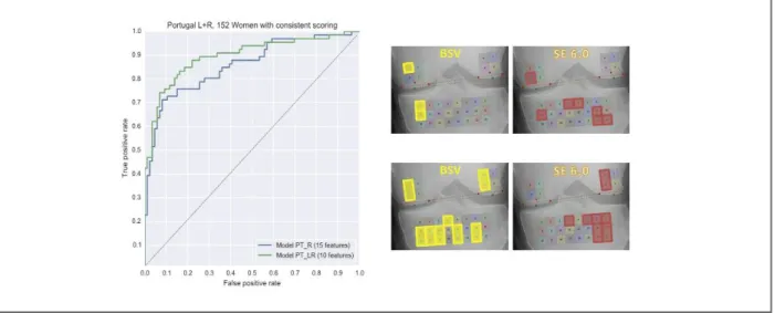
Objective: Texture information of the subchondral bone area of 2D radiographs represents a promising possibility for evaluating the state of osteoarthritis (OA) in addition to traditional clinical means such as visual and semi-quantitative assessments. Algorithms based on fractal analysis have shown to be capable of identi-fying differences in trabecular bone structure. Howe ver such features are likely to vary within the subchondral bone area and therefore the appropriate selection of the region of interest (ROI) plays a crucial role for the re-sult of the analysis. Thus, a feature selection algorithm is being applied in order to determine ROIs that enable an optimum discrimination between patients with and without OA.
Methods: The study included 152 standardized knee radiographs from 66 female patients with OA, and 86 controls. Subchondral bone micro architecture was as-sessed by using both fractal analysis and a Shannon Entropy (SE) algorithm at predefined regions of the pro -ximal tibia and the distal femur. For fractal analysis the distinct parameter Bone Structure Value (BSV) was de-fined. The selected area of the proximal tibia involved a matrix of 3x8 ROIs, whereas a 2x2 matrix was de-fined for each condyle of the distal femur. SE and the BSV were calculated for each of the 32 ROIs,
tively. Based on these 64 variables, a feature selection algorithm was applied to determine the variables that showed the best discrimination power between Case and Control subjects.
Results: By combining the BSV and SE, the odds ratio increased significantly from 3.08 (95% CI: 1.78-5.30) to 14.82 (95% CI: 6.69-32.83) when using 15 features, and to 39.75 (95% CI: 15.41-102.51) based on 10 fea-tures. By using the selected 10 features the accuracy was found to be 0.86. This showed to be a significant improvement compared to the accuracy achieved when calculating a single mean value for the 3x8 ROIs of the proximal tibia alone (0.62 vs. 0.86).
Conclusions: The application of a feature selection al-gorithm in accordance with the combination of the two bone micro architecture analysis methods shows a signi ficant improvement with respect to the discrimi-nation power between subjects with and without OA. The high odds ratios confirm that reliable results can be achieved by combining the BSV and the SE. This novel algorithm for the assessment of bone micro ar-chitecture may not only be useful in osteoarthritis sub-jects but also for the early prediction and assessment of other degenerative bone diseases like osteoporosis and rheumatoid arthritis.
GRUPO 2
P169 – RHEUMA SPACE: STANDARD
PRACTICE AIMING CLINICAL EXCELLENCE IN RHEUMATOLOGY
Carla Macieira1, Luís Cunha-Miranda2,
Alexandre Lourenço3, Anabela Santos4, Joana Sousa5,
Monica Bogas6, Pedro Almeida Laires7,
Pedro Lucas5, Sara Farinha8, JE Fonseca1,
José António Canas da Silva9
1. Rheumatology Department, Hospital Santa Maria, CHLN, Lisbon Academic Medical Center, Lisboa, Portugal
2. Rheumatology Department, Instituto Português de Reumatologia, Lisboa, Portugal
3. Hospital Administrators, Associação Portuguesa de Administradores Hospitalares, Lisboa, Portugal 4. Medical Department, Pfizer Biofarmacêutica, Lisboa, Portugal
5. Healthcare, IMS Healthcare, Lisboa, Portugal 6. Medical Department, Roche Farmacêutica, Amadora, Portugal
7. Outcomes Research, MSD, Lisboa, Portugal 8. Market Access, Abbvie, Lisboa, Portugal
9. Rheumatology Department, Hospital Garcia de Orta, Almada, Portugal
Introduction: The evaluation of the quality of medical practice and the implementation of corresponding measures to improve it are crucial steps for the deve lopment of Rheumatology in Europe. Herein we des -cribe the implementation of a national program, Rheuma Space, aiming at quality improvement in the field of Rheumatology.
Objectives: To develop standards for the quality of care accepted by most of the rheumatology services in Por-tugal.
Methods: Quality indicators were obtained through a four-step RAND-modified Delphi methodology. The first step involved a literature search for international benchmarking of quality of care initiatives and indica-tors, followed by a preselection of a initial set of crite-ria by a task force. The final steps, aiming at defining a smaller set of criteria that could best analyse rheuma-tology quality of care, encompassed an online Delphi round with all Portuguese rheumatologists and a con-sensus meeting with a panel of invited experts repre-senting all the Portuguese Rheumatology Departments of the National Health Service.
Results: A total of 412 different indicators were col-lected throughout the first project phase and the final set of 26 quality indicators was defined, within the three Donabedian dimensions of healthcare quality: nine “structure”, eleven “processes” and seven “out-come” indicators. These criteria cover eleven domains of quality of care: personnel and organizational struc-ture, training and research, facilities, equipment and IT, budgeting and financial resources, access to care, clinical records, patient communication, multidisci-plinary management, clinical outcomes, and patient and personnel satisfaction.
Conclusions: The 26 quality indicators set constitutes the basis for a quality management tool, which is now being implemented in all the Portuguese Rheumatolo-gy Departments of the National Health Service. Direct surveys derived from the 26 quality indicators are be-ing applied to health professionals and patients allow-ing to benchmark departments and to identify strengths and weakness for future improvement. This initiative is deemed to improve the process of care for Portuguese rheumatic patients, thereby ensuring qua -lity standards of structure and process criteria, for a pa-tient oriented clinical practice, favouring desirable con-tinuous quality improvement on health outcomes.
ACkNOWLEDGEMENT
Portuguese Rheumatologists participating in the online Delphi and all the participants in the expert consensus meeting
P38 – PREVALÊNCIA E CORRELATOS DE FIBROSE PULMONAR NUMA COORTE DE MIOPATIAS INFLAMATÓRIAS IDIOPÁTICAS Carlos Costa1, Marília Rodrigues1, Mariana Santiago1,
Pedro David Carvalho1, Alexandra Daniel1,
Gisela Eugénio1, Jorge Silva1, JAP da Silva1,
Armando Malcata1
1. Rheumatology Department, Centro Hospitalar Universitário de Coimbra, Coimbra, Portugal
Introdução: A prevalência do envolvimento pulmonar nos doentes com Miopatias Inflamatórias Idiopáticas (MII) tem sido estimada entre 5-65 %. A alteração mais comum é a doença pulmonar intersticial (DPI), da qual a Fibrose Pulmonar (FP) constitui a fase mais avança-da. O diagnóstico do envolvimento pulmonar é esta-belecido através da tomografia axial computorizada to-rácica de alta resolução (TAC-AR).
Objetivos: O objetivo desde estudo foi avaliar a pre-valência de Fibrose Pulmonar num grupo de doentes com MII seguidos durante os últimos 20 anos no nos-so serviço e apreciar as características clínicas e imu-nológicas que se relacionem com a mesma.
Métodos: Este estudo transversal incluiu pacientes com MII seguidos na consulta de Reumatologia do Centro Hospitalar e Universitário de Coimbra, nos úl-timos 20 anos. O diagnóstico de MII foi efetuado ba-seando-se nos critérios de Bohan e Peter. Foram ex-cluídos os doentes com miopatia tóxica, miopatia ami-loide, distrofia muscular ou neuropatias motoras. As características clínicas da MMI, o envolvimento de ór-gão-alvo, o perfil imunológico e as comorbilidades fo-ram analisadas. As variáveis categóricas são apresenta-das como proporções/percentagens e as variáveis con-tínuas como média ± desvio-padrão. As diferenças sig-nificativas entre os grupos foram estimadas pelos testes de Chi-quadrado e Mann-Whitney, respetivamente. A FP foi definida pela presença na TAC-AR torácica, de espaços aéreos císticos (padrão em “favo de mel”), bronquiectasias de tração, áreas em “vidro despolido” e/ou espessamento dos septos interlobulares envol-vendo mais de 20% do parênquima pulmonar. Na aná-lise multivariada, incluímos todas as variáveis identifi-cadas com valor de p<0,1 na análise univariada. O va-lor de p<0,05 foi considerado como sendo estatistica-mente significativo. Na análise estatística foi utilizada a versão 20.0 de SPSS® para Windows.
Resultados: O grupo de estudo incluiu 40 doentes com MII, dos quais 29 (72,5%) eram mulheres, com uma idade média de 54,1 ± 16,9 anos e com uma du-ração média de doença de 8,2 ± 6,3 meses. Dezoito (45%) dos pacientes apresentavam Dermatomiosite, 13 (32,5%) Polimiosite, 4 (6,2%) síndrome de sobreposi-ção Polimiosite/Esclerose Sistémica, 3 (4,6%) Derma-tomiosite Juvenil e 2 (3,1%) Miopatia por Corpos de In-clusão. Dezanove (47,5%) doentes apresentaram FP. A FP era mais frequente (p<0,1) nos doentes que asentavam rash heliotropo, rash da zona do decote, pre-sença de padrão restritivo nas provas de função respi-ratória (PFR), maior tempo de evolução de doença e valores mais elevados de lactato desidrogenase. Não se detetaram diferenças estatisticamente significativas en-tre o perfil imunológico e a presença de FP. Apenas o tempo de duração da doença manteve relação signifi-cativa (direta) com a presença de FP, após análise de re-gressão logística.
Conclusão: A FP é relativamente frequente nos doen-tes com MII, traduzindo doença mais agressiva. No nosso grupo de doentes com MII, após análise de re-gressão logística, apenas o tempo de duração de doen-ça se relacionou diretamente com a presendoen-ça de FP. P157 – CONSULTA DE ARTRITE INICIAL: VALOR DOS CRITÉRIOS DE REFERENCIAÇÃO
Filipa Farinha1, 2, Gisela Eugénio2,
Mary Lucy Marques2, Diogo Jesus2,
Alexandra Daniel2, Joana Ferreira2,
Marília Rodrigues2, Carlos Costa2,
Pedro David Carvalho2, Armando Malcata2, 3,
JAP da Silva2, 3, Cátia Duarte2, 3
1. Rheumatology Department, Hospital Infante D. Pedro (CHBV), Aveiro, Portugal
2. Rheumatology Department, Centro Hospitalar Universitário de Coimbra, Coimbra, Portugal 3. Faculdade de Medicina, Universidade de Coimbra, Coimbra, Portugal
Introdução: O diagnóstico e tratamento precoces das doenças reumáticas inflamatórias, nomeadamente a Ar-trite Reumatoide, permitem alcançar maiores taxas de remissão, melhorando o prognóstico dos doentes. A identificação e referenciação precoces destes doentes são cruciais neste processo.
O nosso objetivo foi avaliar, para os diversos crité-rios, a concordância entre o médico referenciador e o médico reumatologista. Como objetivo secundário pre-tendeu-se avaliar o valor preditivo positivo e negativo
matologista, sendo esta concordância menor nos crité-rios clínicos do que nos laboratoriais. De acordo com o referenciador, a presença de artrite (VPP=0,91) e os critérios laboratoriais (VPP=0,92-0,93) foram os que apresentaram maior VPP para doença reumática infla-matória (Tabela I).
Discussão e conclusão: Verificou-se uma concordân-cia relativamente baixa entre referenconcordân-ciadores e reuma-tologistas, especialmente no que se refere ao caracter in-flamatório das artralgias e a duração da rigidez matinal. Os critérios clínicos subjetivos apresentaram menor va-lor preditivo positivo para doença reumática inflama-tória.
Estes resultados sugerem a necessidade de reforçar a formação aos médicos que referenciam doentes à con-sulta de Artrite Inicial. Por outro lado, sugerem que os critérios de referenciação poderão ser reequacionados de modo a melhorar a referenciação de doentes com artrite.
P71 – THE USE OF ANALGESIC AND OTHER PAIN RELIEF DRUGS TO MANAGE CHRONIC LOW BACk PAIN – RESULTS FROM A NATIONAL SURVEY
Gouveia N1, 2, Rodrigues AM2, 3, Sofia Ramiro1, 2, 4,
Mónica Eusébio2, Pedro Machado2, 5, Helena Canhão2,3,
Jaime C. Branco1, 2, 6
1. CEDOC, NOVA Medical School, Nova University, Lisboa, Portugal
2. Sociedade Portuguesa de Reumatologia, Lisboa, Portugal 3. Rheumatology Research Unit, Instituto de Medicina Molecular, Faculdade de Medicina da Universidade de Lisboa, Lisboa, Portugal
4. Rheumatology, Leiden University Medical Center, dos vários critérios.
Métodos: Estudo observacional retrospetivo
unicên-trico, no qual se incluíram os doentes observados em primeira consulta de Artrite Inicial entre 24/11/2010 e 13/01/2016, que apresentavam sintomas com menos de 12 meses de evolução. Constituíram-se dois grupos: grupo 1 – com doença reumática inflamatória; grupo 2 – sem doença reumática inflamatória. Através da con-sulta dos processos clínicos e do Registo Nacional de Doentes Reumáticas, Reuma.pt, foram obtidos dados demográficos, proveniência e critérios de referenciação e diagnóstico final. A concordância nos critérios de re-ferenciação, entre o médico que referencia e o médico que avalia o doente na primeira consulta foi determi-nada através do coeficiente Kappa de Cohen, atribuin-do-se significância estatística a valores de p <0,05. Con-siderou-se existir concordância fraca se k<0,40, mo-derada a boa se k=0,41-0,75 e excelente se k>0,76. Para cada um dos critérios, foram calculados os valores pre-ditivos positivo (VPP) e negativo (VPN) para doença reumática inflamatória.
Resultados: Dos 185 doentes observados em consul-ta de Artrite Inicial, foram incluídos 145 (excluídos 40 doentes que apresentavam sintomatologia com >12 meses de evolução), 61% do sexo feminino, com ida-de média ida-de 51±17,6 anos à data da primeira consul-ta. O maior grupo de doentes foi referenciado a partir dos Cuidados de Saúde Primários (49%), seguindo-se o Serviço de Urgência (26,9%) e a consulta externa de outras especialidades (11%). Dos 145 doentes, 123 (84,8%) foram incluídos no grupo 1 e os restantes 22 doentes no grupo 2.
Observou-se baixa concordância entre os critérios referidos pelo referenciador e os objetivados pelo
reu-TABELA I. CONCORDÂNCIA NOS CRITÉRIOS DE REFERENCIAÇÃO, ENTRE O MÉDICO REFERENCIADOR E O MÉDICO REUMATOLOGISTA qUE AVALIA O DOENTE NA PRIMEIRA CONSULTA DE ARTRITE INICIAL; VALORES PREDITIVOS POSITIVOS (VPP) E NEGATIVOS (VPN), DOS VÁRIOS CRITÉRIOS, PARA DOENÇA REUMÁTICA INFLAMATÓRIA
N Ref/ VPP Ref/ VPN Ref/
Critério de referenciação Reum kappa IC 95% p /Reum Reum
Artrite 91/111 0,24 0,07; 0,42 0,05 0,91/0,98 0,27/0,59 Squeeze 17/46 0,13 -0,04; 0,29 0,13 0,88/0,89 0,15/0,26 Artralgias inflamatórias 81/133 0,07 -0,05; 0,19 0,26 0,79/0,85 0,04/0,17 Rigidez matinal > 30' 46/100 0,17 0,06; 0,29 0,01 0,87/0,91 0,15/0,41 Fator Reumatoide 28/41 0,55 0,38; 0,71 0,00 0,93/0,98 0,16/0,20 Velocidade de Sedimentação 60/81 0,34 0,18; 0,50 0,00 0,92/0,95 0,19/0,29 Proteína C Reativa 61/96 0,32 0,17; 0,46 0,00 0,92/0,95 0,20/0,35
gesic/pain relief therapeutic groups (in separate mo -dels), adjusted for confounders.
Results: Among 1,487 subjects with ACLBP, 18.7% were using analgesic/pain relief drugs. Estimated prevalences were: anxiolytics, 14.1%; NSAIDs, 12.3%; antidepres-sants, 10.1%; analgesic antipyretic, 6.6%; anticonvul-sants, 3,4%; central muscle relaxants, 2.6%; opioids, 1.6%. Most subjects with severe pain were in the 1st step of the WHO analgesic ladder: NSAIDs plus anxiolytics, sedatives & hypnotics (4.6%); NSAIDs plus antidepres-sants (3.2%); NSAIDs plus central muscle relaxants (2.5%). The presence of ACLBP was significantly associa -ted with the intake of all therapeutic groups (Table). Conclusions: ACLPB was associated with the intake of drugs belonging to all analgesic drug classes. Ne -vertheless, this drug intake was very low, even for those with severe pain. The WHO analgesic ladder was care-fully followed with an extremely conservative use of analgesic opioids even for those with severe pain. There is clearly an unmet need in what concerns pain ma -nagement in patients with ACLBP, particularly taking into account the well-know burden of LBP.
P231 – VITAMINE D STATUS: A TRANSVERSAL EVALUATION IN RHEUMATIC PATIENTS
Joana Borges1, Susana Fernandes1, Vera Vieira2,
Alexandra Cardoso3, Helena Madeira1,
Maria Jesus Mediavilla1, Rui Leitão1, Cândida Silva1,
Augusto Faustino1
1. Rheumatology Department, Instituto Português de Reumatologia, Lisboa, Portugal
Leiden, Netherlands
5. Centre for Rheumatology Research & MRC Centre for Neuromuscular Diseases, University College London, London, United Kingdom
6. Rheumatology Department, Hospital Egas Moniz (CHLO), Lisboa, Portugal
Background: Managing chronic pain is challenging due to the long-term safety profile of most drugs. The treatment of chronic Low Back Pain (CLBP) represents a significant medical and financial burden and aims at relieving pain, improving functional ability, and pre-venting recurrence and chronicity. It is therefore im-portant to gain insight into how CLBP is managed in the population.
Objectives: To characterize the intake profile of anal-gesic and other pain relief drugs in the Portuguese adult population with CLBP, taking the World Health Orga-nization (WHO) analgesic ladder and pain intensity into account. To assess the relationship between ha -ving CLBP and the intake of analgesic and other pain relief drugs.
Methods: EpiReumaPt was a cross-sectional Por-tuguese population-based study (10,661 subjects). Self-reported active CLBP (ACLBP) was considered: LBP on the day of enrollment and for =90 days. Preva-lence and profile of analgesic intake was characterized among those self-reporting ACLBP, taking into account the intensity of pain and the WHO analgesic ladder. To understand whether the presence of ACLBP was a fac-tor associated with drug intake, multivariable logistic regressions were conducted for each of the
anal-TABLE. ASSOCIATION OF THE PRESENCE OF ACLBP WITH THE INTAkE OF EACH CLASS OF ANALGESIC/PAIN RELIEF DRUGS (SEPARATE MULTIVARIABLE MODELS)
Active CLBP (yes/no)
Dependent variables* OR (95% CI) p-value
Anxiolitics, sedatives & hypnotics§ 8.86 (6.08-12.90) <0.001†
NSAIDs± 8.56 (5.84-12.53) <0.001†
Antidepressants§ 12.56 (7.96-19.81) <0.001†
Analgesics, Antipyretics± 7.68 (3.38-17.48) <0.001†
Anticonvulsants 9.27 (4.16-20.69) <0.001†
Analgesic Opioids 8.13 (2.78-23.77) <0.001†
Central Muscle Relaxants 12.01 (5.88-24.51) <0.001†
*All the analysis were adjusted for: age, gender, geographic area, education level, number of self-reported comorbidities, and self-report of rheumatic and musculoskeletal diseases
§Additionally adjusted for: anxiety and depressive symptoms; ±Additionally adjusted for: heart disease and gastrointestinal diseases.
2. Centro Hospitalar de Leiria, Leiria, Portugal 3. Nutrition Department, Instituto Português de Reumatologia, Lisboa, Portugal
Background: Vitamine D deficiency is a condition re-ported both in young adults and in elderly and institutionalized patients. It has been increasingly reco -gnized and, although conflicting data subsists, hy-povitaminosis D has been related to a variety of rheumatic conditions from osteoporosis (OP) to in-flammatory di seases and generalized pain syndromes. Objectives: To assess vitamine D status in a rheuma tic outpatient setting.
Methods: Observational, transversal, retrospective study encompassing rheumatic outpatients with at least one 25-hidroxyvitamin D determination since 2014. Data ascertained included gender, age, parathyroid hor-mone (PTH) and vitamin D levels, rheumatic condi-tions (<3/patient), co morbidities and therapeutics. Bone mineral density was assessed by densitometry (DXA) and results classified as normal (Tscore >-1), os-teopenia (-2.5 < Tscore <-1) and osteoporosis (T score <-2.5). Statistics: Mann-Whitney, Kruskal-Wallis, ANOVA and Chi-Squared tests and Pearson’s correla-tion; p<0.05. Software: SPSS 17.
Results: 370 patients included, 87.3% female, mean age 64.9 (SD 13.9 years), 151 patients <65years (y). Most prevalent rheumatic conditions: osteoarthritis (OA; 47.8%), OP (43.5% - 33.5% of which with re-ported fractures), fibromyalgia (FM; 14.6%) and rheumatoid arthritis (RA; 13.2%); 43.9% patients had inflammatory diseases. Most common co morbidities: arterial hypertension (38.6%), dyslipidemia (32.4%) and depression (22.9%). 38.4% were on synthetic Di -sease Modifying Anti-Rheumatic Drugs (DMARD); 34.1% on corticosteroids (CTC), 49.2% on bisphos-phonates (BF) and 69.5% on calcium/vitamin D (Ca/D) supplements. As for patients with available DXA, 41.4% were classified as having osteopenia and 43.6% as having osteoporosis. Mean vitamin D was 29.7 (SD 24.5 g/L); 65.4% patients had vitamin D levels <30 g/L. Mean PTH was 56.6 (SD 31.3ng/L) and alkaline phos-phatase (ALP) was 73.8 (SD 31.8U/L). In the group >65y, mean PTH was higher than in the <65y group (61.3+/-33.6 vs 49.7+/-26.1, p=0.015); 70.9% of the patients <65y and 61.6% of the patients >65y had vi-tamin D<30 – not significant. We compared vivi-tamin D in OP vs OP with fractures vs OA vs associated OP/OA and found lower vitamin D levels in patients with iso-lated OA (38.7+/-32.5 vs 38.1+/-33.9 vs 25.9+/-23.3 vs
30.6+/-20.7, p=0.012); 76.6% with isolated OA had vitamin D<30. 55.7% of the patients on BF >5y, 61.2% on BF <5y and 72.3% without BF had vitamin D<30 (p=0.013). 68.7% in the group with inflammatory con-ditions and 62.9% in the group with non-inflammato-ry conditions had vitamin D <30 – not significant. We further compared vitamin D levels in OA vs AR, AR vs FM and OA vs FM, DMARD and CTC use and Ca/D supplementation and found no difference. There was a positive correlation between age and vitamin D (r=0.111, p=0.033), age and PTH (r=0.208, p=0.010) and vitamin D and ALP levels (r=0.311, p<0.0001). Conclusions: In this study, vitamin D levels <30 g/L were found in nearly 2/3 of patients, across disease groups. There was a tendency for higher vitamin D le vels in elderly patients, in the group with OP compa -ring with OA and in the group on BF, which may be re-lated to previous supplementation. A potential bias is the fact rheumatologists request vitamin D dosing in patients with risk factors/clinical features of hypovita-minosis.
P225 – ENVOLVIMENTO CARDÍACO NAS DOENÇAS REUMÁTICAS – DOIS CASOS CLÍNICOS
Lídia Teixeira1, Inês Cruz2, Raquel Roque1, Luís Lopes2,
Filipe Vinagre1, José António Canas da Silva1 1. Rheumatology Department, Hospital Garcia de Orta, Almada, Portugal
2. Serviço de Cardiologia, Hospital Garcia de Orta, Almada, Portugal
Introdução: O envolvimento cardíaco é uma causa sig-nificativa de morbilidade e mortalidade nas doenças reumáticas. Pode surgir em doentes com diagnóstico de doença reumática conhecida ou ser a primeira ma-nifestação clínica. O envolvimento pode ser do mio-cárdico, do pericárdio, valvular ou do sistema de con-dução e cursar com aterosclerose precoce, frequente nas doenças reumáticas, quer pelo próprio processo in-flamatório, quer pelas terapêuticas. A evolução dos meios de diagnóstico associado ao reconhecimento clí-nico mais frequente tem permitido a detecção atempa-da atempa-da doença cardíaca, terapêutica dirigiatempa-da e prevenção de complicações.
Objectivo: Descrição de dois casos clínicos de doentes com envolvimento cardíaco no contexto de doenças reumáticas.
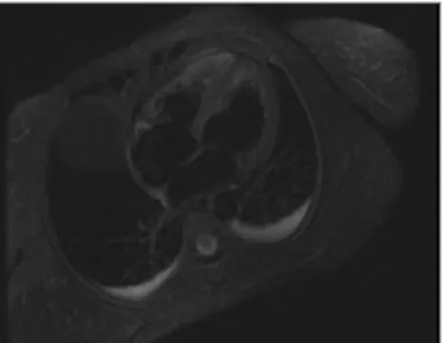
Caso Clínico 1: Mulher de 42 anos internada por quadro de febre, aftas orais recorrentes, lesões de pseudofo
-liculite e duas extensas lesões de pioderma gangreno-so cuja investigação conduziu ao diagnóstico de doen-ça de Behçet. Ao 8º dia de internamento desenvolve quadro de precordialgia associado a instabilidade he-modinâmica. O electrocardiograma não revela altera-ções dinâmicas de ST-T sugestivas de isquemia aguda, mas laboratorialmente havia uma elevação dos marca-dores de necrose miocárdica (Troponina Ths 450>> 778>>1520) e ecograficamente um ligeiro derrame pe-ricárdio, sem alterações da cinética segmentar. Peran-te esPeran-tes achados, foi assumido o diagnóstico de peri-miocardite, posteriormente confirmado em ressonân-cia magnética (fig1). Com ajustamento da imunossu-pressão (ciclosporina 3mg/Kg e prednisolona 1mg/Kg), houve melhoria clínica e analítica. Actualmente, 2 anos após o diagnóstico, a doente apresenta queixas de can-saço inespecífico e alterações ecocardiográficas – ven-trículo esquerdo com hipocinésia difusa das paredes e com função sistólica global diminuída (fracção de ejec-ção Simpson biplano de 49%), apesar de apresentar re-dução das áreas de edema na ressonância magnética (traduz menor atividade da doença inflamatória). Caso Clínico 2: Mulher de 38 anos, hipertensa, com lúpus eritematoso sistémico (envolvimento mucocutâ-neo e hematológico) e síndrome dos anticorpos anti-fosfolípidos secundário, é internada por quadro de ce-faleias e cansaço para médios esforços. Durante a mar-cha diagnóstica é pedido ecocardiograma transto rácico que mostrou uma hipocinésia difusa do ventrículo es-querdo com uma fracção de ejecção de 40%, de novo (em 2013 tinha um ecocardiograma normal). Analiti-camente apresentava valores normais de biomarcado-res cardíacos e de NT ProBNP, bem como um eletro-cardiograma normal. Iniciou como terapêutica um diu-rético, IECA e beta-bloqueante e aumentou-se dose de
prednisolona para 20mg/dia, sem melhoria significati-va do cansaço para médios esforços, pelo que, e para exclusão de doença coronária, fez coronariografia que não mostrou lesões significativas. Embora a doente es-teja a aguardar a realização de ressonância magnética cardíaca, a hipótese de diagnóstico mais provável é a de envolvimento cardíaco secundário à doença reumática de base.
Conclusão: O envolvimento cardíaco nas doenças reu-máticas pode ser consequência da própria doença, da terapêutica ou de comorbilidades associadas. A expo-sição destes casos serve para relembrar que o envolvi-mento cardíaco pode surgir mesmo em doentes jovens, ter um curso mais agudo ou mais indolente e que do ponto de vista clínico podem ter sintomatologia mais florida ou mais frustre. O seu reconhecimento precoce e tratamento dirigido são fundamentais para permitir a recuperação funcional, prevenção de complicações e diminuição da mortalidade.
P234 – ECOGRAFIA NA SÍNDROME DO CANAL CÁRPICO
Lídia Teixeira1, Sandra Sousa1, Filipe Vinagre1,
Pedro Gonçalves1, José António Canas da Silva1 1. Rheumatology Department, Hospital Garcia de Orta, Almada, Portugal
Introdução: A síndrome do canal cárpico (SCC) é a neuropatia compressiva mais comum, atingindo cerca de 2,7% da população geral1. O seu diagnóstico é
ba-seado na história clínica, exame objectivo e estudo ele-trofisiológico1,2. Contudo a ecografia, com medição área
da secção transversal do nervo mediano a nível do tú-nel do carpo, tem vindo a ganhar importância como exame para diagnóstico desta síndrome. Em estudos recentes o uso da ecografia como ferramenta para dia gnóstico, mostrou ter uma sensibilidade e especificida -de que se aproximam -de estudos -de eletrofisiológicos3,4. Objectivo: Caracterização dos doentes com síndrome do canal cárpico referenciados à consulta de Reumato-logia e analisar o papel da ecografia no seu diagnóstico. Métodos: Estudo retrospectivo que incluiu doentes re-ferenciados à consulta de Reumatologia por síndrome do canal cárpico submetidos a ecografia com medição da área transversal do nervo mediano. Foram regista-dos daregista-dos clínicos, informações de ecografias do punho realizadas no nosso centro por dois operadores inde-pendentes, tendo-se estabelecido como aumento pato-lógico do nervo mediano uma área igual ou acima de 10mm2 e os resultados do estudo eletrofisiológico FIGURE 1.Imagem RM - edema do miocárdio nos segmentos
(quando realizado e disponível).
Resultados: Incluídos 58 doentes, dos quais 54
(93,1%) apresentavam parestesias no território do ner-vo mediano,18 (31%) queixas álgicas no punho e 11 (19%) diminuição da força muscular, 15 (25.9%) ti-nham um teste de phalen e de tinel positivos, sendo que, 19 (32.8%) tinham qualquer um dos testes posi-tivos. Apenas 8.6% tinham atrofia da eminência tenar. De todas as ecografias, 89.7% apresentava uma área entre 10 e 19 mm2, e 10.3% com pelo menos um ner-vo mediano com 20 ou mais mm2 de área. De todos os doentes apenas 11 realizaram estudo electrofisiológico e apenas numa situação houve discordância entre o achado deste exame e a ecografia, havendo portanto uma concordância em 90,9% dos casos.
Comparando os sintomas com os achados ecográfi-cos, 98,2% dos doentes com parestesias tinham eco-grafia positiva.
Discussão/Conclusão: Embora os nossos resultados tenham por base uma pequena amostra, é possível ver a correlação entre a sintomatologia mais frequente da SCC (as parestesias seguindo trajecto do nervo media-no) e o resultado de ecografia., mais de 90% apresen-tavam ecografia positiva. Houve também na maioria dos casos uma concordância nos achados do estudo eletrofisiológico (ainda o gold standard para o diagnós -tico da SCC) e os achados da ecografia, o que demons-tra a importância da ecografia, nomeadamente como um dos exames que pode substituir o estudo eletrofi-siológico no diagnóstico do SCC. São necessários es-tudos para determinar a correlação entre os achados electrofisiológicos e a ecografia
P160 – YEARS OF WORkING LIFE LOST CAUSED BY OSTEOARTHRITIS IN PORTUGAL – ANALYSIS FROM THE EPIREUMAPT STUDY
Pedro A Laires1,2, Miguel Gouveia3, Helena Canhão1,4,
Rodrigues AM1,4,5, Gouveia N1,5, Mónica Eusébio1,
Jaime C Branco1, 5, 6
1. Sociedade Portuguesa de Reumatologia, Lisboa, Portugal
2. Centro Académico de Medicina de Lisboa, Lisboa, Portugal 3. Catolica Lisbon School of Business, Lisboa, Portugal 4. Rheumatology Research Unit, Instituto de Medicina Molecular, Faculdade de Medicina da Universidade de Lisboa, Lisboa, Portugal
5. CEDOC, NOVA Medical School, Nova University, Lisboa, Portugal, 6Rheumatology Department, Hospital Egas Moniz (CHLO), Lisboa, Portugal
Objectives: The aim of this study was to calculate the years of working life lost (YWLL) from early exit from work associated with Osteoarthritis (OA).
Methods: We used data from the population-based EpiReumaPt study (Sep2011-Dec2013). 10,661 inhabitants were surveyed to capture all cases of RD withi -n a represe-ntative sample of the Portuguese po pulatio-n. We analyzed all aged 50-65 years old (yo), near the of-ficial retirement age. YWLL were determined for OA cases with premature exit from work caused by RD (self-reported) estimated as the difference between each par-ticipant’s age and the respective age (“observed stock”), while the potential YWLL (PYWLL: YWLL+“expected stock” of YWLL still to occur if there is no return-to-work) was the difference between official and actual re-tirement ages. We also calculated the percentage of time in inactivity (inactivity ratio=YWLL/Active age--range[15-65yo]). We excluded all OA participants with concomitant inflammatory rheumatic diseases with pos-sible impact on retirement, such as rheumatoid arthri-tis and spondylarthriarthri-tis. All cases with retirement prior to onset of OA symptoms were also excluded. All re-sults were based on weighted data.
Results: The estimated prevalence of OA in the Por-tuguese population (50-64 yo) is 29.7% (knee:18.6%; hand:12.6%; hip:3.6%). Among these, 61.8% were out of work versus 47.6% for those without OA (p=0.004), which potentially led to 162,735 YWLL (95 per 1000 inhabitants). Early retirement contributed the most for these YWLL (58%; 94,432 YWLL), followed by un-employment (35%; 57,209 YWLL) and disability pen-sions (7%; 11,094 YWLL). Women accounted for 80% of these YWLL (153 per 1000 female inhabitants). A to-tal of 161,621 PYWLL were estimated if early retire-ment is considered and 369,839 PYWLL for all forms of exit from work. The mean YWLL and PYWLL inac-tivity ratios were 16% and 30%, respectively.
Conclusions: We estimated a considerable amount of working life years losses associated with OA in Portu-gal.
P45 – BIÓPSIA DE GLÂNDULAS SALIVARES MINOR – SETE ANOS DE EXPERIÊNCIA DE UM SERVIÇO DE REUMATOLOGIA
Sofia Silvério Serra1, Teresa Pedrosa1, Tiago Costa1,
João Lagoas Gomes1, Carina Lopes1, Inês Silva1,
Walter Castelão1, Jaime C. Branco1, 2
1. Rheumatology Department, Hospital Egas Moniz (CHLO), Lisboa, Portugal
2. CEDOC, NOVA Medical School, Nova University, Lisboa, Portugal
Objectivos: A síndrome de Sjögren (SS) é uma doença reumática sistémica cujos critérios de classificação in-cluem achados histopatológicos típicos na biópsia de glândulas salivares minor (BGSM) do lábio. O objectivo do trabalho foi fazer uma revisão destas biópsias realiza-das no serviço de Reumatologia nos últimos sete anos, e concluir sobre a sua importância para o diagnóstico da SS. Métodos: Foram revistos os registos clínicos dos doen-tes que foram submetidos a BGSM pelos reumatologistas do nosso serviço entre Janeiro 2009 e Dezembro de 2015. Resultados: Realizaram-se 100 BGSM no lábio infe-rior num total de 100 doentes sob anestesia local no bloco de cirurgia de ambulatório. Não houve registo de intercorrências e obtiveram-se em média 3,3 peças de biópsia. Noventa doentes eram do sexo feminino, com uma média de idades de 54,5 ± 13,5 anos. O tem-po de seguimento médio em consulta de Reumatologia até à data da técnica foi de 11,5 meses. Oitenta e seis por cento dos doentes não tinham diagnóstico prévio, 4% tinham artrite reumatóide e 3% lúpus eritematoso sistémico. Em 90% dos doentes a indicação para bió-psia foram os sintomas secos: 83% apresentavam xe-rostomia e 68% xeroftalmia (11 com Keratoconjunctivitis sicca). Dezoito doentes (18%) apresentaram cin -tigrafia de glândulas salivares positiva. Nove doentes (9%) apresentaram achados histopatológicos de SS (fi-brose do estroma, atrofia glandular e infiltrado infla-matório crónico focal). Nos restantes doentes a histo-patologia foi inconclusiva: 41 com processo inflama-tório inespecífico, 1 com presença de substância ami-lóide, 11 amostras inadequadas ou insuficientes para análise do tecido glandular e 38 sem alterações. Do es-tudo imunológico salientam-se: 34 (34%) doentes com anti-SSA e/ou anti-SSB positivos, 10 com factor reu-matóide positivo (10%) e 65 com anticorpos anti-nu-cleares positivos (65%; 45 dos quais com título =1/320 com maior prevalência do padrão mosqueado). O diag-nóstico de SS primário foi feito em 21 doentes (21%, 13 destes cumpriam critérios de classificação do Revi-sed American-European Consensus Group- AECG e/ou do American College of Rheumatology- ACR), e o de SS se-cundário em 6 doentes (6%, 2 destes cumpriam crité-rios de classificação AECG e/ou ACR).
Conclusões: A BGSM é uma técnica segura que apre-sentou uma rentabilidade de 89%. A identificação do padrão histológico de SS pela BGSM contribuiu nesta amostra para 8% dos diagnósticos de SS.
P111 – HAEMOPHILUS INFLUENZAE SEPTIC ARTHRITIS IN THE ERA OF IMMUNIZATION
Vítor Teixeira1, Catarina Resende1, Luís Lito2,
José Carlos Romeu1
1. Rheumatology Department, Hospital de Santa Maria (CHLN), Lisbon Medical and Academic Centre, Lisboa, Portugal
2. Clinical Pathology Department, Hospital de Santa Maria (CHLN), Lisbon Medical and Academic Centre, Lisboa, Portugal
Introduction: Haemophilus influenzae (Hi) is a
gram-negative coccobacilli. Strains of Hi that possess a polysac-charide capsule are serotyped into 6 different types (a-f) based on their antigenically distinct capsules. Some Hi strains however have no capsule and are termed nonencapsulated or nontypeable. Hi is an un-common cause of bacterial arthritis in adults. We report what we believe to be the first Portuguese case of sep-tic arthritis with isolation of non-capsulated Hi in the synovial fluid.
Clinical Case: A 39 years old healthy man was admit-ted in our department with fever, night sweats and nine days duration left knee pain with swelling. At physical examination there was tenderness at knee palpation with joint effusion and limited motion, with a <20º arch of flexion. Left knee X-rays detected a soft tissue swelling, a marked reduction of the lateral compart-ment and a bipartite patella. The left knee ultrasound revealed extensive synovial proliferation in the sub-quadricipital recess with a small amount of synovial ef-fusion. Lab tests presented leukocytosis (16.55x 10^9/L) and neutrophilia (74.9%), reactive thrombo-cytosis (626 x10^9/L) and elevated C-reactive protein (32.2 mg/dl). The synovial fluid was purulent.
He was treated empirically for septic arthritis with flucloxacilin and ceftriaxone and submitted to closed joint lavage and physiotherapy. At the 6th day of hos-pitalization, a Hi ampicillin resistant (ß-lactamase producer) was isolated in the synovial fluid. Antibiothe -rapy was switched to amoxicillin and clavulanic acid (2.2 g 8/8h) according to the antibiogram. Blood cultures were sterile. Because of persistent synovitis, li -mited recovery of joint flexion and delay of the decline of acute phase reactants, he was proposed for arthro-scopic lavage. He completed a total of 3 weeks of en-dovenous and 3 weeks of oral amoxicillin and acid clavulanic with normalization of acute phase reactants and symptoms improvement. Synovial fluid was sent
to the National Institute of Health Ricardo Jorge for ty -ping tests, which revealed to be a nonencapsulated strain of Hi.
Discussion: Nonencapsulated Hi is responsible for most invasive Hi infections across all age groups in countries where immunization programmes include Hi serotype b (Hib) vaccine (Gkentzi D, 2012). In Portu-gal, Hib conjugate vaccination was included in the Na-tional Immunization Program in the year 2000. Data from the Portuguese Group for Study of Haemophilus influenzae Invasive Infection obtained between 2002 and 2010 informed that 77.1% isolates were nonca -psulated, representing a significant increase compared to the pre-vaccination era (19.0%), and this was asso-ciated with a large decrease in serotype b strains (from 81.0 to 13.2%) (Bajanca-Lavado MP, 2014). However, none of the isolates sent to the Portuguese National Ins -titute of Health Ricardo Jorge between the years 1989 and 2010 (Bajanca-Lavado MP, 2014; Bajanca P, 2004), were obtained from the synovial fluid, making our case likely the first case report in Portugal. Those data also revealed that resistance was associated to non-capsu-lated Hi isolates when compared with serotypeable iso-lates, making the frequently applied empirical treat-ment for septic arthritis ineffective against ß-lactamase-producing Hi.
GRUPO 3
P142 – COMBINATION THERAPY OF MYCOPHENOLATE MOFETIL AND CYCLOSPORINE A FOR INDUCTION TREATMENT OF REFRACTORY LUPUS NEPHRITIS: A CASE SERIES
Diogo Jesus1, Marília Rodrigues1, JAP da Silva1, Ines L1 1. Rheumatology Department, Centro Hospitalar
Universitário de Coimbra, Coimbra, Portugal
Background: Cases of resistance of lupus nephritis (LN) to induction treatment with mycophenolate mofetil (MMF) or cyclophosphamide (CYC) regimens are common and difficult to manage. There is lack of evidence for alternative rescue therapy. Combination of MMF and calcineurin inhibitors was suggested but studies on this strategy are scarce.
Objective: To report efficacy and tolerability of com-bined immunosuppression with MMF and cy-closporine A (CsA) for induction treatment of LN who failed to respond to standard induction therapy (CYC and/or MMF).
Methods: We report efficacy and tolerability on all pa-tients with biopsy-proven LN (class III/IV/V of ISN/RPS 2003 classification) from a single tertiary Lupus Cli nic that since 2012 received combined MMF+CsA induction treatment after failure of at least one standard re -gimen (CYC and/or MMF) for 6 months.
Results: Six patients with lupus nephritis (100% female, 100% Caucasian, age 2352 years) were evalua -ted. The LN was class IV in 3, class V in 2 and class III in 1. Four patients first received i.v. pulse CYC and all 6 received MMF as first or second induction regimen in association with ACE inhibitor or ARB and predni -sone. After failure of achieving response, CsA was added to MMF. Mean proteinuria was 2407 mg/24h and serum creatinine was 0.53 mg/dl at start of MMF+CsA regimen. Daily dose of MMF was 2-3 g and CsA was dosed up to 2.63.7mg/kg/day. Mean predni -sone dosage was 17.5 mg/day and 6 mg/day at baseline and after 6 months of MMF+CsA, respectively. Mean proteinuria at 3 and 6 months of MMF+CsA was 1484 mg/day and 544 mg/day, respectively. Mean serum crea tinine at 6 months was 0.68 mg/dl. Four patients achieved complete renal remission, one patient had a 48.8% reduction in proteinuria and one failed to res -pond. The mean time to remission was 5 months. One patient presented hirsutism and another stopped CsA after 36 months due to nausea and vomiting. No patients presented raised blood pressure or serum creatinine with CsA or other adverse events with this regimen. Conclusion: Combination of MMF and CsA was ef-fective and well tolerated as a rescue induction treatment of class III/IV/V LN after failure of standard thera -py with MMF and/or CYC in this small series.
P61 – CONTRIBUTION OF ULTRASONOGRAPHY TO ASSESS DISEASE ACTIVITY IN PATIENTS WITH INFLAMMATORY MYOPATHIES
Joana Sousa Neves1, Daniela Santos Faria1,
Marcos Cerqueira1, José A. Costa1, Carmo Afonso1,
Filipa Teixeira1
1. Rheumatology Department, Hospital Conde de Bertiandos (ULSAM), Ponte de Lima, Portugal
Background: Inflammatory myopathies (IM) comprise a group of rare and heterogeneous diseases, challen -ging to diagnose, treat and follow-up. Monitoring pa-tients with IM is difficult as there is a lack of precise measures for assessing disease activity. Ultrasonogra-phy (US) is a non-invasive, relatively inexpensive tech-nique that is increasingly being used to evaluate
mus-cular system.
Objective: To assess the utility of US evaluation in the
monitoring of patients with IM.
Methods: Patients diagnosed of IM from 2005 to 2015 followed up at a Rheumatology Department were in-cluded. Fourteen patients (11 female) with a mean age of 50.1 ± 21.4 years and mean disease duration of 4.8 ± 3.2 years were evaluated. The different diagnosis were as follows: 6 dermatomyositis (DM), 3 polymyositis (PM), 2 myositis associated with Systemic Lupus Ery-thematosus, 1 inclusion body myositis (IBM), 1 recur-rent focal myositis (RFM) and 1 undifferecur-rentiated IM. Physical examination, including muscle strength tests, laboratorial tests (comprising levels of serum muscle enzymes) and a selective muscle US assessment were performed to each patient. For US evaluation a Gene ral Electric LOGIQ S8 equipment with a 615 MHz li -near transducer was used. Muscles were subjectively evaluated for echogenicity (determined by gray-scale) and muscle morphology. Power Doppler (PD) was used to assess vascularity.
Results: Nine of the 14 patients evaluated were on clini cal remission, with absence of symptoms or signs of disease, preserved muscle strength, normal muscle enzyme levels and conserved muscle pattern on US as-sessment. One female with DM presented with an acute exacerbation of disease, with proximal muscular weak-ness of the upper limb and US findings of increased muscle echogenicity of biceps revealing a muscle oede-ma pattern. The patient with IBM also experienced a flare which was associated with proximal muscle weak-ness and asthenia; US assessment showed focal oedema through right biceps muscle but also proxioedemal lo -wer limb muscle atrophy characterized by increased echogenicity with diminished volume of the quadri-ceps compartment and change of normal architecture of tibialis anterior muscle of both legs. These findings are consistent with a long term disease with acute flare. Another patient with longstanding PM in remission re-vealed no signs or symptoms of active disease, normal muscle strength but low creatine phosphokinase levels and symmetrical proximal muscle atrophy pattern in US evaluation. On the other hand, the patient with shorter duration of disease, recently diagnosed as myositis (undifferentiated IM), revealed proximal weak-ness and elevated serum muscle enzymes; in this case, US assessment showed diffuse increased echogenicity of proximal muscle compartment and increased PD sig-nal. Finally, the patient with RFM presented with left arm swelling and weakness and US evaluation showed
enlargement and diffuse increased echogenicity within the left carpal extensor compartment.
Conclusion: US findings in patients with IM seem to correlate well with disease activity suggested by clinical and laboratory data. As a result, US imaging may be a useful tool to assess disease activity and damage in these patients as well as to monitor disease progression.
REFERENCES
Ronald S. Adler and Giovanna Garofalo. Ultrasound in the evalua-tion of the inflammatory myopathies. Curr Rheumatol reports. 2009. 11:302-308.
P49 – FROM BURSITIS TO FASCIITIS Magda Varelas Alves1, Pedro Campos1,
Naida Gonçalves1, Gloria Magalhaes1
1. General Surgery, Hospital Vila Franca de Xira, Vila Franca de Xira, Portugal
Introduction: We report a case of olecraneon bursitis in an immunocompromized patient which evolved to extensive cellulitis, fasceitits and an upper limb com-partment syndrome. Haematogenous infection was due to S. Pyogens, a very uncommom pathogen. Antibio -tics, fasciotomies, negative pressure dressings and skin flaps were required to treat this severe lesion.
Olecranon bursitis a pathology accounting for 0.01--0.1% of overall hospital admissions and is more fre-quent in males aged 30-60. It can be classified as acute, chronic and acute on chronic, either septic or nonsep-tic. Post-traumatic etiology is the most frequent.
Rheumatoid arthritis seems to predispose to non-septic olecranon bursitis, nevertheless the real cause-ef-fect relation is not yet established.
Objective: Alert to an unusual and serious complica-tion of olecranon bursitis.
Results: We report a patient admitted with a non-trau-matic bursitis which revealed to be infected without apparent point of departure. Regardless empiric an-tibiotherapy for 3 days, it became an upper limb necro-tizing fasciitis and forearm compartment syndrome. Multiple revision surgeries were performed each 24--48h negative pressure dressings in between. A skin graft was applied 16 days after the first procedure, with closure achieved. He maintained the functional capaci -ty and limb sensitivi-ty in few days post-operatively. Discussion/Conclusion: The development of septical bursitis depends on several predisposing factors, such as history of trauma, immunocompromised state, Dia-betes and Rheumatoid Arthritis. It is usually associa ted with reduced morbidity, however, in rare cases,