FACULDADE DE CIÊNCIAS
DEPARTAMENTO DE BIOLOGIA VEGETAL
Characterization of Argonaute-related small RNA
pathways in Caenorhabditis elegans.
Pedro Jorge de Oliveira Rodrigues Batista
DOUTORAMENTO EM BIOLOGIA
(Genética)
FACULDADE DE CIÊNCIAS
DEPARTAMENTO DE BIOLOGIA VEGETAL
Characterization of Argonaute-related small RNA
pathways in Caenorhabditis elegans.
Tese Orientada por:
Professor Doutor Rui Gomes e
Professor Doutor Craig C. Mello
Pedro Jorge de Oliveira Rodrigues Batista
DOUTORAMENTO EM BIOLOGIA
(Genética)
artigos científicos já publicados. Uma vez que o trabalho publicado nos referidos artigos foi realizado em colaboração com outros investigadores, e de acordo com o disposto no n°1 do Artigo 41° do Regulamento de Estudos Pós-Graduados da Universidade de Lisboa, publicado in Diário da Republica 2a série – N.o 209 – 30 de Outubro de 2006, esclareço que participei integralmente na concepção e execução do trabalho experimental, na intrepretação dos resultados e na redacção dos manuscritos.
Os trabalhos apresentados nesta tese foram realizados com o apoio financeiro da Fundação para a Ciência e Tecnologia (bolsa de referencia SFRH/BD/11803/2003), NIH grant GM58800 e Howard Hughes Medical Institute.
In this thesis I have included scientific articles already published as chapters. Because the data published in the articles was generated in collaboration with other researchers, I declare, in accordance with ‘disposto no n°1 do Artigo 41° do Regulamento de Estudos Pós-Graduados da Universidade de Lisboa, publicado in Diário da Republica 2a série – N.o 209 – 30 de Outubro de 2006’ that I have participated in the design and execution of the experimental work, in the analysis of the results and writing of the manuscripts.
The work presented in this thesis was funded by Fundação para a Ciência e Tecnologia (SFRH/BD/11803/2003), NIH grant GM58800 and the Howard Hughes Medical Institute.
Capítulo II
Yigit, E.*, Batista, P.J.*, Bei, Y., Pang, K.M., Chen, C.C., Tolia, N.H., Joshua-Tor,L., Mitani, S., Simard, M.J., Mello, C.C (2006). Analysis of the C. elegans Argonaute family reveals that distinct Argonautes act sequentially during RNAi. Cell 17;127(4):747-57.
*These authors contributed equally.
Capítulo III
Batista, P. J. *, Ruby, J. G. *, Claycomb, J. M., Chiang, R., Fahlgren, N., Kasschau, K. D., Chaves, D. A., Gu, W., Vasale, J. J., Duan, S., et al. (2008). PRG-1 and 21U-RNAs interact to form the piRNA complex required for fertility in C. elegans. Mol Cell 31, 67-78.
*These authors contributed equally.
Capítulo IV
Claycomb, J. M.*, Batista, P. J.*, Pang, K. M., Gu, W., Vasale, J. J., van Wolfswinkel, J. C., Chaves, D. A., Shirayama, M., Mitani, S., Ketting, R. F., Conte, D. J., and Mello, C. C. (2009). The Argonaute CSR-1 and its 22G-RNA cofactors are required for holocentric chromosome segregation. Cell 139, 123-134.
Capítulo II
Yigit, E.*, Batista, P.J.*, Bei, Y., Pang, K.M., Chen, C.C., Tolia, N.H., Joshua-Tor,L., Mitani, S., Simard, M.J., Mello, C.C (2006). Analysis of the C. elegans Argonaute family reveals that distinct Argonautes act sequentially during RNAi. Cell 127, 747-757.
*These authors contributed equally.
Capítulo III
Batista, P. J. *, Ruby, J. G. *, Claycomb, J. M., Chiang, R., Fahlgren, N., Kasschau, K. D., Chaves, D. A., Gu, W., Vasale, J. J., Duan, S., et al. (2008). PRG-1 and 21U-RNAs interact to form the piRNA complex required for fertility in C. elegans. Mol Cell 31, 67-78.
*These authors contributed equally.
Capítulo IV
Claycomb, J. M.*, Batista, P. J.*, Pang, K. M., Gu, W., Vasale, J. J., van Wolfswinkel, J. C., Chaves, D. A., Shirayama, M., Mitani, S., Ketting, R. F., Conte, D. J., and Mello, C. C. (2009). The Argonaute CSR-1 and its 22G-RNA cofactors are required for holocentric chromosome segregation. Cell 139, 123-134.
Acknowledgments i
List of figures iii
List of tables vi
Abbreviations vii
Resumo ix
Abstract xvii
CHAPTER I: GENERAL INTRODUCTION 1
Caenorhabditis elegans as a model organism 3
From anti-sense to RNAi 7
RDE-1, the argonaute link 10
The RNA-induced silencing complex. 11
The microRNAs 15
Biogenesis of miRNA in animals and plants 16
The PIWI interacting small RNAs 19
Biogenesis of piRNAs 20
The Ping-Pong cycle 21
Piwi dependent, Aub- and Ago3-independent pathways 23
Function of PIWI in the nucleus 24
Endogenous small RNAs 25
The RNA interference pathway in C. elegans 31
The C. elegans Endogenous siRNA pathway 36
The Germ granules of C. elegans 39
Summary of thesis 43
that distinct Argonautes Act Sequentially During RNAi. 69
Summary 71
Introduction 73
Results 77
RDE-1 interacts with trigger-derived single-stranded RNA 77
RDE-1 does not interact with secondary siRNAs 80
Genetic analysis of AGO mutants in C. elegans 81
Multiple AGOs contribute incrementally to RNAi 84
AGOs required for RNAi exhibit qualitatively distinct activities 85
SAGO-1 and SAGO-2 interact with Secondary siRNAs 87
An endogenous small RNA pathway requires ERGO-1 and the SAGO
proteins 89
Discussion 91
Intersecting RNAi pathways in C. elegans 95
AGOs and transcriptional gene silencing 97
Experimental Procedures 99
Acknoledgments 103
References 105
Supplemetal Information 111
CHAPTER III: PRG-1 and 21U-RNAs interact to form the piRNA
complex required for fertility in C. elegans. 119
Summary 121
Introduction 123
21U-RNAs are expressed in the C. elegans germline 127 PRG-1 is expressed in the germline and required for 21U-RNA
accumulation 129
21U-RNAs depend on and interact physically with PRG-1 132
prg-1 mutants exhibit a broad spectrum of germ-line defects 134
prg-1 mutants exhibit surprisingly subtle changes in gene expression 137
Discussion 141
piRNAs in worms, flies and mammals 141
Piwi-AGO complexes exhibit a conserved localization in germ-line nuage
143
A potential role for 21U-RNAs in Tc3 silencing 144
A conserved function for piRNA complexes in maintaining pluripotency 145 Experimental Procedures 149 Acknoledgments 153 Accession Numbers 153 References 155 Supplemental Information 161
CHAPTER IV: The Argonaute CSR-1 and its 22G-RNA co-factors target germline genes and are required for holocentric chromosome segregation. 175
Summary 177
Introduction 179
Results 183
and alignment of metaphase chromosomes 186 Expression studies reveal localization to P Granules and mitotic
chromosomes 189
CSR-1 associates with small RNAs that are antisense to
germline-expressed genes 194
CSR-1 targets are not mis-regulated in csr-1 mutants 197
CSR-1 is bound to chromatin at 22G-RNA target loci 199
Discussion 201
How does CSR-1 influence chromosome segregation? 202
P granules and 22G-RNA biogenesis 204
Distinct roles for Argonautes in RNAi and 22G-RNA pathways 205
Experimental Procedures 209
Acknoledgments 213
Accession Numbers 213
References 215
Supplemental Information 223
CHAPTER V: GENERAL DISCUSSION 247
The origin of small RNA pathways 249
The small RNAs of C. elegans. 250
Generation of 22G-RNAs by the RNA dependent RNA polymerases 255
Biogenesis and function of the ts22G-RNAs 257
The primary small RNA pathways trigger generation of d22G-RNAs 258 Aberrant RNAs are used as templates in the biogenesis of i22G-RNAs 262
C. elegans transcriptome. 268
Biogenesis and function of cs22G-RNAs 270
Dicer independent biogenesis of 22G-RNAs? 271
The downstream step – the WAGO argonautes 272
micro-RNAs and 21U-RNAs 273
Germ granules, germline function and small RNA pathways 275
PRG-1 and 21U-RNAs 275
22G-RNA pathways in the P granules 278
Sperm branch of the 26G-RNA pathway 281
Future Experiments 283
Concluding remarks 287
I consider myself to be extremely lucky, for I have extraordinary colleagues, a supporting family and the best of friends. I am afraid I will never be able to translate into words how grateful I am to all of them.
Rui Gomes was my first mentor and the one who first taught me what going to lab everyday to do research is all about. I wouldn’t have chosen RNAi as a research field if I hadn’t been given the opportunity to join the Gulbenkian Ph.D. program in BioMedicine. Being a part of PGDB3 allowed me to expand my horizons and gave me the opportunity to join Craig’s laboratory. And it was a really fun year. I wish to thank all my colleagues of PGDB3, the teachers and organizers of PGDB and the alumni that built the Gulbenkian program reputation.
One of the things I love about science is how the answer to a question always turns into multiple new questions. If you ever doubt this is really true, just step into Craig’s office. Craig’s has been an extraordinary advisor, who has provided a wealth of problems to solve, the freedom to choose which ones to look at and all the tools required to tackle them.
During these seven years, far more important than the equipments and reagents I always had available, was the priceless help and patient tutoring I got from my colleagues. Countless borrowed reagents, worms rescued from drying to death or enzymes saved from spoiling in melting ice are just the tip of the iceberg. I knew I could always rely on you, and that makes everything much easier.
ii ‘my’ co-authors, for making it all be possible.
Our collaborators for the knowledge and invaluable reagents they shared with us.
My parents for the many invaluable lessons they taught me. As I grow up I realize they always made sure I never missed anything important. My brother who is always ready to participate, to travel, to help. My ‘adopted’ brothers and sisters, with whom it never feels like I have been away. I am grateful for all you did, and do for me. I miss you.
Sinzu and Minik for the all the affection I get in exchange for food. Efsun for making me try new things, for helping me go outside my comfort zone, for making me grow, for making me better.
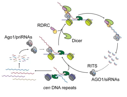
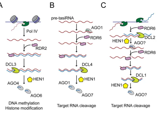
Figure I-1 Biogenesis of miRNAs. 18 Figure I-2 Biogenesis of piRNAs in Drosophila melanogaster. 22 Figure I-3 Biogenesis of centromeric DNA repeat associated small RNAs. 26 Figure I-4 Biogenesis of different classes of endogenous siRNAs in Plants. 28
Figure I-5 RNAi pathway in C. elegans. 32
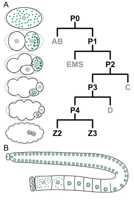
Figure I-6 Localization of P granules during germline development and
embryogenesis 41
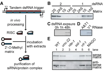
Figure II-1 Sequence specificity and genetics of RDE-1/RNA affinity matrix
binding 78
Figure II-2 RDE-1 does not interact with secondary siRNAs. 81 Figure II-3 AGO Genes are Required for RNAi and Development. 83 Figure II-4 GFP::SAGO-1 and GFP::SAGO-2 rescue the MAGO strain and
interact with secondary siRNAs 86
Figure II-5 ergo-1(tm1860) and the MAGO strain are deficient in
endo-siRNA expression 90
Figure II-6 Model 92
Figure II-7 Secondary AGOs lack key catalytic residues. 96
Figure II-S1 Argonaute deletion alleles. 112
Figure II-S2 Phenotypic analysis of Argonaute genes. 114 Figure II-S3 RNAi sensitivity in Argonaute multiple mutants. 116 Figure III-1 21U-RNAs can be distinguished from other RNA species by their
lengths and upstream motifs matches. 126
iv
RNA accumulation. 130
Figure III-4 PRG-1 interacts with and is required for the accumulation of all
21U-RNAs. 132
Figure III-5 PRG-1 exhibits a broad spectrum of germline defects. 136 Figure III-6 prg-1 mutants exhibit surprisingly subtle changes in gene
expression. 138
Figure III-7 Models for 21U-RNA function. 147
Figure III-S1 Analysis of small RNA reads associated with the conserved
21U-RNA motif. 164
Figure III-S2 Proteins involved in the RNAi are not required for the
accumulation of 21U-RNAs. 165
Figure III-S3 Specificity of the PRG-1 antibody. 166
Figure IV-1 csr-1, ego-1, ekl-1 and drh-3 mutants display chromosome
segregation defects in mitosis and meiosis. 184 Figure IV-2 csr-1, ego-1, ekl-1 and drh-3 RNAi-depleted embryos display
defects in chromsome organization. 188
Figure IV-3 CSR-1, DRH-3, EKL-1 and EGO-1 are expressed in the
germline. 190
Figure IV-4 CSR-1, DRH-3, EKL-1 and EGO-1 localize to chromosomes. 192 Figure IV-5 Analysis of small RNAs enriched in CSR-1 IP complexes. 194 Figure IV-6 CSR-1 22G-RNA complexes bind to target genomic loci. 198 Figure IV-7 Model for the activity of the CSR-1 22G-RNA pathway in
Figure IV-S2 Fluorescence in situ hybridization with probes against
chromosome V. 225
Figure IV-S3 Localization of outer kinetochore, condensin, and cohesin
proteins in wild type and RNAi depleted embryos. 226 Figure IV-S4 Quantitative real-time RT-PCR analysis of csr-1 transcripts. 227 Figure IV-S5 Localization of CSR-1, EGO-1, EKL-1 and DRH-3 is ablated in
respective mutant or RNAi-depleted embryos. 228 Figure IV-S6 Addition of untemplated uridine to the 3’ ends of CSR-1
22G-RNAs. 229
Figure IV-S7 Analysis of csr-1(tm892), ego-1(om97), and DA1316 small RNA
libraries. 230
Figure IV-S8 CSR-1 22G-RNA target mRNA and protein levels are not
changed in drh-3(ne4253) or cde-1(tm1021) mutants. 231 Figure IV-S9 CSR-1 association with chromatin is 22G-RNA dependent. 232 Figure IV-S10 CSR-1 22G-RNAs are expressed at low levels in wild-type small
RNA libraries. 233
Figure V-1 Model of the small RNA pathways of C. elegans 254
vi
Table II-S1a Genetic analysis of Argonaute deletion mutants 117
Table II-S1b Strains generated in this study 118
Table IV-S1 Localization summary of the patterns of CSR-1, EKL-1, EGO-1
and DRH-3 in each RNAi-depleted background 234
AGO Argonaute
C. briggsae Caenorhabditis briggsae C. elegans Caenorhabditis elegans
CDE cosupression defective
Chr Chromosome
CSR chromosome segregation and RNAi defective
D. melanogaster Drosophila melanogaster
DCR C. elegans Dicer protein
DNA deoxyribonucleic acid
DRH Dicer related helicase
dsRNA double stranded RNA
eft Elongation Factor
EGO Enhancer of Glp-One
EKL Enhancer of Ksr-1 lethality endo-siRNA endogenous small interfering
ERGO endogenous RNAi deficient Argonaute
ERI enhancer of RNAi
exo-RNAi exogenous RNAi
glp abnormal germ line proliferation
hcp Holocentric chromosome binding protein
him high incidence of males
IP Immunoprecipitation
IR Inverted repeats
viii
miRNA micro RNA
mRNA messanger RNA
piRNA piwi interacting RNA pre-miRNA miRNA precursor
PRG Piwi related gene
pri-miRNA primary miRNA transcript
PTGS Post-transcriptional Gene Silencing rasiRNA repeat associated small interfering RNA
rde RNAi defective
rDNA ribosomal deoxyribonucleic acid
RDRC RNA-dependent RNA polymerase complex RdRP RNA dependent RNA polymerase
RISC RNA-induced silencing complex RITS RNA induced transcriptional silencing RNAi RNA interference
rrf RNA-dependent RNA polymerase family
RSD RNA spreading defective
sago synthetic secondary-siRNA defective Argonaute
sid Systemic RNA Interference defective siRNA Small Interfering RNA
stRNA short temporal RNA
tncRNA tiny-noncoding RNA
Os pequenos RNAs1 estão presentes em múltiplos organismos, onde desempenham um papel fundamental na manutenção da homeostase do organismo. Estas vias influenciam a expressão de genes, protegem o genoma contra transposões e outros elementos ‘egoístas’2, participam no combate à replicação de vírus e promovem modificações ao nível da cromatina.
No centro das vias de regulação por pequenos RNAs encontram-se complexos compostos por um pequeno RNA, em cadeia simples, e uma proteína pertencente à família de proteínas Argonauta. A família de proteínas Argonauta divide-se em três ramos: as proteínas semelhantes a AGO1 da planta Arabidopsis thaliana, as proteínas semelhantes à proteína PIWI da mosca Drosophila melanogaster e as proteínas específicas do filo Nematoda, ao qual pertence C. elegans.
Os primeiros complexos deste tipo a serem caracterizados estão envolvidos no silenciamento de genes através da degradação de moléculas de RNA mensageiros, e são conhecidos como Complexos Silenciadores Induzidos por RNA (RISC3). Actualmente, outros complexos de composição semelhante, mas com funções distintas foram identificados. Exemplos disso são complexos compostos por proteínas Argonauta e pequenos RNAs, como o RITS4 e o miRISC5. O complexo RITS caracterizado inicialmente na levedura Schizosaccharomyces pombe, promove o silenciamento genico ao nível da cromatina, através do recrutamento de enzimas capazes de modificar
1
Em Inglês: small RNAs.
2
Sequências de DNA que agem como parasitas moleculares, aumentando o seu numero de cópias no genoma hospedeiro.
3
Em Inglês, RNA Induced Silencing Complex.
4
Em Inglês: RNA induced transcriptional silencing complex. Em Português: Complexo de Silenciamento transcripcional induzido por RNA.
5
x
complexos compostos por proteínas Argonauta e pequenos RNAs, pela presença de pequenos RNAs da família dos miRNA, executam as suas funções reguladorar inibindo a tradução dos RNAs mensageiro alvo. .
As proteínas da família Argonauta caracterizam-se pela presença de um domínio PAZ e um domínio PIWI. Os pequenos RNA permitem aos complexos efectores encontrar as moléculas alvo através do emparelhamento de bases entre o pequeno RNA e a molécula alvo. Nos casos em que o complexo formado entre o Argonauta e o pequeno RNA levam a degradação do RNA alvo, o domínio PIWI é responsável pela actividade enzimática que leva ao corte endonucleolítico do RNA alvo.
O foco da minha pesquisa incide sobre a caracterização de membros da família de proteínas denominada Argonautas, e das classes de pequenos RNAs que com elas interagem, no organismo modelo Caenorhabditis elegans. Este nematóide não-parasitico foi utilizado na descoberta de vários aspectos das vias reguladoras dependentes de pequenos RNAs, incluindo a descobertas dos miRNAs e da técnica de Interferencia por RNA (RNAi).
Em C. elegans foram identificadas, até hoje, cinco classes de pequenos RNAs: os miRNA, os pequenos RNAs primários, os 21U-RNAs, os 22G-RNAs e os 26G-RNAs. Cada tipo de pequeno RNA interage com proteínas Argonauta específicas. Nalguns casos, os Argonautas funcionam de forma redundante, pelo que algumas classes de pequenos RNAs interagem com mais do que uma proteína. Em C. elegans, existem 27 genes que codificam proteínas da família Argonauta, sendo que três deles são prováveis pseudogenes6.
6
Genes que pela acumulação de mutações levam a producao de RNAs mensageiro que não são traduzidos em proteinas.
uma resposta específica e potente, que leva a destruição dos RNAs endógenos com sequências semelhantes ao RNA em cadeia dupla utilizado. Esta resposta é conhecida como Interferência de RNA (RNAi).
Este fenómeno foi identificado após a observação de que a injecção, na linha germinal de C. Elegans, de moléculas de RNA com sequências, em ambas as polaridades possíveis (sense e antisense), correspondentes a RNAs expressos, levava ao silenciamento dos RNAs mensageiros endógenos. Apesar da capacidade dos RNAs de polaridade inversa à dos RNAs mensageiros (antisense) de interferir com a expressão de genes ter sido atribuída ao mecanismo de inactivação por emparelhamento complementar7, o mecanismo responsável pelo silenciamento do gene alvo por RNAs com a mesma polaridade que os RNAs mensageiros (sense) não podia ser explicada por nenhum dos mecanismos de regulação de genes conhecidos. Ao tentarem compreender como é que a injecção de moléculas de RNA levava ao silenciamento de genes com sequência homologa ao RNA exógeno em cadeia dupla, Craig Mello e Andrew Fire identificaram os RNAs de cadeia dupla como os agentes responsáveis pelo silenciamento dos RNAs mensageiros endógenos. Quantidades vestígiais de RNAs de cadeia dupla teriam contaminados as preparações de RNA de polaridade única utilizadas até então nas experiências de silenciamento por anti-polaridade, dado que constituem produtos secundários raros das reacções de síntese de RNA, e seriam eles os verdadeiros responsáveis pelo silenciamento observado.
No trabalho apresentado nesta tese, demonstra-se que o silenciamento induzido por RNA de cadeia dupla envolve duas fases distintas. Numa primeira fase, as moléculas longas de RNA em cadeia dupla são processadas pela enzima Dicer, em pequenos RNAs
7
xii
complexo primário de silenciamento. Pensa-se que este complexo identifica então o RNA alvo e inicia a segunda fase da via de silenciamento, ao promover a geração de pequenos RNAs secundários, pequenos RNAs que pertencem à classe dos 22G-RNAs. Os pequenos RNAs secundários são essenciais para silenciar o gene alvo, uma vez que mutantes incapazes de os produzir são resistentes à interferência por RNA.
Este trabalho demonstra também que os pequenos RNAs secundários interagem com um grupo de proteínas Argonautas que funcionam de forma redundante8. Este grupo de Argonautas, que pertencem ao ramo da família Argonauta específico do filo nematoda, não possui os resíduos de aminoácidos necessários à actividade enzimática do domínio PIWI. Como tal, é pouco provável que estas proteínas sejam capazes de degradar directamente o RNA alvo. Até hoje ainda não foi determinado como é que os pequenos RNAs secundários promovem a degradação dos seus alvos, mas uma possibilidade interessante é a de que estes Argonautas conduzam os RNAs alvo para o complexo do exossoma, conhecido pelo seu papel central nas vias de degradação de RNAs na célula.
Apesar de se desconhecer ao certo qual é a função da via de Interferência por RNA em C. elegans, sabe-se, através do trabalho de vários grupos de investigação, que existem várias classes de pequenos RNAs endógenos que desempenham papéis essenciais na manutenção da linha germinal. Consequentemente, são várias também as proteínas Argonauta necessárias ao desenvolvimento da linha germinal de C. elegans.
Uma destas proteínas é PRG-19, que pertence ao ramo PIWI dos Argonautas. Os nossos estudos identificaram os 21U-RNAs como os pequenos RNAs associados a PRG-1 durante o desenvolvimento da linha germinal de C. elegans. Esta classe de RNAs
8
Ou seja, proteínas que se podem substituir entre si para executar a mesma função. Neste caso particular, levar ao silenciamento de RNAs alvo através da interacção com os mesmos pequenos RNAs.
9
predominante é o Uracilo, e pela modificação do ultimo nucleotido da extremidade 3´. No genoma existem acima de 15000 loci que codificam 21U-RNAs, estando todos eles concentrados em duas regiões do cromossoma IV.
Estes loci são compostos pela sequência correspondente ao pequeno RNA e um pequeno motivo a montante. Apesar do motivo associado aos 21U-RNAs ser conservado entre diferentes espécies de nemátodes, as sequências dos pequenos RNAs não exibem conservação evolutiva. Os 21U-RNAs e o Argonauta PRG-1 são expressos exclusivamente na linha germinal, onde se associam directamente. Estas observações estabelecem os 21U-RNAs como membros da família de pequenos RNAs conhecida como piRNAs10. Os piRNAs estão presentes em todos os metazoa, onde são necessários para o desenvolvimento e manutenção da linha germinal. Apesar de existirem milhares de loci que dão origem a 21U-RNAs, em C. elegans apenas o RNA derivado do transposão TC3 pode ser identificado como alvo dos 21U-RNAs. Os alvos dos restantes 21U-RNAs permanecem desconhecidos. Uma vez que os 21U-RNAs não apresentam homologia perfeita em relação a outros RNAs expressos, pensa-se que os 21U-RNAs possam funcionar através de emparelhamento imperfeito com os seus alvos, à semelhança do que acontece com os miRNAs.
Tal como as proteínas do ramo PIWI noutros metazoários, a proteína PRG-1 localiza-se em estruturas especializadas conhecidas como grânulos da linhagem P11, estruturas na região perinuclear, presentes especificamente em células que dão origem a linha germinal de C. elegans. Sabe-se que vários RNAs mensageiros expressos durante o desenvolvimento da linha germinal são retidos nestas estruturas. Uma hipótese interessante é a de que os 21U-RNAs desempenharem um papel importante na retenção
10
Em Inglês: piwi interacting small RNAs.
11
xiv
um complexo que, através da elevada diversidade de sequências dos pequenos RNAs, seria capaz de interagir através de homologia parcial, com os RNA mensageiros que transitam pelos grânulos de linhagem P e levar a sua retenção nestas estruturas.
A classe mais abundante de pequenos RNAs endógenos em C. elegans é a classe dos 22G-RNAs. Os RNAs desta classe desempenham um papel essencial em várias vias de regulação na linha germinal. Estes pequenos RNAs têm características únicas que os diferenciam das classes de pequenos RNAs presentes noutros organismos. Os 22G-RNAs são sintetizados directamente por polimerases de RNA dependentes de RNA e sem intervenção da enzima Dicer. Devido ao mecanismo único de biogénese, os 22G-RNAs são tri-fosforilados na extremidade 5´, posição na qual apresentam uma forte tendência para incorporar o nucleósido guanidina.
Todos os 22G-RNAs interagem com proteínas do ramo especifico dos nemátodes. Uma das vias reguladoras em que os 22G-RNAs funcionam como determinantes de especificidade funcional é necessária para a correcta segregação dos cromossomas e depende exclusivamente do argonauta CSR-112. Esta via reguladora utiliza 22G-RNAs gerados a partir de genes que codificam proteínas expressas na linha germinal, de modo a promover a organização apropriada da cromatina nos cromossomas holocêntricos de C.
elegans. Ao contrário do que foi observado na maioria das vias reguladoras dependentes
de pequenos RNAs, os complexos de CSR-1 não parecem promover à degradação dos seus alvos. Em vez disso, a proteína CSR-1 parece utilizar estes 22G-RNAs para interagir com RNAs mensageiros nascentes, recrutando enzimas capazes de modificar as histonas e proteínas associadas para regiões específicas da cromatina.
12
silenciamento de moléculas de RNA aberrantes, moléculas cuja expressão tem potencialmente efeitos nefastos. Esta via reguladora identifica RNAs com características ‘aberrantes’ e promove o seu silenciamento através da geração de pequenos RNAs homólogos. Esta via reguladora é capaz de identificar dois grupos de RNAs.
Um dos grupos de RNAs silenciados por esta via reguladora possui características de RNA aberrantes inerentes a sua biogenese ou obtidas durante a maturação das moléculas de RNA. Um exemplo de moléculas pertencentes a este grupo são os transposões, que levam a geração de RNA de cadeia dupla, e pseudogenes, RNA que não completam as reacções de maturação.
O segundo grupo de RNA regulados por esta via são os RNAs que são alvo de outras vias reguladoras dependentes de pequenos RNAs, tais como a via de interferência por RNA. Neste caso, apesar do RNA alvo não possuir características de RNAs aberrantes, a interacção do RNA com o complexo de RDE-1, leva a que o RNA seja utilizado na geração de 22G-RNAs, que consequentemente, levam ao silenciamento do RNA. Além da via de interferência por RNA, as vias de pequenos RNAs que levam ao silenciamento do RNA através da geração de 22G-RNAs, incluem as vias dependentes de pequenos RNAs conhecidos como 26G-RNAs.
Os 26G-RNA são pequenos RNAs com uma guanidina na extremidade 5´, e, à semelhança dos RNAs, são modificados na extremidade 3´. Ao contrário dos 21U-RNAs e dos 22G-21U-RNAs, a produção de 26G-21U-RNAs requer a enzima Dicer. A via dos 26G-RNAs divide-se em dois ramos distintos, o ramo embrionário e o ramo da espermatogénese. Os parceiros Argonauta para os 26G-RNAs são ERGO-1 no ramo embrionário e ALG-3 e ALG-4 no ramo da espermatogénese.
xvi
Argonautas de C. elegans, e das classes de pequenos RNAs com eles associadas, sugerem que a escala e o espectro das vias reguladoras por pequenos RNAs vai muito alem daquilo que inicialmente se presumia. Os nossos estudos sugerem que os pequenos RNAs, alem de estarem envolvidos na regulação de genes durante o desenvolvimento, funcionam à escala do genoma inteiro, sendo essenciais no controlo de qualidade de todos os RNA expressos na linha germinal e na formação das estruturas necessárias a segregação dos cromossomas.
Palavras Chave: Caenorhabdites elegans; Interferência de RNA; pequenos RNAs; Argonauta; Polimerase de RNA dependent de RNA; Dicer
In Small-RNA-mediated pathways, small RNAs engage a protein of the Argonaute family and utilize base-pairing interactions to identify and regulate complementary genetic information. My research has focused on understanding how diverse classes of small RNAs in the model organism Caenorhabditis elegans interact with specific members of the Argonaute protein family to carry out unique biological functions.
During RNA interference (RNAi), functionally and structurally distinct Argonaute proteins act sequentially to silence target mRNAs. In the first step, the Argonaute RDE-1 interacts with primary siRNAs, and interaction of this complex with the target mRNA triggers a secondary amplification step. In this second step, RNA dependent RNA polymerases (RdRPs) use the targeted mRNA as a template to generate an abundant pool of small RNAs (22G-RNAs), which interact multiple Argonaute proteins to mediate target silencing.
Several endogenous small-RNA-mediated pathways are essential for germline development. One of these pathways is required for chromosome segregation and relies exclusively on the Argonaute CSR-1, which utilizes 22G-RNAs generated from protein-coding genes to promote the proper organization of chromatin domains. A distinct 22G-RNA germline pathway utilizes ‘aberrant’ 22G-RNAs as templates and is essential in maintaining genome stability.
Proper germline development also requires the 21U-RNA class of small RNAs. RNAs specifically interact with the Piwi Argonaute PRG-1, thus establishing 21U-RNAs as members of the piRNA family, which is important for germline integrity in all metazoans. With only one known exception, 21U-RNAs fail to exhibit sequence complementarity or evidence for direct regulation of other expressed sequences.
xviii
that small RNA pathways function on a genome-wide scale, to regulate many aspects of cell biology and organismal homeostasis, from chromosome structure to gene expression.
KEYWORDS: Caenorhabditis elegans; RNA interference; Small RNAs;
CHAPTER I
CAENORHABDITIS ELEGANS AS A MODEL ORGANISM.
Caenorhabditis elegans was originally described, as Rhabditis elegans, in the
early 20th century by Maupas (Riddle, 1997). This species belongs to the phylum Nematoda (Nema for thread and Eidos for form), one of the most universally distributed groups of animals on the planet. Nematodes, also known as roundworms, are cylindrical pseudocoelomate worms with a thick, multilayered cuticle, which is shed and secreted four times during the animal life cycle. Reproduction is usually sexual, and in most species, the two sexes are separated (Riddle, 1997).
Nematodes have successfully adapted to a wide range of ecological niches and exist both as free-living and parasitic animals. Roundworms can be found in most aquatic habitats, wet soils, moist tissues of plants and in the body fluids or tissues of animals. Nematode diet is varied and includes bacteria, fungi, protozoans, and in some cases other free-living nematodes. Nematodes play an important role in decomposition and nutrient cycling, and virtually every animal or plant important in human activities (including humans themselves), are hosts of parasitic nematode species. As such, nematodes have a tremendous impact on human civilization (Kiontke and Sudhaus, 2006).
The Caenorhabditis (Caeno, recent; rhabditis, rod; elegans, nice) genus is a branch of the Rhabditidae family, a group composed of free-living nematodes. All
Caenorhabditis species are colonizers of nutrient- and microorganism- rich organic
material. Caenorhabditis elegans, as well as the related species C. briggsae and C.
remanei, can be found in anthropogenic habitats such as compost and garden soil
(Kiontke and Sudhaus, 2006). Although their natural diet is not known, all
Caenorhabditis species studied in the laboratory can be cultured on an Escherichia coli
diet. In the wild, dauer juveniles (see below) from many Caenorhabditis species associate with other invertebrates. The dauer juveniles embark onto the associated animal
4
and either use the animal as a means of transportation (phoresy), or wait for the carrier to die, resuming development in the decomposing cadaver (necromeny).
Caenorhabditis elegans was established as a model organism in the early 1970s
through the efforts of Sidney Brenner and his co-workers, who wanted to explore the genetics of complex traits such as behavior. To understand the link between genes and behavior it was essential to establish the structure of the nervous system, and to define how this system is constructed. Thus, a model organism suitable for both genetics and anatomical studies was necessary. So, as Sidney Brenner would put it later: “After some
searching, my choice finally settled on the small nematode, Caenorhabditis elegans. This was a self-fertilizing hermaphrodite with rare spontaneous males. The adults are about 1 mm in length and the life cycle is completed in 3 1/2 days. The animals live in a two-dimensional world feeding on E. coli on the surface of agar plates. They are easy to grow in bulk, each animal producing about 300 progeny during a cycle.” (Brenner,
2003).
Under laboratory conditions, C. elegans animals can be cultured at temperatures ranging from 13°C to 25°C. At higher temperatures, animals develop faster and have a smaller brood size. At 20°C, the C. elegans life cycle is completed in 60 hours. During the last larval stage, hermaphrodites produce sperm cells that are stored in the spermatheca. As hermaphrodites molt into adult animals, sperm development ceases and oocyte production ensues. The number of sperm cells produced before the gonads switch to oocyte production (around 300), determines the number of self-fertilized progeny a single hermaphrodite can generate. Self-fertilization occurs during ovulation as oocytes pass through the hermaphrodite spermatheca. When males fertilize hermaphrodites, male sperm displaces the hermaphrodite sperm from the spermatheca, ensuring that the new progeny will result from cross-fertilization with the male sperm. Embryonic development
begins shortly after fertilization and continues within the uterus of the hermaphrodite. After the egg is laid it hatches and develops through 4 larval stages: L1, L2, L3 and L4 before reaching adulthood. Under unfavorable or stressful conditions, C. elegans larvae can go through an alternative third larval stage termed the dauer stage. Dauer (from the german – enduring) larvae are highly resistant to several forms of stress.
Adult males can be distinguished from hermaphrodites through the presence of a specialized mating tail (Riddle, 1997).
The complete cell lineage in C. elegans has been determined, meaning that the fate of every somatic cell throughout development is known (Sulston and Horvitz, 1977). The body of C. elegans is transparent, and every cell is both visible and accessible to laser microsurgery. C. elegans animals can be recovered after freezing, making it possible to easily store strains over long periods of time. A large collection of mutant strains, collected over several years of research, is stored and shared throughout the C. elegans research community by the Caenorhabdits Genetic Center (CGC). In addition, if no alleles for the gene of interest are available, gene deletions can be requested from two, independent gene knock-out consortiums. In addition, RNA interference (Fire et al., 1998), which was discovered using C. elegans, has facilitated the study of loss-of-function of C. elegans genes.
The C. elegans genome is composed of five pairs of autosomes and one pair of sex chromosomes. Males are XO while females have two X chromosomes. Dosage compensation in hermaphrodites is achieved by the reduction of gene expression in both X chromosomes. With the first version of its genome published in 1998, C. elegans became the first animal to have a fully sequenced genome. The genome is approximately 100 million base pairs long and encodes for approximately 19,000 protein-coding genes (1998). The annotated genome, along with a wealth of associated information, is
6
available through the wormbase website (www.wormbase.org).
In summary, several characteristics make C. elegans a powerful model organism: (1) rapid life cycle, (2) small transparent body, (3) ease of laboratory cultivation and storage, (4) high number of progeny, (5) possibility of inbreeding by self-fertilization-fertilization or crossing with rare males, (6) small genome (initially estimated to be 20x the E. coli genome), and (7) amenability to forward and reverse genetics.
FROM ANTISENSE TO RNAi.
Disrupting the wild type function of a gene is one of the most powerful approaches to understand the role of different genes in biological pathways. Unfortunately, loss-of-function mutations are not available for every gene of interest, and the methods to generate loss-of-function mutations are not available to all experimental systems. In an early attempt to disrupt an endogenous gene by homologous recombination in C. elegans, Fire and colleagues injected plasmids containing a variety of fragments of the unc-22 gene and screened in the next generation for animals with the
unc-22 phenotype (Fire et al., 1991). Although the authors did not detect any evidence of
homologous recombination in the progeny of the injected animals, they did observe a high incidence of the unc-22 phenotype, which could be transmitted to subsequent generations. In the majority of lines with the unc-22 phenotype, no abnormalities were found at the endogenous unc-22 gene. Instead, the unc-22 phenotype correlated with the presence of an extrachromosomal transgene1 array containing unc-22 sequences.
The authors demonstrated that antisense transcripts derived from transgenes had the ability to interfere with the expression of the endogenous gene. Although the observations collected in this study suggested that genes were silenced through an antisense mechanism, the authors noted that in some cases, transgenes that generated transcripts sense to the targeted mRNA also led to silencing of the targeted gene. The ability of transgenes that generate transcripts in the same orientation as the endogenous mRNA to silence the chromosomal locus was attributed to the presence of antisense
1
A transgene is a gene, or genetic material, used to transform an organism. Transgenes can be transferred naturally or by any of a number of genetic engineering techniques. Often, but not always, the transgene is derived from a different species than that of the recipient organism. Across this thesis, the majority of transgenes used originates from the
C. elegans genome, but are engineered to produce proteins compatible with a wide range
8
transcripts generated through indiscriminate transcription from the transgene array. The authors proposed that the hybridization of the antisense RNA to the sense mRNA disrupted either RNA transport or translation. This proposal was similar to a mechanism proposed earlier by Izant and Weintraub, who had proposed that expression of a given gene could be disrupted by the presence of excess amounts of a homologous nucleic acid (Izant and Weintraub, 1984).
Fire and colleagues proposed that antisense studies could be used to “yield hypothesis about null/or hypomorphic phenotypes for a gene of interest”. In a later study, Guo and Kemphues used antisense inhibition to confirm that a predicted cDNA corresponded to the genetically defined par-1 gene (Guo and Kemphues, 1995). Since
par-1 was expected to be a germline-expressed transcript, antisense RNA was injected
into the gonads of wild type animals. As a result of antisense RNA injection into gonads of wild type animals, most of the resulting progeny exhibited phenotypes characteristic of the par-1 loss-of-function mutant, confirming that the candidate open reading frame corresponded to the par-1 gene. Although injection of water or unrelated RNAs did not result in par-1 like phenotypes, the injection of par-1 sense RNA also resulted in a high frequency of par-1 phenotypes among the progeny of injected worms.
These two studies demonstrated that the antisense method could be used in C.
elegans to generate loss-of-function phenotypes for genes of interest. Although several
studies demonstrated that injection of RNA recapitulated loss-of-function phenotypes for several maternally expressed genes (Lin et al., 1995; Guo and Kemphues, 1996; Mello et al., 1996; Powell-Coffman et al., 1996; Guedes and Priess, 1997; Rocheleau et al., 1997), several questions remained as how this process functioned. The observation that sense RNA could also be used to interfere with gene expression could not be explained by a simple antisense mechanism, which depends on hybridization between the injected RNA
and endogenous mRNA transcripts. Therefore, Rocheleau and colleagues proposed that this technique should be referred to as RNA interference (RNAi) (Rocheleau et al., 1997). To understand how injection of RNA could interfere with the function of an endogenous gene, Fire and colleagues investigated the requirements for structure and delivery of the interfering RNA (Fire et al., 1998). The fact that both sense and antisense RNAs could interfere with gene function, and the observation that the interfering agent could persist into the next generation, even though endogenous RNA transcripts are normally degraded in the early embryo, suggested to the authors that a fundamental difference existed between the endogenous mRNA transcripts and the interfering agent.
Since aberrant transcripts were likely present as unwanted side products of the synthesis of the RNA molecules used in the previous studies that utilized antisense silencing, the authors tested the hypothesis that the difference between mRNA transcripts and the interfering agent were related to differences in RNA structure. It was therefore possible that the interfering agent included some RNA molecules with double stranded RNA (dsRNA) character.
Indeed, while injection of a mixture of sense and antisense RNA resulted in a strong interference with an endogenous gene, injections of either purified strand caused only marginal interference activity. Double-stranded RNA was substantially more effective at producing interference than was either strand individually. The effects of double stranded RNA injection were evident in both the injected animals and their progeny. The authors noted that only a few molecules of injected double-stranded RNA were required per affected cell, arguing against the stochiometric interference with endogenous mRNA proposed for antisense silencing mechanisms. Instead, it suggested that there could be a catalytic or amplification component in the interference process (Fire et al., 1998).
10
RDE-1, THE ARGONAUTE LINK.
The ability of dsRNA to interfere with gene expression is not restricted to C.
elegans. Soon after the initial description of dsRNA as the trigger for RNA interference
in C. elegans, several reports extended the range of organisms sensitive to gene interference by dsRNA. Tobacco plants (Waterhouse et al., 1998), Trypanosoma brucei (Ngo et al., 1998), Drosophila melanogaster (Kennerdell and Carthew, 1998; Misquitta and Paterson, 1999), and planaria (Sanchez Alvarado and Newmark, 1999) were shown to be ‘sensitive’ to gene silencing triggered by dsRNA. The fact that gene expression could be disrupted by dsRNA in organisms from different phyla suggested the existence of a conserved gene regulatory mechanism that could be triggered by dsRNA. Interestingly, this interference mechanism could be explored experimentally to abrogate gene expression.
To understand how such a mechanism worked, Tabara and colleagues performed a genetic screen to isolate RNAi deficient (Rde) mutants (Tabara et al., 1999), which led to the identification of several genes required for the RNAi response in C. elegans.
One of the mutants identified in this study, rde-1 (RNAi deficient), was strongly deficient for RNAi, but exhibited no other apparent phenotype. Interestingly, the rde-1 gene was identified as a member of the conserved gene family piwi/sting/argonaute/zwille/eIF2C, a protein family that had already been implicated in silencing phenomena in other organisms (reviewed in (Benfey, 1999)). This protein family is characterized by the presence of the PAZ, the MID and the PIWI domains.
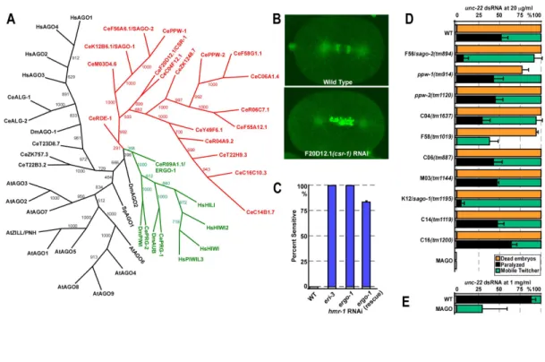
Phylogenic analysis divides the Argonaute family into three paralogous groups: Argonaute-like proteins (based on their similarity to AtAgo1), Piwi-like proteins (based on their similarity to DmPiwi) and the C. elegans specific expansion group (see Figure
II-3). Argonaute-like and Piwi-like proteins are present in bacteria, archaea and eukaryotes. Although plants encode only Argonaute-like paralogues and Amoebozoa phylum members have retained only Piwi-like paralogues, it is likely that the last common ancestor of eukaryotes encoded both Argonaute-like and Piwi-like proteins. The plant and Amoebozoa specific patterns of Argonaute protein paralogues are likely to result from a lineage-specific loss of Piwi-like and Argonaute-like families, respectively. Animal genomes encode representatives of both protein groups (reviewed in (Hutvagner and Simard, 2008) and (Tolia and Joshua-Tor, 2007)).
THE RNA-INDUCED SILENCING COMPLEX.
Argonaute proteins interact with small RNAs to form the core of the RNA-induced silencing complex (RISC), multiprotein complexes that interact with their target RNA transcripts through complementary hybridization with the small RNA.
Small antisense RNAs were first identified in plants (Hamilton and Baulcombe, 1999) as the specificity determinants of post-transcriptional gene silencing (PTGS). PTGS, originally named cosuppression, was first observed in plants when the introduction of an extra copy of an endogenous gene resulted in the degradation of RNAs encoded by both the transgene and the homologous endogenous gene (Napoli et al., 1990; Smith et al., 1990; van der Krol et al., 1990). In Neurospora crassa, a similar phenomenon known as quelling had also been described (Romano and Macino, 1992; Cogoni et al., 1996). Both PTGS and Quelling shared characteristics and appeared to be related to RNAi, suggesting the existence of a conserved sequence-directed gene silencing mechanism. The development of a cell-free system from syncytial blastoderm Drosophila embryos capable of recapitulating many of the features of RNAi (Tuschl et
12
al., 1999) led to several key discoveries on the mechanism of the RNAi pathway. Two independent groups demonstrated that the silencing intermediate in RNAi, similarly to the silencing intermediate of PTGS, was a small RNA approximately 25nt long (Zamore et al., 2000; Hammond et al., 2000). Additionally, chemically synthesized 21-22nt dsRNA was shown to be capable of eliciting gene silencing (Elbashir et al., 2001). Hammond and colleagues demonstrated that RNAi involved cleavage of target RNA by a sequence-specific nuclease activity and named the enzyme responsible for this activity RISC, for RNA-induced silencing complex (Hammond et al., 2000). These early studies also demonstrated that the RISC activity and the generation of siRNAs from dsRNA depended on distinct complexes. Through a candidate approach Dicer was identified as the enzyme responsible for the production of siRNAs (Bernstein et al., 2001).
The details on the mechanisms of RISC activity started to emerge with a series of studies that combined crystallography and biochemistry to study the role of Argonaute proteins. These studies demonstrated that the PAZ domain, also present in proteins of the Dicer family, binds in a sequence-independent manner the 2 nucleotide 3´-end overhang of a small RNA duplex2 (Ma et al., 2004; Lingel et al., 2004). Therefore, the PAZ domain, through its ability to bind the characteristic 3´-end overhangs of siRNA duplexes, can specifically recognize siRNA duplexes, and may play a role in the transfer of siRNAs between Dicer and Argonaute proteins. These studies also demonstrated that the PIWI domain has an RNase-H-like fold (Song et al., 2004; Parker et al., 2004; Ma et al., 2005; Yuan et al., 2005), suggesting that the Argonaute protein was responsible for the enzymatic activity of the RNA-induced silencing complex (RISC). RNase-H-like enzymes are endo-ribonucleases that cleave the 3´-O-P-bond of RNA in a DNA/RNA duplex to produce 3´-hydroxyl and 5´-phosphate products. Similarly to RNase-H, RISC
cleavage products feature a 3´-OH and a 5´-phosphate (Martinez and Tuschl, 2004; Schwarz et al., 2004). Structural studies, in which the protein was crystallized in the presence of ssRNA or a siRNA-like molecule, also provided important insights into target recognition and the cleavage activity of Argonaute proteins. These studies showed that the 5´-end of the small RNA is anchored by a divalent cation at the interface between the PIWI and the MID domain (Parker et al., 2005; Ma et al., 2005). The structural data demonstrated that the catalytic motif of the PIWI domain is positioned adjacent the scissile phosphate of the target RNA, between the tenth and eleventh nucleotide of the small RNA, thus explaining why the site of small RNA cleavage occurs near the center of the region spanned by the siRNA (Haley and Zamore, 2004; Elbashir et al., 2001).
The catalytic activity of the Argonaute proteins was later linked to the enzymatic activity of RISC. These biochemical studies demonstrated that, in humans, only AGO2 can cleave a target mRNA (Meister et al., 2004; Liu et al., 2004) and that disruption of the amino acids in the PIWI catalytic site abrogates RISC activity (Liu et al., 2004). Indeed, the PIWI domain bound to small RNA is sufficient to assemble a minimal RISC (Miyoshi et al., 2005; Rivas et al., 2005). Cleavage-competent Argonaute proteins have a conserved catalytic center (Asp-Asp-Asp/Glu/His/Lis) and require the binding of a divalent cation for activity (reviewed in (Tolia and Joshua-Tor, 2007)). The presence of the Asp-Asp-His motif is necessary, but not sufficient, for slicer activity, as exemplified by HsAgo3 (Meister et al., 2004; Liu et al., 2004). Many Argonaute proteins, such as members of the nematode-specific branch, do not have a complete Asp-Asp-His motif and are unlikely to have slicer activity (Tolia and Joshua-Tor, 2007).
After the initial discovery of siRNAs and miRNAs, several additional classes of small RNAs have been identified, revealing a large diversity of pathways that rely on RISC-like complexes to regulate a myriad of biological processes.
THE MICRO-RNAs.
The first small RNAs to be detected in animals, even before siRNAs were identified as intermediates in the RNAi pathway, were the C. elegans small temporal RNAs (stRNAs)3 lin-4 and let-7. Considering that miRNAs were similar in size to siRNAs, and that it had been proposed that the let-7 miRNA was cleaved from a longer, structured dsRNA precursor (Pasquinelli et al., 2000), one interesting possibility was that miRNAs and siRNAs were generated trough similar mechanisms.
Indeed, several groups independently demonstrated that Dicer was required for the biogenesis of miRNAs (Hutvagner et al., 2001; Grishok et al., 2001; Ketting et al., 2001). In addition, members of the Argonaute family, a gene family also involved in RNAi, were necessary for the maturation and activity of miRNAs (Grishok et al., 2001).
The first miRNA to be identified was lin-4, a gene involved in the normal temporal control of postembryonic development in C. elegans (Lee et al., 1993). The
lin-4 gene does not encode for a protein. Instead, two transcripts of lin-lin-4, around 22 and 61
nucleotides long, were identified as the gene product. These small RNAs contained sequences complementary to a repeated sequence element in the 3´unstranslated region (UTR) of lin-14 mRNA. Remarkably, the lin-4 gene was shown to be conserved in at least 4 species in the Caenorhabditis genus. The second miRNA gene to be identified was let-7 (Reinhart et al., 2000), another gene involved in the temporal control of postembryonic development in C. elegans. Surprisingly, the let-7 gene is conserved not only within the Caenorhabditis genus, but also in a wide range of animals, including vertebrates, ascidians, hemichordates, molluscs, annelids and arthropods. Interestingly, the expression pattern of let-7 was also conserved, suggesting that this small RNA could
3
stRNAs are now known as miRNAs. I will be using miRNA throught out the text to avoid confusion.
16
control development across animal phylogeny (Pasquinelli et al., 2000). The conservation of let-7 across species, in addition to the partial overlap between the miRNA and siRNA pathways, suggested that miRNAs are components of an ancient regulatory mechanism. Taking advantage of characteristics shared by Dicer products (such as the 20 to 22 nucleotide length, the 5´-monophosphate and the 3´-hydroxylgroup) and the characteristics of lin-4 and let-7 miRNAs (such as the location in intragenic regions, the sequence similarity between species and the existence of a stem-loop precursor), several groups identified additional miRNAs from C. elegans, D. melanogaster and human cells trough cDNA cloning of small RNAs and in silico predictions of candidate small RNAs (Lau et al., 2001; Lee and Ambros, 2001; Lagos-Quintana et al., 2001).
The abundance, complex expression patterns and conservation across species suggested that regulation through miRNAs was more complex than initially appreciated.
Biogenesis of miRNAs in animals and plants.
To date, thousands of miRNAs have been identified in plants, animals and viruses. These small RNAs are thought to silence gene expression post-transcriptionally through sequence-directed binding to the 3´ untranslated regions of target mRNAs. miRNAs are processed from a pri-miRNA by RNAse III endonucleases. These precursor transcripts can include more than one miRNA hairpin and are typically transcribed by RNA polymerase II (Lee et al., 2002; Lee et al., 2004; Cai et al., 2004). Processing of the pri-miRNA occurs in two sequential steps (Figure I-1A and I-1C). In animals (Figure I-1A), the pri-miRNA is cleaved in the nucleus into a 60-70 nucleotide long pre-miRNA by Drosha (Lee et al., 2002; Lee et al., 2003; Denli et al., 2004; Gregory et al., 2004; Han et al., 2004; Landthaler et al., 2004). pre-miRNAs are transported to the cytoplasm by
Exportin-5, a nuclear export protein that binds correctly processed pre-miRNAs (Yi et al., 2003; Bohnsack et al., 2004; Lund et al., 2004; Yi et al., 2003). In the cytoplasm, the pre-miRNA is processed by Dicer (Hutvagner et al., 2001; Grishok et al., 2001; Ketting et al., 2001; Forstemann et al., 2005; Chendrimada et al., 2005; Jiang et al., 2005; Lee et al., 2006; Saito et al., 2005). Processing of the pre-miRNA by Dicer generates a duplex containing two strands (miRNA and miRNA*), one of which (from either the 5´ or 3´ arm of the pre-miRNA) is loaded in the RISC complex. The choice of miRNA strand is influenced by the thermodynamic properties of the duplex (Schwarz et al., 2003; Khvorova et al., 2003). Some pre-miRNAs can generate mature miRNA from both arms. In at least 4 nematode species, including C. elegans, there is a bias for the mature miRNA to be located on the 3´ arm of the hairpin (de Wit et al., 2009). In addition, pre-miRNA can be generated in a Drosha-independent manner in at least C. elegans, D. melanogaster and mammals. In a few cases, the pre-mRNA splicing pathway generates the pre-miRNA (Figure I-1B); these pre-miRNA-like introns, named mirtrons, are spliced out of mRNA precursors and, after the initial lariat product is processed by a debranching enzyme, enter the standard miRNA biogenesis pathway to yield an authentic pre-miRNA (Okamura et al., 2007; Ruby et al., 2007; Berezikov et al., 2007).
Although miRNAs are also present in plants, differences between plants and animals in the miRNA biogenesis pathway suggest that miRNA genes arose independently in these multicellular lineages (Bartel, 2004). In plants (Figure I-1C), both pri-miRNAs and pre-miRNAs are processed by DCL1 (Kurihara and Watanabe, 2004) (Figure I-1C). Conversion of pri-miRNA into pre-miRNA requires the function of the DCL1-interacting proteins HYPONASTIC LEAVES1 (HYL1) and the C2H2-zinc finger protein SERRATE (SE) (Kurihara et al., 2006; Fang and Spector, 2007)(Figure I-1C).
18
Figure I-1. Biogenesis of miRNAs in plants and animals.
The majority of miRNA genes are transcribed by RNA polymerase II and generate a structured transcript, the pri-miRNA. (A) In animals, the pri-miRNA is sequentially processed by DROSHA and DICER to generate a mature miRNA. Additionally, in animals (B), certain introns can be processed into a pre-miRNA that is processed by Dicer to generate a mature miRNA. In Plants (C), DCL-1 is involved in both steps of miRNA maturation.
At least some miRNAs are exported to the cytoplasm in a pathway that involves HASTY, the plant homolog of Exportin-5 (Park et al., 2005). In plants, all silencing small RNAs, including miRNAs, are modified at the 3´ end by the S-adenosyl methionine-dependent methyltransferase Hua Enhancer 1 (HEN1) (Yu et al., 2005; Li et al., 2005)(Figure I-1C). The methylation of miRNA protects them from uridylation and subsequent degradation. Unlike animal miRNAs, most plant miRNAs are perfectly complementary to their targets and are thought to regulate their targets through mRNA cleavage (Tang et al., 2003; Llave et al., 2002; Rhoades et al., 2002).
THE PIWI INTERACTING SMALL RNAs.
piRNAs are small RNAs characterized by their interaction with members of the PIWI branch of the Argonaute protein family. In addition, piRNAs typically have a monophosphorilated 5´-end nucleotide with an overwhelming bias for Uracil and carry a 2´-O-methyl modification at the 3´-terminal ribose. In flies and mammals, piRNAs are longer than siRNAs, with a size ranging from 25 to 30 nucleotides, whereas piRNAs in the nematode C. elegans are 21 nucleotides long.
The founding member of the PIWI clade of Argonaute proteins, the Piwi protein, was isolated in a screen for genes that affect germline stem cell division in Drosophila
melanogaster (Lin and Spradling, 1997). The piwi gene was shown to encode a highly
basic, well-conserved protein required for germline stem cell division in diverse organisms, including C. elegans (Cox et al., 1998). In addition to Piwi, the Drosophila genome encodes two more members of this clade of Argonautes: Aubergine (Aub) (Harris and Macdonald, 2001) and Ago3 (Williams and Rubin, 2002). C. elegans contains three members of the Piwi clade: prg-1, prg-2 and ergo-1 (Cox et al., 1998; Yigit et al., 2006). The zebrafish genome encodes at least two members of the Piwi clade: ziwi and zili (Houwing et al., 2007). In mammals, MILI, MIWI and MIWI2 (Kuramochi-Miyagawa et al., 2008; Carmell et al., 2007) have been described in mice and HILI, HIWI, HIWI2 and HIWI3 (Sasaki et al., 2003) in humans.
piRNAs were initially identified in Drosophila as rasiRNAs (repeat-associated small interfering small RNAs), small RNAs derived from transposable elements, satellite and microsatellite DNA and suppressor of Stellate [Su(ste)] repeats. These small RNAs were abundantly detected in testes and early embryos (Aravin et al., 2003). Small RNAs cognate to Su(ste) are longer than siRNAs and require the activity of Aubergine and
20
Spindle-E (a DEAD-box helicase) (Aravin et al., 2001; Aravin et al., 2003). Although it was initially proposed that rasiRNAs were generated from long dsRNA triggers by Dicer (Kalmykova et al., 2005; Aravin et al., 2001), loss-of-function of the Drosophila Dicer enzymes, (Dcr-1 and Dcr-2), their dsRBD partners (R2D2 and loquacious) or Ago2 did not disrupt rasiRNA production or function in the silencing of transposable elements in the germline (Vagin et al., 2006). These observations suggested that rasiRNAs were distinct from miRNAs and siRNAs and participated in a separate small RNA pathway to control selfish genetic elements in the germline.
Biogenesis of piRNAs.
The mechanism of piRNA biogenesis is not yet fully understood. In Drosophila
melanogaster, the system where piRNAs have been most extensively studied, two distinct
piRNA pathways have been identified. In gonadal somatic cells only the Piwi-dependent, Aub- and Ago-3-independent pathway is present, while in the germline both Piwi and Aub/Ago3-dependent pathways function to control transposable elements (Li et al., 2009; Malone et al., 2009). piRNAs that function in the germline, generated through both pathways, are maternally deposited and within a single generation provide full immunity against some repetitive elements. Thus, maternally loaded piRNAs act as epigenetic factors essential to achieve full immunity (Brennecke et al., 2008).
The Ping-Pong cycle.
One of the pathways that functions in the Drosophila melanogaster germline to control tranposons depends on the Piwi proteins Aubergine and Ago3. Both proteins localize to the Nuage4, and require each other to properly localize to this structure (Li et al., 2009). Although piRNA are predominantly antisense to transposons, piRNAs of both polarities are present in the germline and play a role in this piRNA pathway. The sense piRNAs are typically bound to Ago3, while Aubergine is loaded with antisense piRNAs. Primary piRNAs, generated from piRNA clusters, are loaded in Aubergine and guide the cleavage of a transposon transcript or a precursor RNA derived from a cluster, creating the 5´-end of an Ago3-bound sense piRNA. The Ago3-bound sense piRNA will then direct a reciprocal reaction and generate additional antisense piRNAs. A yet unidentified nuclease is responsible for the generation of the 3´-end of piRNAs. These new antisense piRNAs will then contribute to transposon silencing both as part of the silencing effector complex and by reinforcing the feed-forward loop. As a consequence of this biogenesis mechanism, piRNAs that participate in this cycle show a ping-pong signature. Aubergine- and Ago3-bound piRNAs overlap by 10 base pairs at their 5´ ends. The Ago3-bound piRNAs show a bias for A at position 10, while Aubergine-bound piRNAs exhibit a bias for U at position 1 (Brennecke et al., 2007; Gunawardane et al., 2007) (Figure I-2). piRNAs generated exclusively through the ping-pong cycle target two distinct groups of transposons.
4
Nuage are electron-dense perinuclear structure present in animal germ cell-lines. In C.
elegans these structures are known as P granules. (See: Chapter I; The germ granules of C. elegans).
22
Figure I-2. Biogenesis of piRNAs in Drosophila melanogaster.
The argonaute protein Aubergine (Aub), loaded with a primary piRNA cleaves a sense transposon transcript generating the 5´-end of a new piRNA of the opposite polarity. A yet unidentified nuclease trims the 3´-end of the cleaved transcript to generate Ago3 bound mature piRNA. The Ago3 bound piRNA guides the cleavage of a piRNA precursor transcript to generate a new piRNA, in a process similar by the one driven by Aubergine. In germline cells Piwi can also participate in the ping-pong cycle, while in somatic cells Piwi functions in a linear pathway, directly targeting transcripts with homology to piRNAs.
Group I transposons show a strong ping-pong signature and Ago3 associates almost exclusively with sense piRNAs (as described above). Group II transposons (composed of only 5 families) behave “backwards” to group I, since Ago3 is required for the generation of sense piRNAs. In addition to Aubergine and Ago3, spindle-E, vasa and Krimper specifically function in the ping-pong cycle (Li et al., 2009; Malone et al., 2009).