0103 - 5053 $6.00+0.00
A
r
ti
c
le
* e-mail: [email protected]
Alkaloids from
Annona dioica
Paulo R. D. dos Santosa, Anselmo A. Moraisa and Raimundo Braz-Filhob
a
Curso de Pós-graduação em Química Orgânica, Departamento de Química, ICE, UFRRJ, 23851-970 Seropédica - RJ, Brazil
b
Setor de Química de Produtos Naturais, LCQUI, CCT, Universidade Estadual do Norte Fluminense, 28015-620 Campos - RJ, Brazil
Do extrato etanólico da madeira de Annona dióica foram isoladas as conhecidas 1-aza-4-metilantraquinona, lasiodiplodina, liriodenina, uma mistura de 1-aza-5,9,10-trimetoxi-4-metil-2-oxo-1,2-diidroantraceno e 1-aza-8,9,10-trimetoxi-4-metil-2-oxo-1-aza-5,9,10-trimetoxi-4-metil-2-oxo-1,2-diidroantraceno (geovanina) eo novo alcalóide 1,2-metilenodioxi-6α,7-desidroaporfina-4(S )-(4-hidroxi-3,5-dimetoxifenil)-3,4-diidro-2(1H)-piridinona. As estruturas destes produtos naturais foram elucidadas com base em seus dados espectrais, inclusive experiências de NOE e espectros de RMN 2D de correlação homonuclear 1 H-1H-COSY e heteronuclear 1H-13C-COSY-nJ
CH (HMQC, n=1 e HMBC, n=2 e 3).
From the ethanolic extract of the wood of Annona dioica were isolated the known 1-aza-4-methylanthraquinone, lasiodiplodin, liriodenine, a mixture of 1-aza-5,9,10-trimethoxy-4-methyl-2-oxo-1,2-dihydroanthracene and 1-aza-8,9,10-trimethoxy-4-methyl-2-1-aza-5,9,10-trimethoxy-4-methyl-2-oxo-1,2-dihydroanthracene (geovanine) and the new alkaloid 1,2-methylenedioxy-6α,7-dehydroaporphine-4(S
)-(4-hydroxy-3,5-dimethoxyphenyl)-3,4-dihydro-2(1H)-pyridinone. The structures of these natural products were elucidated on the basis of their spectral data, including NOE experiments and homonuclear 1H-1
H-COSY e heteronuclear 1H-13C-COSY-nJ
CH (HMQC, n=1 and HMBC, n=2 and 3) 2D-shift-correlated
NMR spectra.
Keywords: Annona dioica, Annonaceae, quinoline alkaloids, lignoaporphine, lasiodiplodin
Introduction
Annona dioica, Annonaceae, is a shrub distributed throughout the States of São Paulo, Minas Gerais, Paraná and Mato Grosso, Brazil, commonly called “ceraticum (do campo, or grande)”, “arixicum” and “ariticum”. The fruits and leaves are used against rheumatism and the seeds to heal diarrhea.1
This paper reports the isolation of the known 1-aza-4-methylanthraquinone (1), lasiodiplodin (2), liriodenine (3), along with a mixture of 1-aza-8,9,10-trimethoxy-4-methyl-2-oxo-1,2-dihydroanthracene (4, geovanine) and 1-aza-5,9,10-trimethoxy-4-methyl-2-oxo-1,2-dihydro-anthracene (5), and the new alkaloid 1,2-methylenedioxy-6α,7-dehydroaporphine-4(S
)-(4-hydroxy-3,5-dimethoxy-phenyl)-3,4-dihydro-2(1H)-pyridinone (6). The structures were established by spectral analysis, mainly 1H and 13C
NMR including homonuclear 2D 1H-1H-COSY and
heteronuclear 2D 1H-13C-COSY-nJ
CH (HMQC, n=1 and
HMBC, n=2 and 3) and NOE experiments.
To the best of our knowledge, the compound 6 is
hitherto unreported in the literature.
Results and Discussion
Comparative analysis of HBBD- and DEPT-13C NMR
spectra of each natural product (1 to 6) was used to identify signals corresponding to quaternary, methine, methylene
and methyl carbon atoms. Compound 1 was previously
isolated from Cleistopholis patens,2,32 from Lasiodiplodia theobromae4 and
Euphorbia splendens,5 3 from Atherosperma moschatum, Liriodendron tulipifera,6Fusea longifolia, Siparuna guianensis,7
Thalictrum sessile8 and
many other plants species, and 4 (geovanine) from Annona ambotay.9 The structural identification of these compounds
was based on their spectral data, including homonuclear 2D 1H-1H-COSY and heteronuclear 2D 13C-1H-COSY-nJ
(n=1; n=2 and 3, COLOC) experiments and differential NOE data, together with comparison with literature values of 1H and 13C NMR (vide supra).
The mixture of 4 and 5 showed IR absorption for conjugated carbonyl function (ν
max 1650 cm
-1) and aromatic
ring (ν
max 1560 and 1520 cm
-1). The multiplicity of each
carbon signal of the two components 4 and 5 was deduced by comparative analysis of the HBBD- and DEPT-13C NMR
spectra (Table 1). This analysis in combination with GC-EIMS {Rt=2.41min (40%): m/z 299 ([M]+ of 5, 65%;
Rt=3.08 min (60%): m/z 299 ([M]+ of 4, 63%)} and 1D 1H
and 2D 1H-1H-COSY NMR spectra (Table 1) allowed the
deduction of the same molecular formula C17H17NO4 for 4 and 5. Comparison of 1H NMR spectral data of 4 and of
geovanine (isolated from Annona ambotay9) was used to
characterize it as the same compound (Table 1) and consequently to establish the structure of 4 as 1-aza-8,9,10-trimethoxy-4-methyl-2-oxo-1,2-dihydroanthracene. This structure was confirmed by NOE difference experiments performed with irradiation at MeO-10 (δ
H 3.90) which
resulted in signal enhancements at δ
H 2.75 (Me-4) and
7.75 (H-5) and irradiation at MeO-8 (δ
H 4.02) which showed
NOE at δ
H 6.88 (H-7). The remaining signals observed in
the 1H (δ
H 7.82, t, J 8.2 Hz, H-7, position conjugated with
carbonyl group; partial superimposition with signal at 7.75 of the H-5 of 4), 6.59 (s, H-3), 2.65 (s, Me-4), 4.04 (s, MeO)] and 13C (δ
C 63.79, MeO)] NMR and GC/MS (vide supra)
spectra were speculatively used to propose the structure 5 (1-aza-5,9,10-trimethoxy-4-methyl-2-oxo-1,2-dihydro-anthracene) for the other component present in the mixture, probably a new alkaloid.
The lignoaporphine alkaloid 6 showed IR absorption bands for a carbonyl group (ν
max 1660 cm
-1) and for
aromatic ring (ν
max 1630, 1610, 1520 and 1500 cm -1). The
number of hydrogen atoms bound to each carbon atom was deduced by comparative analysis of the HBBD - (25 signals corresponding to 28 carbon atoms) and
DEPT-13CNMR (Table 2) in combination with 1H NMR spectral
data (1D and 2D 1H-1H-COSY) and low resolution mass
spectrum (m/z 469 [M]+, 100%, C
28H23NO6), allowing to
establish the formula (C)13(C=O)(CH)8(CH2)3(OCH2O) (MeO)2(OH)= C28H23O6 (455 daltons) + N = C28H23NO6 (m/z
469, [M]+). This molecular formula is compatible with an
aporphine skeleton (C16H9N) sustaining one methylene-dioxy group and one 4’-hydroxy-3’,5’-dimethoxy-7’,8’-dihydrocinnamoyl moiety in a lactam ring. In fact, the presence of this cinnamoyl system was evidenced by the chemical shifts of 2H-8’ [δ
H 3.20 (dd, J 14.5; 6.2 Hz); 3.07
(br d, J 14.5 Hz)], H-7’ [δ
H 4.80 (br d, J 6.2 Hz)], 2H-2’,6’
[δ
H 6.38 (s)] and 2MeO-3’,5’ [δH 3.74 (s)] in addition to the
chemical shifts of C-1’ (δ
C 131.77), 2CH-2’,6’ (δC 103.82),
2C-3’,5’ (δ
C 147.35), C-4’ (δC 133.96), CH-7’ (δC 37.96),
CH2-8’ (δ
C 39.90) and C-9’ (δC 167.65). The location of the
cinnamoyl unit, involving carbon C-7 and the nitrogen atom, was defined by heteronuclear 2D 1H-13C long-range
couplings (2J CH and
3J
CH) between: C-7 (δC 115.85) and
H-7’ (δ
H 4.80, 2J
CH), 2H-8’ (δH 3.20; 3.07, 3J
CH) and H-8 (δH
7.85, 3J
CH); C-6a (δC 132.22) and H-5 (δH 5.29, 3J
CH) and
H-7’ (δ
H 4.80, 3J
CH); C-9’ (δC 167.65) and H-5(δH 5.29, 3J
CH),
H-7’ (δ
H 4.80, 3J
CH) and 2H-8’ (δH 3.20 and 3.07, 2J
CH). The
remaining signals observed in the 1H and 13C NMR spectra
(Table 2) were used to define the 2,3-methylene-dioxyaporphine [(C)9(CH)5(CH2)2(OCH2O)] unit, being the homo- and heteronuclear correlations obtained from 2D
1H-1H-COSY and 1H-13C-COSY-nJ
CH (n=1, HMQC; n=2 and
3, HMBC) spectra summarized in Table 2. The 2D shift-correlated spectra were also used to complete 1H and 13C
chemical shift assignments of 6. The following cross-peaks observed in the NOESY spectrum were also utilized for additional confirmation of the structure proposed to 6: a)
NOE between H-8 (δ
H 7.85) and H-7’ (δH 4.80) and
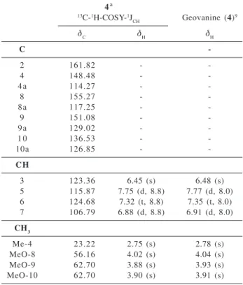
2H-Table 1.1H (200 MHz) and 13C (50 MHz) NMR for 4, in CDCl
3 and TMS as internal standard, compared with geovanine in CDCl3.9 Chemical shifts in δ(δ
H and δC) and coupling constants (J, in paren-thesis)
4a
13C-1H-COSY-1J
CH Geovanine(4) 9
δ
C δH δH
C
-2 161.82 -
-4 148.48 -
-4a 114.27 -
-8 155.27 -
-8a 117.25 -
-9 151.08 -
-9a 129.02 -
-1 0 136.53 -
-10a 126.85 -
-C H
3 123.36 6.45 (s) 6.48 (s)
5 115.87 7.75 (d, 8.8) 7.77 (d, 8.0) 6 124.68 7.32 (t, 8.8) 7.35 (t, 8.0) 7 106.79 6.88 (d, 8.8) 6.91 (d, 8.0)
CH3
Me-4 23.22 2.75 (s) 2.78 (s)
MeO-8 56.16 4.02 (s) 4.04 (s)
MeO-9 62.70 3.88 (s) 3.93 (s)
MeO-10 62.70 3.90 (s) 3.91 (s)
aNumber of hydrogen bound to carbon atoms deduced by
com-parative analysis of HBBD- and DEPT-13C NMR spectra. Homo-nuclear 2D 1H-1H-COSY spectrum and {1H}-1H-NOE difference spectra were also used in these assignments. Chemical shifts (δ
2’,6’ (δ
H 6.38); b) NOE between H-7’(δH 4.80) and 2H-2’,6’
(δ
H 6.38); c) NOE between 2H-2’,6’ (δH 6.38) and
2MeO-3’,5’ (δ
H 3.74); d) NOE between H-3 (δH 7.13) and
pseudoequatorial H-4 (δ
H 3.19).
Additional peaks observed in the low resolution EIMS at m/z 316 (96%), 314 (22%) and 286 (26%) attributed to fragments 6a, 6b and 6c, respectively, are also consistent with structure 6.
Thus, the structure of the new lignoaporphine alkaloid 6 was established as 1,2-methylenedioxy-6a,7-dehydro- aporphine-4-(4-hydroxy-3,5-dimethoxyphenyl)-3,4-dihydro-2(1H)-pyridinone. The [α]
D of + 326 o (CHCl
3; c
2.50) indicated that it was not a racemic mixture, ruling out the possibility of 6 being an artifact. The assignment of the absolute stereochemistry at the chiral centre CH-7’ of the 3-(4-hydroxy-3,5-dimethoxyphenyl)propanoyl
moiety of 6 was postulated by comparison of its optical rotation [α]
D + 326 o (CHCl
3; c 2.50) with those reported for
structures 7-10, which contain an asymmetric carbon with nearly identical substituents. In those compounds the S
configuration gave an optical rotation about 200o higher
than the R configuration.10 Consequently, the[α] D + 326
o
(CHCl3; c 2.50) is consistent with (S)-stereochemistry of the chiral carbon CH-7’ present in the 3-(4-hydroxy-3,5-dimethoxyphenyl)propanoyl unit of 6.
Thus, the structure of new lignoaporphine alkaloid was
established as 1,2-methylenedioxy-6α
,7-dehydro-aporphine-4(S )-(4-hydroxy-3,5-dimethoxyphenyl)-3,4-dihydro-2(1H)-pyridinone, IUPAC name 5-(4-hydroxy-3,5-m e t h o x y p h e n y l ) - ( 5S) - 6 , 7 , 9 , 1 0 - t e t r a h y d r o - 5H -benzo[g][1,3]dioxolo[4’,5’:4,5]benzo[de
]pyrido[3,2,1-ij]quinolin-7-one (6).
Table 2. 1H (500 MHz) and 13C (125 MHz) NMR spectral data of 6, in CDCl
3 and TMS as internal standard. Chemical shifts in δ (δH and δC) and coupling constants (J, in parenthesis)a
1Hx13C-HMQC-1J CH
1Hx13C-HMBC-nJ CH
δ
C δH
2J CH
3J CH
1Hx1H-NOE
C
1 142.42 - H-3; OCH2O
1a 117.17 - H-11
1 b 117.07 - H-3
2 145.79 - H-3 OCH2O
3a 126.97 - H-4 H-5
6a 132.22 - H-5; H-7’
7 115.85 - H-7’ H-8; 2H-8’
7a 130.34 - H-7’
11a 126.09 - H-8
1 ’ 131.77 - 2H-2’,6’; H-7’ 2H-8’
3’,5’ 147.35 - 2H-2’,6’ 2MeO-3’,5’
4 ’ 133.96 - 2H-2’,6’
9 ’ 167.65 - 2H-8’ H-5; H-7’
C H
3 108.82 7.13 (s)
8 122.99 7.85 (d, 8.7) H-10
9 127.70 7.55 H-11
1 0 125.09 7.54
1 1 127.55 9.14 (dd, 8.0, 1.9) H-9
2’,6’ 103.82 6.38 (s) H-7’ 2MeO-3’,5’; H-7’
7 ’ 37.96 4.80 (d, 6.2) 2H-8’ 2H-2’,6’ H-8; 2H-2’,6’
CH2
4 29.93 3.32 (dt, 15.8, 3.8) 3.19 H-5 H-3
5 38.41 5.29 (dd, 12.3, 3.8) 3.11 (dt, 12.3, 2.8) H-4 8 ’ 39.90 3.20 (dd, 14.5, 6.2) 3.07 (br d, 14.5) H-7’ OCH2O 101.43 6.32 (br s) 6.31 (br s)
CH3
2MeO-3’,5’ 56.27 3.74 (s)
a Number of hydrogen bound to carbon atoms deduced by comparative analysis of HBBD- and DEPT-13C NMR spectra. Superimposed 1H signals are described without multiplicity and chemical shifts were deduced by 1H-13C-COSY-nJ
CH (n=1, HMQC; n=2 and 3, HMBC) NMR spectra. Homonuclear 2D 1H-1H-COSY spectrum was also used in these assignments. Chemical shifts (δ
Experimental
General experimental procedures
Mps are uncorr. NMR spectra were run on Bruker
AC-200 and Advance 500 spectrometers in CDCl3 or
CD3COCD3 using TMS as internal standard or by reference to the solvent signal (CHCl3 at δ
H 7.24 or CD2HCOCD3 at
δ
H 2.08 and CDCl3 at δC 77.00 or CD3COCD3 at δC 24.8 and
206.0). EIMS were obtained at 70 eV on a Hewlett Packard spectrometer model 5987. The IR spectra were obtained on a Perkin-Elmer FT-1500 spectrometer. Column
chroma-tography was carried out with silica gel 0.063–0.2 mm and TLC was done employing silica gel Kieselgel 60 from Merck and spots were visualized by UV (λ
max 259 and 360
nm) and exposure to I2 vapour.
Plant material
Extraction and isolation of constituents
Dried and powdered wood (5.2 kg) was extracted successively with n-hexane and EtOH at room temp. and the solvent removed under vacuum to yield 81.79 g of residue. This residue was chromatographed on a silica gel column using CH2Cl2, CHCl3, n-hexane-EtOAc (4:1, 3:2, 2:3 and 1:4), EtOAc and EtOAc-MeOH (4:1 and 1:1). Fractions 52-71, eluted with CH2Cl2, were rechromato-graphed on a silica gel column (70-230 mesh) using CHCl3 and CHCl3-MeOH (97:3) as eluents to furnish 15 fractions: fractions 6-10 yielded 1 (22 mg) and 2 (20 mg) after chromatography on a silica gel column using CHCl3-MeOH
(97:3). Preparative TLC (CHCl3-MeOH, 97:3) of the
fractions 98-115 (eluted with CHCl3-MeOH 97:3) afforded 3 (15 mg) and 6 (83 mg). Fractions 135-229 (4.7 g, eluted with CHCl3-MeOH 97:3) were chromatographed on a silica gel (100 g) column using CH2Cl2, CH2Cl2-CHCl3 (1:1 and 1:4), CHCl3, n-hexane-EtOAc (1:4) and MeOH to furnish 4+5 (18.8 mg) as amorphous yellow solid after treatment with acetone.
1-Aza-4-methylanthraquinone (1). Oil. Spectral data in agreement with literature values.2,3
12-Hydroxy-14-methoxy-3-methyl-3,4,5,6,7,8,9,10-octahydro-1H-benzo[c]oxa-cyclododecin-1-one (2, Lasidioplodin). Amorphous yellow solid, mp 183-186 oC;
spectral data in agreement with lit. values.4,5
8H-Benzo[g][1,3]dioxolo[4’,5’:4,5]benzo [de ]quinolin-8-one (3,Liriodenine). Amorphous yellow solid, mp 284-286 oC; spectral data in agreement with lit. values.6-8
Mixture of 1-Aza-8,9,10-trimethoxy-4-methyl-2-oxo-1,2-dihydroanthracene (4, geovanine) and 1-aza-5,9,10-trimethoxy-4-methyl-2-oxo-1,2-dihydroanthracene (5).
Amorphous yellow solid, mp 189-191 oC; IR (KBr) ν max/
cm-1: 1650 (C=O), 1560 and 1520 (aromatic ring); GC Rt =
2.418 (5)/3.086 (4) min; EIMS (rel. int., 4/5) m/z: 299 ([M]+,
63/65), 284 (M-Me., 100/100), 269 (M-CH
2O, 76/72), 268
(M-MeO, 7/7), 252 (M-CH2O-OH, 9/9), 240 (M-MeO.-C=O,
17/18); 1H and 13C NMR of 4: Table 1.
1,2-methylenedioxy-6α,7-dehydroaporphine-4(S
)-(4-hydroxy-3,5-dimethoxyphenyl)-3,4-dihydro-2(1H
)-pyridinone (6). Amorphous yellow solid, mp 211-213 oC;
IR (KBr) ν
max/cm
-1: 3600, 3500, 1660, 1630, 1610, 1520,
1500, 1260, 1050, 850, 720 cm-1;1H and 13C NMR: Table
2.
Acknowledgements
This work was supported by a fellowship from the Conselho Nacional de Desenvolvimento Científico e Tecnológico (CNPq) and grants from Financiadora de Estudos e Projetos (FINEP)/Programa de Apoio ao Desenvolvimento Científico e Tecnológico (PADCT), Coordenação de Aperfeiçoamento de Pessoal Nível Superior (CAPES) and Fundação de Amparo à Pesquisa do Estado do Rio de Janeiro (FAPERJ). The authors also wish to thank Professor José Badini of the Universidade Federal de Ouro Preto, Minas Gerais, Brazil for the plant identification.
References
1. Pott, A.; Pott, V. J.; Plantas do Pantanal; Centro de Pesquisa Agropecuária do Pantanal, CPAP, Serviço de Produção de Informação: Brasília, Brazil, 1994, p. 34
2. Goulart, M. O. F.; Santana, A. E. G.; de Oliveira, A. B.; Maia, J. G. S.; Phytochemistry 1986, 25, 1691.
3. Dragara, T.; Cassels, B. K.; Leboeuf, M.; Cavé, A.; Phytochem-istry 1987, 26, 537.
4. Aldrige, D. C. S.; Galt. S.; Turner, W. B.; J. Chem. Soc. C
1971, 1623.
5. Lee, K.-H.; Hayashi, N.; Okano, M.; Hall, I. H.; Wu, R.-Y.; Mc Phail, A. T.; Phytochemistry 1982, 21, 1119.
6. Bick, I.R.C.; Douglas C.K.; Tetrahedron Lett. 1964, 1629. 7. Braz-Filho, R.; Gabriel, S. J.; Gomes, C. M. R.; Gottlieb, O. R.;
Bichara, M. das G. A.; Maia, J. G. S.; Phytochemistry 1976, 15, 1187.
8. Wu. Y.-G.; Lu. S. T.; Chang, J. J.; Lee, K.-H.; Phytochemistry
1988, 27, 1563.
9. de Oliveira, A. B.; de Oliveira, G. G.; Carazza, F.; Maia, J. G. S.; Phytochemistry 1987, 26, 2650.
10. Foo, L. Y.; Phytochemistry 1987, 26, 2825.
Received: April 5, 2002