133 Pró-Fono Revista de Atualização Científica. 2008 abr-jun;20(2).
Síndrome do aqueduto vestibular alargado: uma causa de disacusia neurossensorial.
Síndrome do aqueduto vestibular alargado: uma causa de
disacusia neurossensorial*****
The large vestibular aqueduct syndrome: a cause of neurosensory
dysacusia
*Fonoaudióloga. Mestranda do Programa de Bases Gerais da Cirurgia da Faculdade de Medicina de Butucatu. Fonoaudióloga do Hospital das Clínicas da Faculdade de Medicina de Botucatu - Universidade do Estado de São Paulo. Endereço para
correspondência: Departamento de Oftalmologia e Otorrinolaringologia Faculdade de Medicina de Botucatu Distrito de Rubião Júnior s/n. SP -CEP 18618-970.
**Médico Otorrinolaringologista. Professor Titular da Faculdade de Medicina de Botucatu - Universidade do Estado de São Paulo.
*** Fonoaudióloga. Mestranda do Programa de Bases Gerais da Cirurgia da Faculdade de Medicina de Butucatu. Voluntária do Hospital das Clínicas da Faculdade de Medicina de Botucatu -Universidade do Estado de São Paulo.
****Fonoaudióloga. Mestre Faculdade de Medicina de Botucatu -Universidade do Estado de São Paulo.
*****Trabalho Realizado na Faculdade de Medicina de Botucatu
-Universidade Estadual Paulista.
Artigo de Estudo de Caso
Artigo Submetido a Avaliação por Pares
Conflito de Interesse: não
Recebido em 19.04.2007.
Revisado em 10.07.2007; 25.03.2008; 17.04.2008; 13.05.2008.
Aceito para Publicação em 13.05.2008. Daniela Polo Camargo da Silva* Jair Cortez Montovani** Danielle Tavares Oliveira*** Marisa Portes Fioravanti**** Ivanira Ayako Tamashiro****
Abstract
Background: the large vestibular aqueduct syndrome (LVAS) is characterized by the enlargement of the vestibular aqueduct associated with sensorioneural hearing loss. The level of hearing loss varies and may be fluctuant, progressive or sudden. Vestibular symptoms may be present. The diagnosis is reached by imaging methods. Aim: To report an LVAS case. Method: a female infant was submitted to a computerized tomography of the ears and to audiologic tests. Results: enlargement of the vestibular aqueduct of more than 1.5mm and sensorioneural hearing loss in the right ear were observed. Conclusion: with an early hearing evaluation it is possible to diagnose hearing loss, even in children were this loss is unilateral. Although the literature indicates that the diagnosis of LVAS occurs at a later age, in this case the etiologic diagnosis was enabled by computerized tomography.
Key Words: Vestibular Aqueduct; Malformation; Hearing; Hearing loss.
Resumo
Tema: a síndrome do aqueduto vestibular alargado (SAVA) é caracterizada pelo alargamento do aqueduto vestibular associada a disacusia. O grau da perda auditiva é variável, podendo ser flutuante, progressiva ou súbita. Sintomas vestibulares podem estar presentes. O diagnóstico é realizado por exames de imagem. Objetivo: relatar um caso de SAVA. Método: lactente, gênero feminino, realizou tomografia computadorizada de ouvidos e exames de audição. Resultado: constatou-se alargamento do aqueduto vestibular maior que 1,5mm de diâmetro e perda auditiva neurossensorial à direita. Conclusão: com a avaliação auditiva precoce é possível o diagnóstico da disacusia, mesmo em crianças com disacusias unilaterais. Embora a literatura consultada mostre que o diagnóstico da SAVA ocorra tardiamente, no presente caso, o diagnóstico etiológico foi possibilitado pela tomografia computadorizada.
Palavras-Chave: Aqueduto Vestibular; Malformação; Audição; Deficiência Auditiva.
Referenciar este material como:
Pró-Fono Revista de Atualização Científica. 2008 abr-jun;20(2).
Silva et al. 134
Introduction
The large vestibular aqueduct syndrome (LVAS) was first described by Valvassori e Clemis1 in 1978. The LVAS is characterized by an enlargement of the vestibular aqueduct (EVA) associated with neurosensory dysacusia. The syndrome might be manifested from birth and present possible genetic etyology2.
Described in 14% of the children with progressive neurosensorial dysacusia, it is more frequent in females, presenting descendent audiometric configuration and it is generally bilateral3. The hearing fluctuation can occur in the beginning, with occasional vestibular symptoms4. The distinguishment of the diagnosis with other illnesses is carried through by means of image examination.
The treatment is clinical or surgical. Lin et al.5 observed significant auditory improvement in individuals treated with corticoid, after recurrent episodes of sudden deafness. However, Willing et al.6 demonstrated that the ELA surgery does not provide significant benefit. However, other authors reported benefits in patients with LVAS and deafness acquired after language development, submitted to cochlear implantation7. Another possibility is to adapt hearing aids.
Objective
The aim of the present study was to report the case of an infant with unilateral dysacusia and LVAS.
Method
This research was submitted to the Committee of Ethics in Research of the FMB - UNESP, protocol 03/10/05 and legal responsible of the subject read and signed a consent form, allowing the spreading of the present results.
The subject was an infant, one year of age, female, without familiar antecedents of hypoacusia. She was born full-term, 3380g, without any neonatal intercurrence. The mother presented diagnosis of Diabetes Mellitus type II. Serological tests were negative.
Results
Examinations of transient otoacoustic emissions (TOAE) and Auditory brainstem response (ABR) were carried through. At the first evaluation, the TOAE were present to the left and absent to the
right. At the second evaluation, the results were maintained, and a conductive alteration was discarded by the tympanometry. On the behavioral auditory evaluation, at seven months of age, response to sonorous localization to the left was evidenced even when the stimulation was to the right.
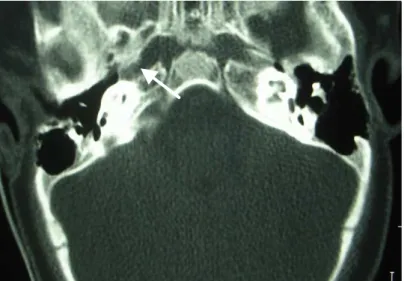
On the ABR, the electrophysiological threshold at 20 dBnHL to the left and the absence in 100 dBnHL to the right was observed. At one year of age, a computerized CAT scan (CT) of the temporal bone was carried through evidencing EVA only to the right, measuring 2,5mm (Figure 1).
FIGURE 1. CT of temporal bone showing EVA to the right.
Discussion
The vestibular aqueduct is an osseous canal that extends from the medial vestibule wall to an opening in the posterior surface of the squamous portion of the temporal bone. The duct and the endolymphatic sacs filled with endolympha course through this aqueduct. Some authors report that the EVA results from an abnormal development of the endolymphatic duct during the embryological period8.
135 Pró-Fono Revista de Atualização Científica. 2008 abr-jun;20(2).
Síndrome do aqueduto vestibular alargado: uma causa de disacusia neurossensorial.
With regards to the etiology, some authors suggest autossomal recessive inheritance2, explaining, as in our case, the lack of familial history for hypoacusia. Other authors had described mutation of gene SLC26A4 associating isolated deafness and LVAS10.
In the bilateral case the diagnosis is generally precocious given to the characteristics of the dysacusia, however, when the case is unilateral, such the dysacusia as the LVAS can remain without diagnosis as a result of the non-accomplishment of image examinations.
The treatment of this disorder consists of amplification, speech-language therapy and the prevention of cranium trauma. Occasionally, the cochlear implantation must be considered, mainly in the progressive cases.
Conclusion
Even so consulted literature reports that the diagnosis of LVAS is delayed in the majority of the cases, in the present case study, the etiological diagnosis was carried through precociously. The precocious diagnosis was made possible through the use of the CT in order to search for a unilateral dysacusia of unknown etiology.
References
1. Valvassori GE, Clemis JD. The large vestibular aqueduct syndrome. Laryngoscope. 1978;88:723-8.
2. Berrettini S, Forli F, Bogazzi F, Néri E, Salvatori L, Casani AP, Franceschini SS. Large vestibular aqueduct syndrome: audiological, radiological, clinical, and genetic features. Am J Otolaryngol. 2005:26:363-71.
3. Madden C, Halsted M, Benton C, Guinwald J, Choo D. Enlarged vestibular aqueduct syndrome in the pediatric population. Otol Neurotol. 2003;24:625-32.
4. Grimmer JF, Hedlund G. Vestibular symptons in children with enlarged vestibular aqueduct anomaly. Int J Pediatr Otorhinolaryngol. 2007; 71: 275-82.
5. Lin CY, Lin SL, Kao CC, Wu JL. The remediation of hearing deterioration in children with large vestibular aqueduct syndrome. Auris Nasus Larynx. 2005;32:99-105. 6. Willing, DB, Martín MD, Miles BA, Oehler M, Schmalbrok P. Endolymphatic sac occlusion for the enlarged vestibular aqueduct syndrome. Am J Otol. 1998;19:145-51.
7. Loundon N, Rouillon I, Munier N, Marlin S, Roger G, Garabedian EN. Cochlear implantation in children with internal ear malformations. Otol Neurotol. 2005;26:668-73. 8. Ramirez-Camacho R, Ramón GBJ, Arellano B, Trinidad A. Familial isolated unilateral large vestibular aqueduct syndrome. J Otorhinolaryngol Relat Spec. 2003;65:45-8. 9. Sugiura M, Naganawa S, Sato E, Nakashima T. Visualization of a high protein concentration in the coclea of a patient with a large endolymphatic duct and sac, using three-dimensional fluid-attenuated inversion recovery magnetic resonance imaging. J Laryngol Otol. 2006;120:1084-6.