from Antarctic Ice
Marymegan Daly
1*, Frank Rack
2, Robert Zook
21 Department of Evolution, Ecology & Organismal Biology, The Ohio State University, Columbus, Ohio, United States of America, 2 Antarctic Geological Drilling Science Management Office, University of Nebraska-Lincoln, Lincoln, Nebraska, United States of America
Abstract
Exploration of the lower surface of the Ross Ice Shelf in Antarctica by the Submersible Capable of under-Ice Navigation and Imaging (SCINI) remotely operated vehicle discovered a new species of sea anemone living in this previously undocumented ecosystem. This discovery was a significant outcome of the Coulman High Project’s geophysical and environmental fieldwork in 2010-2011 as part of the ANDRILL (ANtarctic geologic DRILLing) program. Edwardsiella andrillae n. sp., lives with most of its column in the ice shelf, with only the tentacle crown extending into the seawater below. In addition to being the only Antarctic representative of the genus, Edwardsiella andrillae is distinguished from all other species of the genus in the number of tentacles and in the size and distribution of cnidae. The anatomy and histology of Edwardsiella andrillae present no features that explain how this animal withstands the challenges of life in such an unusual habitat.
Citation: Daly M, Rack F, Zook R (2013) Edwardsiella andrillae, a New Species of Sea Anemone from Antarctic Ice. PLoS ONE 8(12): e83476. doi: 10.1371/journal.pone.0083476
Editor: Erik V. Thuesen, The Evergreen State College, United States of America
Received September 4, 2013; Accepted November 2, 2013; Published December 11, 2013
Copyright: © 2013 Daly et al. This is an open-access article distributed under the terms of the Creative Commons Attribution License, which permits unrestricted use, distribution, and reproduction in any medium, provided the original author and source are credited.
Funding: The ANDRILL 200 Coulman High Project (CHP) fieldwork in Antarctica during the austral spring and summer of 2010-2011 was funded by the U.S. National Science Foundation, Office of Polar Programs, and in New Zealand by the NZ Foundation for Research, Science and Technology. Any opinions, findings, and conclusions or recommendations expressed in this material are those of the author(s) and do not necessarily reflect the views of the National Science Foundation. The funders had no role in study design, data collection and analysis, decision to publish, or preparation of the manuscript. Competing interests: The authors have declared that no competing interests exist.
* E-mail: daly.66@osu.edu (corresponding author)
Introduction
The biota associated with glacial ice is poorly documented because the habitat is largely inaccessible and is technologically difficult to access. As part of the multi-national ANtarctic geological DRILLing (ANDRILL) program, a remotely operated vehicle called the Submersible Capable of under-Ice Navigation and Imaging (SCINI) [1] was deployed from the Ross Ice Shelf (Figure 1) through a 30-cm hole drilled by a hot water drill at two distinct locations [2]. This provided an unexpected and astonishing glimpse into this subsurface world, discovering an unusual and likely unique marine biological community dominated by anemones living inside burrows in the lower surface of the ice shelf.
At 77° 31.6’ S 171° 20.1’ E (Figure 1, site A), the upward-facing cameras on SCINI captured images of a field of approximately 100 m2 inhabited by small, tentaculate animals
living with most of their body inside the ice shelf, with tentacles dangling into the water below. A second field of animals was discovered approximately 6 km away at 77° 28.03’ S 171° 36.28’ E (Figure 1, site B). In both places, the animals appear similar in size and are spaced more or less uniformly (Figure 2A, B). The ice shelf is approximately 250-260 m thick at these
sites; mean sea level below the ice shelf surface is approximately 40 m.
These animals are sea anemones of a new species, here described as Edwardsiella andrillae. Edwardsiella is a genus of Edwardsiidae, a family of burrowing anemones reported from habitats ranging from the deepest trenches [3] to hypersaline [4] and hyposaline [5,6] coastal estuaries. All previously described species belonging to Edwardsiella are from coastal waters.
This is the first species of sea anemone reported to live in ice. Previously described species of sea anemones from Antarctica are reported from hard [7-9] or soft [10-12] substrates, but always below the anchor ice.
[14]. Annelid worms living in glaciers have physiological mechanisms including novel strategies for producing and using energy [16] and for stabilizing tubulin [17] to facilitate life at extremely low temperatures. As is the case in the ice worm
Mesenchytraeus solifugus [17], the morphology of Edwardsiella andrillae does not suggest any adaptation to the unusual environment it inhabits.
Materials and Methods
Specimens were removed from the ice using an improvised suction sampler mounted on the outside of the SCINI remotely operated vehicle. The sampler consists of a plastic tube with an opening positioned within the SCINI forward camera’s field of view that is connected through a one-way valve to a water filter and chamber where the samples are collected and stored until the vehicle is recovered to the surface. An external, inverted tunnel thruster powered by the vehicle is connected to the distal end of the plastic tube and sampling chamber to provide
water suction. The SCINI vehicle was flown under the ice shelf and positioned so that the tube opening was close to the seawater-ice interface and thus able to capture the organisms as they floated by or were extracted from their ice shelf burrows. Hot water from the drill system was pumped down from the surface of the ice shelf and used to flood the basal ice to stun the organisms and assist with the extraction process. Once the vehicle was recovered, the suction sampler was disassembled and the specimens were placed in ethanol for the helicopter trip back to McMurdo Station, where some samples were transferred to formalin for long-term preservation and further study. More than 20 samples were collected using this device mounted on the SCINI vehicle during a series of dives through the ice shelf.
The samples were collected through the U.S. Antarctic Program (USAP) by Event G
75 049-M (PI = F. Rack) based on a permit request that was processed by the U.S. National Science Foundation (NSF)
Figure 1. Known localities of Edwardsiella andrillae, n. sp. The site labeled A is at 77° 31.6’ S 171° 20.1’ E ; this corresponds to “Site 3” for the 2010-2011 SCINI dive series. The site labeled B is at 77° 28.03’ S 171° 36.28’ E ; this corresponds to “Site 4 (CH-1)” for the for the 2010-2011 SCINI dive series (Rack et al., 2012).
doi: 10.1371/journal.pone.0083476.g001
pursuant to the Antarctic Conservation Act as amended by the Antarctic Science, Tourism and Conservation Act (NSF Form 1078). NSF determined that no specific permit was required to collect marine anemones from under the Ross Ice Shelf at this location.
Whole formalin-fixed specimens were examined and photographed under a dissecting microscope. Four formalin-fixed specimens were dehydrated and embedded in paraffin, serially-sectioned at 10 µm, and stained in Heidenhain’s Azan [18]. Nematocyst preparations were made by cutting a small (>0.5 mm2) piece of tissue from each of two formalin-fixed
specimens, floating this tissue in water on a microscope slide and then smashing and smearing the tissue with a coverslip. Because of the small size of specimens, sampling for cnidae was destructive and thus the number of samples examined is limited. Nematocyst measurements were made following [19] and capsules were identified following [20,21].
Nomenclatural acts
The electronic edition of this article conforms to the requirements of the amended International Code of Zoological Nomenclature, and hence the new names contained herein are available under that Code from the electronic edition of this article. This published work and the nomenclatural acts it contains have been registered in ZooBank, the online registration system for the ICZN. The ZooBank LSIDs (Life Science Identifiers) can be resolved and the associated information viewed through any standard web browser by appending the LSID to the prefix "http://zoobank.org/". The
LSID for this publication is:
urn:lsid:zoobank.org:pub:BB12B7B1-89F4-4DE3-ADCA-66CE0EA8D149. The electronic edition of this work was published in a journal with an ISSN, and has been archived and is available from the following digital repositories: PubMed Central, LOCKSS. The holotype and paratypes have been deposited in the American Museum of Natural History.
Figure 2. External anatomy and habitus of Edwardsiella andrillae n. sp. A. Close up of specimens insitu. Image captured by SCINI. B. “Field” of Edwardsiella andrillae n. sp. in situ. Image captured by SCINI. Red dots are 10 cm apart.
Taxonomic treatment
Order Actiniaria
Family Edwardsiidae Andres, 1881
Definition. Actiniaria with elongate, vermiform body usually divisible into two or more regions: between long scapus provided with periderm and short capitulum may be short scapulus lacking periderm and ectodermal specializations. Aboral end rounded, may be differentiated into physa. No sphincter or Acontia. Mesenteries divisible into macro- and micro-cnemes; always eight perfect macrocnemes and at least four microcnemes. Macrocnemes comprise two pairs of directives and four lateral mesenteries, two on each side, whose retractors face ventral directives. Retractors diffuse to strongly restricted; parietal muscles always distinct [19].
Genus Edwardsiella Andres, 1883
Definition. Edwardsiidae with column clearly differentiated into capitulum and scapus. Three or more cycles of tentacles. Tentacles hexamerously arranged, those of innermost cycle longest. Capitulum ridged; nematocysts concentrated on ridges. Scapus generally bears periderm, always lacks nemathybomes or tenaculi. Aboral end rounded but not differentiated into a physa. Ciliated tracts of filaments short, discontinuous [22].
Type species. Edwardsia carnea Gosse, 1856 by subsequent designation [23].
Edwardsiella andrillae n. sp.
Figures 2-4; Table 1 urn:lsid:zoobank.org:act: 42A55C7F-86B8-41F0-A6B6-2CD0221A8FF3
Diagnosis. Edwardsiella with tapering, elongate column and 20-24 tapering tentacles (Figure 2B); inner tentacles notably longer. Column and tentacles opaque white, without periderm. Length of column of whole contracted specimens 16-20 mm, column diameter to 6 mm.
External anatomy. Column naked: no periderm or cuticle. In preserved specimens, capitulum short, same yellowish-white color as scapus, ridges faint; scapus long, smooth, tapers from widest point at junction with capitulum to slightly pointed aboral end (Figure 2). Regionation of the column not very pronounced. Capitulum not visible in most specimens because capitulum and base of tentacles contracted and pulled inside scapus. Mesenterial insertions visible as unbroken, straight furrows along length of column; highly contracted animals have deeper furrows than relaxed animals. Aboral end tapered rather than swollen or rounded, not differentiated from scapus; highly contracted individuals may have small pore at tip, suggesting that proximal-most part of aboral end is contracted inside the column. Tentacles in two crowded cycles differentiated by size: 8 tentacles in inner cycle longer, thicker than 12-16 tentacles of outer cycle.
Internal anatomy and histology. Longitudinal muscles of tentacles (Figure 3B) and radial muscles of oral disc (Figure 3C) ectodermal; muscles of disc weaker than those of tentacles. Tentacle with endodermal musculature at junction with oral disc (Figure 3J). Column without marginal sphincter muscle (Figure 3L). Ectoderm of capitulum thicker, more columnar than that of scapus (compare Figure 3L, 3M); ectoderm of aboral end slightly thicker but otherwise not differentiated from scapus (Figure 3N).
Eight macrocnemes of equal size and development (Figure 3A) span length of column. Two pairs directives; ventral pair attaches to single, deep, ventral siphonoglyph (Figure 3A). Siphonoglyph glandular, without nematocysts; ectoderm of actinopharynx columnar, containing glandular cells and nematocysts (Figure 3F). At least four pairs of microscopic microcnemes (Figure 3E); microcnemes without muscles, reproductive tissue, filaments, extend less than 1 mm below tentacle, not visible in every specimen or section because of unequal contraction of specimens.
Parietal muscle strongly restricted, with thick mesoglea and few, globular folds; muscle approximately equally developed on both surfaces but not symmetrical (Figure 3H). Macrocnemic mesenteries very thin between retractor and parietal muscle. Retractor muscles circumscribed, reniform, with many thin, highly branched folds (Figure 3I). Branches of retractor similar in height, widely spaced, with few ramifications. All macrocnemes fertile below region of actinopharynx; sexes apparently separate, only female specimens sectioned (Figure 3D). Eggs large (200-500 µm), yolky, with trophonema (Figures 3D, 3G). Basilar muscles absent; mesentery has microscopic expansion of mesoglea and slight fold at junction with aboral end (Figure 3K).
Cnidom Spirocysts, basitrichs, microbasic p-mastigophores (Figure 4, Table 1).
Material examined. Specimens were observed at 77° 31.6’ S 171° 20.1’ E and 77° 28.03’ S 171° 36.28’ E (Figure 1A, B respectively). Samples collected from these two sites, within 50 m of the drill hole at the lower surface of the Ross Ice Shelf. Holotype: AMNH 5350, whole specimen, from 77° 31.6’ S 171° 20.1’ E (Figure 1, site A). Paratypes: AMNH 5351, two whole specimens, 77° 31.6’ S 171° 20.1’ E (Figure 1, site A); AMNH 5352, 16 cross-section histological slides, 77° 31.6’ S 171° 20.1’ E (Figure 1, site A); AMNH 5353, whole specimen, 77° 28.03’ S 171° 36.28’ E (Figure 1, site B); AMNH 5354, 13 longitudinal-section histological slides, 77° 28.03’ S 171° 36.28’ E (Figure 1, site B).
Etymology. This species is named after the Antarctic
Drilling program that resulted in the collection of the specimens.
Discussion
Although most edwardsiids are burrowers in soft sediments [5,19,22,24], members of Edwardsiella also live in vegetation mats, in crevices, and in skeletons of dead Lophelia corals [23]. Unlike many other groups of anemones whose dispersal potential is limited, some members of Edwardsiella may disperse larger distances because of associations with other animals: members of Edwardsiella carnea (Gosse 1856) and
Edwardsiella lineata (Verrill, 1873) parasitize ctenophores as juveniles, using the host for dispersal and food [5,25,26]. Such associations are not known for all species in the genus.
Most species of Edwardsiella are described from the northern hemisphere [27]; only Edwardsiella ignota Carlgren 1959 has been reported from the southern hemisphere (Chile).
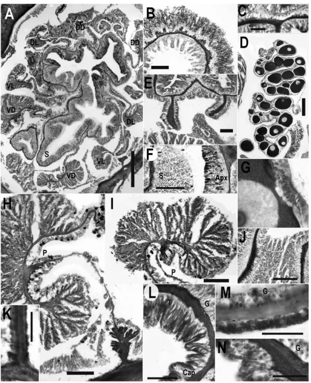
Figure 3. Internal anatomy and histology of Edwardsiella andrillae n. sp. All scale bars =100µm unless otherwise noted. A. Cross section through actinopharynx showing mesenteries and siphonoglyph. Scale = 500µm. B. Cross section through tentacle showing relatively strong ectodermal musculature and abundant spirocysts. C. Longitudinal section through oral disc showing relatively weak ectodermal musculature. Scale bar =20 µm. D. Gametogenic region of mesentery of female specimen. E. Cross section through distal column showing microcnemes. F. Close-up view of actinopharynx, showing histological differentiation of siphonoglyph. G. Trophonema of mature oocyte. Scale bar =30 µm. H. Retractor and parietal muscle of macrocnemic mesentery. I. Retractor muscle of Macrocneme. J. Musculature of base of tentacle. K. Junction between aboral end and mesentery. Note absence of basilar muscles. Scale bar =25 µm. L. Longitudinal section through distal column showing transition between tentacle and capitulum. M. Longitudinal section through scapus. Scale bar =30 µm. N. Longitudinal section through aboral end.
Abbreviations: Apx, actinopharynx; Cap; capituluar ectoderm; DD, dorsal directive mesentery; DL, dorsolateral mesentery; G, gastrodermal side of body wall; P; junction of mesentery and retractor muscle; S, siphonoglyph; T, tentacle; VD, ventrolateral directive mesentery; VL, ventrolateral mesentery.
Figure 4. Cnidae of Edwardsiella andrillae n. sp. Scale at bottom, in µm, applies to all images. See Table 1 for size ranges for each capsule type in each tissue. A. Basitrich. B. Spirocyst. C. Spirocyst. Although this capsule is smaller and has a thinner tubule than the spirocyst in Figure 4A, spirocysts show continuous variation in capsule size and robustness. D. Small basitrich. E. Basitrich. F. Small microbasic mastigophore. The small size of these cnidae precludes distinguishing them as b- or p -mastigophores. G. Spirocyst. H. Basitrich. I. Microbasic p-mastigophore. J. Small basitrich. K. Basitrich. L. Microbasic p -mastigophores.
doi: 10.1371/journal.pone.0083476.g004
includes at least two other species of Edwardsiidae: Edwardsia meridionalis Williams 1981 and Scolanthus intermedius
(McMurrich 1893). These differ from Edwardsiella andrillae in having nemathybomes, small batteries of nematocysts in the column ectoderm. Furthermore, E. meridionalis also has fewer tentacles [10], and S. intermedius has much smaller retractor muscles despite having a generally larger body size [25,26].
All eggs in the sectioned individuals are at approximately the same developmental stage (Figure 3D): some of these appear
Table 1. Size range (in μm) of the cnidae of Edwardsiella andrillae.
Edwardsiella
andrillae S N
Edwardsiella ignota
(from [30]) Tentacles
Small basitrichs none seen 17-19.7 x 3-3.5 Basitrichs (A) (29.5) 33.1-46.6
(48.5) x 3.5-5.3 2:2 58 17-24 x 4.2-5.6 Spirocysts (B, C) 19.5-37.1 (39.9) x
2.9-6.3 2:2 62 none reported
Column
Small basitrichs (D) 15.4-20.2 x 2.2-3.0 2:2 16 14-21.5 x 3.5-4.2 Basitrichs (E) 41.2-46.6 x 4.3-5.1 2:2 6 (22.6) 26.8- 36.7 x
5.6-8.5 Small mi.
mastigophores (F) 9.9-13.7 x 2.4-4.0 2:2 22 none reported Spirocysts (G) 24.0-32.4 x 3.9-6.0 2:2 9 none reported
Filaments
Small basitrichs none seen 15.5-19.7 x 2.5 Basitrichs (H) 16.0-24.7 x 2.3-3.5 3:3 33 22.6-25.4 x 3.5-5 Mi. p-mastigophores (I) 18.3-27.5 x (4.3)
4.3-6.7 3:3 65 18.3-24.0 x 3.5-5.6
Actinopharynx
Small basitrichs (J) 20.0-31.3 x 2.0-3.1 2:2 14 15.5-19.7 x 2.8 Basitrichs (K) 39.6-54.3 x 3.0-5.3 2:2 35 none reported Mi. p-mastigophores (L) 17.3-28.6 x 4.4-6.3 2:2 28 21.0-24.0 x 3.0-3.5 Specimens (S) indicates how many of the sampled specimens contained a particular type of cnida; Number (N) is the total number of capsules measured. The letter in parentheses after each type refers to Figure 4. Where multiple sizes of e.g., basitrichs are reported, the capsules within any specimen show discontinuous rather than overlapping size ranges. The small microbasic mastigophores of the column could not be distinguished as p- or b-mastigophores; these overlap in size with the small basitrichs but differ in capsule shape and tubule morphology (see Figure 4). Spirocysts are more common in the column than the measurement imply: many broken or partially discharged spirocysts were seen in the column samples. Abbreviation: Mi.= microbasic.
doi: 10.1371/journal.pone.0083476.t001
smaller (or larger) in section because the sections are tangential. All have a clearly defined nucleolus and are thus at the “late vitellogenic oocycte” stage as defined in [28]. This suggests that Edwardsiella andrillae undergoes seasonal rather than continuous reproduction, as is common in many Antarctic invertebrates [28].
The means by which Edwardsiella andrillae achieves it relatively large numbers is not clear. Edwardsiella lineata and the edwardsiid Nematostella vectensis Stephenson 1935 are able to reproduce asexually via transverse fission [29]; this can lead to large numbers of coincident individuals. Even edwardsiids that are not known to undergo asexual reproduction can achieve high densities, through high recruitment, low dispersal, or unrecognized asexual reproduction: Edwardsia meridionalis occurs at densities in excess of 10,000 individuals per square meter in waters 20-65 m deep in Antarctica (Cape Bird, New Harbor, and the jetty off McMurdo Station: [8]) and Edwardsia isimangaliso Daly et al. 2012 and Edwardsia elegans Verrill 1869 can achieve densities in the tens to hundreds of individuals per meter [4], (MD pers obs). Although not testable with the present material, these alternatives can be distinguished because they predict different population genetics and demography: Low larval dispersal or high larval recruitment would lead to genetically heterogeneous populations of individuals at many sizes or developmental stages, whereas asexual reproduction would lead to genetically homogenous populations of individuals of the same size or developmental stage.
Acknowledgements
FR and RZ are grateful for the field support received from both the US Antarctic Program and Antarctica New Zealand, and for the use of shared ANDRILL consortium equipment. We acknowledge the contributions of Dustin Carroll and Paul Mahacek, who helped pilot and maintain the SCINI vehicle during the Dives, and the rest of the ANDRILL CHP survey team, especially the drillers, who contributed to the discovery of this unusual environment and these organisms.
This manuscript was improved through comments by Estefania Rodriguez and an anonymous reviewer.
Author Contributions
Conceived and designed the experiments: MD FR. Performed the experiments: MD. Analyzed the data: MD. Contributed reagents/materials/analysis tools: FR RZ. Wrote the manuscript: MD FR.
References
1. Cazenave F, Zook R, Carroll D, Flagg M, Kim S (2011) Development of the ROV SCINI and deployment in McMurdo Sound, Antarctica. J Ocean Tech 6(3): 39-58
2. Rack FR, Zook R, Levy RH, Limeburner R, Stewart C et al. (2012) What lies beneath? Interdisciplinary outcomes of the ANDRILL Coulman High Project site surveys on the Ross Ice Shelf. Journal of Oceanography 25(3): 84–89. doi:10.5670/oceanog.2012.79.
3. Carlgren O (1956) Actiniaria from depths exceeding 6000 meters. Galathea Rep 2: 9-16.
4. Daly M, Perissinotto R, Laird M, Dyer D, Todaro A (2012) Description and ecology of a new species of Edwardsia de Quatrefages, 1842 (Anthozoa, Actiniaria) from St Lucia Estuary, South Africa. Mar Biol Res 8: 233-245. doi:10.1080/17451000.2011.617757.
6. Hand C, Uhlinger KR (1994) The unique, widely distributed, estuarine sea anemone, Nematostella vectensis Stephenson: a review, new facts, and questions. Estuaries 17: 501-508. doi:10.2307/1352679. 7. Dunn DF (1983) Some Antarctic and sub-Antarctic sea anemones
(Coelenterata: Ptychodactiaria and Actiniaria). Ant Res Ser 39: 1–67. 8. Fautin DG (1984) More Antarctic and Subantarctic sea anemones
(Coelenterata: Corallimorpharia and Actiniaria). Ant. Res Ser 41: 1–42. 9. Rodriguez ER, Lopez-Gonzalez PJ (2013). New records of Antarctic
and Sub-Antarctic sea anemones (Cnidaria, Anthozoa, Actiniaria) from the Weddell Sea, Antarctic Peninsula, and Scotia Arc. Zootaxa 3624.1: 1-100
10. Williams RB (1981) A sea anemone, Edwardsia meridionalis, sp. nov., from Antarctica and a preliminary revision of the genus Edwardsia de Quatrefages, 1841 (Coelenterata: Actiniaria). Rec Aust Museum 33: 325-360. doi:10.3853/j.0067-1975.33.1981.271.
11. Ansell AD, Peck LS (2000) Burrowing in the Antarctic anemone, Halcampoides sp., from Signy Island, Antarctica. J Exp Mar Bio Ecol 252: 45-55. doi:10.1016/S0022-0981(00)00232-X. PubMed: 10962064. 12. Rodríguez E (2012) Another bipolar deep-sea anemone: new species
of Iosactis (Actiniaria, Endomyaria) from Antarctica. Helg Mar. Resour 66: 211-218.
13. Ansell AD, Trueman ER (1968) The mechanism of burrowing in the anemone Peachia hastata Gosse. J Exp Mar Biol Ecol 2: 124–134. doi: 10.1016/0022-0981(68)90003-8.
14. Thomas DN, Dieckmann GS (2002) Antarctic sea ice--a habitat for extremophiles. Science 295 : 641-644. doi:10.1126/science.1063391. PubMed: 11809961.
15. Napolitano MJ, Nagele RG, Shain DH (2004) The ice worm, Mesenchytraeus solifugus, elevates adenylate levels at low physiological temperature. Comp Biochem Physiol A Mol Integr Physiol 137: 227-235. doi:10.1016/j.cbpb.2003.10.005. PubMed: 14720608. 16. Tartaglia LJ, Shain DH (2008) Cold-adapted tubulins in the glacier ice
worm, Mesenchytraeus solifugus. Gene 423: 135-141. doi:10.1016/ j.gene.2008.07.025. PubMed: 18718858.
17. Shain DH, Carter MR, Murray KP, Maleski KA, Smith NR et al. (2000) Morphologic characterization of the ice worm Mesenchytraeus
solifugus. J Morphol 246: 192–197. doi: 10.1002/1097-4687(200012)246:3. PubMed: 11077431.
18. Presnell J, Schreibman MP (1997) Humanson’s Animal Tissue Techniques. Baltimore, MD : Johns Hopkins University Press. 600 p. 19. Daly M, Ljubenkov JC (2008) Edwardsiid sea anemones of California
(Cnidaria: Actiniaria: Edwardsiidae), with descriptions of eight new species. Zootaxa 1860: 1-27.
20. Mariscal RN (1974) Nematocysts. In: L MuscatineHM Lenhoff. Coelenterate biology: reviews and new perspectives. New York: Academic Press. pp 129-178.
21. England KW (1987) Actiniaria from the Red Sea and tropical Indo-Pacfic. Bull Brit Mus Nat l History 53: 205-292
22. Daly M (2002) A systematic revision of Edwardsiidae (Cnidaria: Anthozoa). Invert Biol 121: 212-225.
23. Manuel RL (1981) On the identity of the sea anemone Scolanthus callimorphus, Gosse, 1853 (Actiniaria: Edwardsiidae). J Nat Hist 15: 265-276
24. Carlgren O (1921) Actiniaria I. Danish Ingolf Expedition 5(9): 1-241. 25. Crowell S (1965) An anemone parasitic as a larva in a comb jelly. Am
Zool 5: 645.
26. Reitzel AM, Sullivan JC, Brown BK, Chin DW, Cira EK et al. (2007) Ecological and developmental dynamics of a host-parasite system involving a sea anemone and two ctenophores. J Parasitol 93: 1392-1402. doi:10.1645/GE-1250.1. PubMed: 18314686.
27. Fautin DG (2013) Hexacorallians of the World. Available: http:// geoportal.kgs.ku.edu/hexacoral/anemone2/index.cfm. (Accessed January 2013)
28. Rodríguez E, Orejas C, López-González PJ, Gili JM (2013) Reproduction in the externally brooding sea anemone Epiactis georgiana in the Antarctic Peninsula and the Weddell Sea. Mar Biolr 160: 67-80. doi:10.1007/s00227-012-2063-x.
29. Shick JM (1991) A functional biology of sea anemones. London: Chapman & Hall. 395 pp.
30. Carlgren O (1959) Reports of the Lund University Chile Expedition 1948–49, 38. Corallimorpharia and Actiniaria with description of a new genus and species from Peru Ark Zool series NF 71(6): 1–38