A Novel Lipase as Aquafeed Additive for
Warm-Water Aquaculture
Chao Ran☯, Suxu He☯, Yalin Yang, Lu Huang, Zhigang Zhou*
Key Laboratory for Feed Biotechnology of the Ministry of Agriculture, Feed Research Institute, Chinese Academy of Agricultural Sciences, Beijing, P. R. China
☯These authors contributed equally to this work.
*zhou_zg@msn.com
Abstract
A novelAcinetobacterlipase genelipG1was cloned from DNA extracted from intestinal sample of common carp (Cyprinus carpio), and expressed inE.coliBL21. The encoded protein was 406 amino acids in length. Phylogenetic analysis indicated that LipG1 and its relatives comprised a novel group of true lipases produced by Gram-negative bacteria. LipG1 showed maximal activity at 40℃and pH 8.0 whenpNP decanoate (C10) was used as the substrate, and remained high activity between 20℃and 35℃. Activity of the lipase was promoted by Ca2+and Mg2+, and inhibited by Zn2+and Cu2+. Moreover, LipG1 is stable
with proteases, most commercial detergents and organic solvents. Substrate specificity test indicated that LipG1can hydrolysepNP esters with acyl chain length from C2 to C16, with preference for medium-chainpNP esters (C8, C10). Lastly, LipG1was evaluated as an aquafeed additive for juvenile common carp (Cyprinus carpio). Results showed that supple-mentation of LipG1significantly improved the gut and heptaopancreas lipase activity of fish fed with palm oil diet. Consistently, improved feed conversion ratio and growth performance were recorded in the LipG1 feeding group, to levels comparable to the group of fish fed with soybean oil diet. Collectively, LipG1 exhibited good potential as an aquafeed additive enzyme, and deserves further characterization as the representative of a novel group of lipases.
Introduction
China is the largest aquaculture producer in the world, with a total production of 53.94 million tons of fish in 2012, accounting for nearly 60% of the world total [1]. Consequently, there has been a consistent increase in the demand for aquafeed over time. However, the price of aqua-feed is relatively high, which represents one of the main bottlenecks for the development of aquaculture. In aquafeed, dietary lipids provide essential fatty acids and are important source of energy for fish. The optimum dietary lipid levels for tilapia and common carp are as high as 8% and12%, respectively [2]. Fish oil and soybean oil have been used as the lipid source in aquafeed for these fish species. However, due to the high price of both fish oil and soybean oil, as well as the finite nature of fish oil, alternative vegetable oils are prevalent. Among these,
a11111
OPEN ACCESS
Citation:Ran C, He S, Yang Y, Huang L, Zhou Z (2015) A Novel Lipase as Aquafeed Additive for Warm-Water Aquaculture. PLoS ONE 10(7): e0132049. doi:10.1371/journal.pone.0132049
Editor:Maoteng Li, Huazhong university of Science and Technology, CHINA
Received:March 4, 2015
Accepted:June 9, 2015
Published:July 6, 2015
Copyright:© 2015 Ran et al. This is an open access article distributed under the terms of theCreative Commons Attribution License, which permits unrestricted use, distribution, and reproduction in any medium, provided the original author and source are credited.
Data Availability Statement:The nucleotide sequence of the chitinase gene (lipG1) was deposited in GenBank under the accession number KM925083. All other data are within the paper and its Supporting Information files.
palm oil (PO) is the vegetable oil with the highest production volume and lowest price world-wide, and therefore is a very attractive lipid source for reduction of the aquafeed price.
Fatty acids (FA) digestibility in fish decreases with increasing chain length and increases with increasing degree of unsaturation [3,4,5]. Also, the digestibility of FA is influenced by dietary lipid source and the level of dietary saturates [6], i.e., high level of saturated fatty acids in the diet may reduce the digestibility of saturated FA, and to a lesser extent, of the monoenes and PUFA [3,4]. Although research into the use of palm oil in fish diets has shown encourag-ing results [7–10], supplementation of palm oil in aquafeed may reduce the digestibility of lipid and fatty acids, especially when used at high dietary level and low temperature, due to its high level of saturated fatty acids [3,4,8]. The reduction in lipid and fatty acids digestibility could lead to compromised growth and feed utilization efficiency of fish, given a lower level of total lipid in the aquafeed and longer feeding period [4,8]. The reduction of fatty acids digestibility of high PO diet, especially saturates, was partially due to the poor triacylglycerol (TAG) diges-tion, as indicated by the increased TAG content in fecal lipids of fish fed high PO diet [3]. Therefore, a potential way to solve the problem is supplementation of exogenous lipase in the diet to improve the lipolysis of TAGs of palm oil.
The studies on the supplementation of exogenous lipase for improvement of lipid digestion and growth performance have rarely been conducted in fish. Samuelsen et al. reported that lipase addition had no effect on growth performance and related parameters of rainbow trout (Oncorhynchus mykiss) [11]. Moreover, properties of many available lipases were not suitable as aquafeed additive.
Microbial lipases have been widely utilized due to their availability, substrate specificity, and diversity in catalytic activities [12,13]. In particular, many novel lipases have been isolated or cloned fromAcinetobacterspp., showing various beneficial biochemical properties such as sta-bility in organic solvents, broad substrate specificity, cold-adapted asta-bility and stereo-selectivity [14–19]. In this study, a novelAcinetobacterlipase gene was cloned from DNA extracted from intestinal sample of common carp,Cyprinus carpio, and subsequently expressed inE.coli. Bio-chemical properties of the lipase were characterized, and the application of this lipase as aqua-feed additive was evaluated in common carp fed diet with high level of palm oil.
Materials and Methods
Ethics statement
All experimental and animal care procedures were approved by Feed Research Institute of Chi-nese Academy of Agricultural Sciences Animal Care Committee, under the auspices of the China Council for Animal Care (Assurance # 2012-HSX01). MS-222 was used as the anaes-thetic. Field study was conducted on private aquaculture pond under the owner's permission.
Strains and media
E.coliDH5αwas used for cloning of the PCR product.E.coliBL21 was used for expression of the recombinant enzyme.E.colistrains were cultured in LB medium (10 g tryptone, 5 g yeast extract, 10 g NaCl per liter) at 37°C.
Cloning of the lipase gene and sequence analysis
By comparing the amino acid sequences ofAcinetobacterlipases, two highly conserved regions were identified. The degenerate primers were designed based on the sequences of the two regions by CODEHAP primer [20]. The partialAcinetobacterlipase gene sequences between the two conserved motifs were amplified from DNA extracted from intestinal sample of and analysis, decision to publish, or preparation of
the manuscript.
common carp by touchdown PCR. The amplified PCR product was cloned intoE.coliDH5α
using pGEM-T Easy (Promega, Madison, USA). Fifty transformants were randomly picked. Plasmids were extracted with the TIANprep Mini Plasmid Kit (TIANGEN, Beijing, China), and the cloned partial lipase gene sequences were sequenced at Beijing Calculation Centre. One sequence that showed relatively high abundance among the sequenced clones was selected for further study. To obtain the upstream and downstream of the partial sequence of the lipase gene, genome walking PCR was performed with a genome walking kit according to manufac-turer's instructions (Takara, Japan). The primers were designed based on the partial sequence (Table 1). The elongated fragment was sequenced at Beijing Calculation Centre. Sequence assembly was performed using Vector NTI Suite 7.0 (InforMax, Gaithersburg, MD, USA). ORF was found using the alternative initiation codon finder in OR finder (http://www.ncbi. nlm.nih.gov/projects/gorf/). The amino acid sequence was analyzed by SignalP 3.0 server [21] to identify the signal peptide. Alignments of the DNA and protein sequences were carried out using BLASTn and BLASTp, respectively (http://www.ncbi.nlm.nih.gov/BLAST/). Phyloge-netic tree was generated using the neighbor-joining method by MEGA 5.0 [22].
Expression of the recombinant lipase in
E
.
coli
The following primers were used for construction of the lipase expression plasmid: lipG1F (withEcoRI site) and lipG1 R (withNotI site). The PCR products were inserted into pET-28a (+) digested withEcoRI andNotI. The recombinant plasmid, pET28a—lipG1, was sequenced to confirm the insertion, followed by transformation intoE.coliBL21 (DE3) cells. Transformed cells were picked from a single colony and grown overnight at 37°C in shake flask containing 3 ml LB broth supplemented with 100μg/ml kanamycin, followed by inoculation at a dilution of
1:100 in 100 ml fresh LB medium containing kanamycin and aerobic incubation at 37°C. When OD600reached 0.6, isopropyl -β-D-1-thiogalactopyranoside (IPTG) was added to the
growth medium at a final concentration of 0.5 mM. After further incubation overnight at 16°C, the cells were harvested by centrifugation at 8,000×g for 10 min at 4°C.
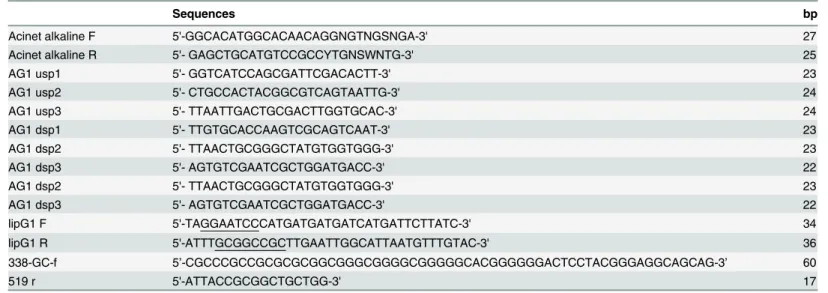
Table 1. Primers used for gene cloning and expression.
Sequences bp
Acinet alkaline F 5'-GGCACATGGCACAACAGGNGTNGSNGA-3' 27
Acinet alkaline R 5'- GAGCTGCATGTCCGCCYTGNSWNTG-3' 25
AG1 usp1 5'- GGTCATCCAGCGATTCGACACTT-3' 23
AG1 usp2 5'- CTGCCACTACGGCGTCAGTAATTG-3' 24
AG1 usp3 5'- TTAATTGACTGCGACTTGGTGCAC-3' 24
AG1 dsp1 5'- TTGTGCACCAAGTCGCAGTCAAT-3' 23
AG1 dsp2 5'- TTAACTGCGGGCTATGTGGTGGG-3' 23
AG1 dsp3 5'- AGTGTCGAATCGCTGGATGACC-3' 22
AG1 dsp2 5'- TTAACTGCGGGCTATGTGGTGGG-3' 23
AG1 dsp3 5'- AGTGTCGAATCGCTGGATGACC-3' 22
lipG1 F 5'-TAGGAATCCCATGATGATGATCATGATTCTTATC-3' 34
lipG1 R 5'-ATTTGCGGCCGCTTGAATTGGCATTAATGTTTGTAC-3' 36
338-GC-f 5’-CGCCCGCCGCGCGCGGCGGGCGGGGCGGGGGCACGGGGGGACTCCTACGGGAGGCAGCAG-3’ 60
519 r 5'-ATTACCGCGGCTGCTGG-3' 17
Purification and identification of the recombinant LipG1
The lipase was purified with protocol described in detail previously [19]. The bacterial pellets were resuspended in 15 ml lysis buffer (50 mM Tris-Cl pH 8.0), disrupted by sonication, fol-lowed by centrifugation at 10,000 × g for 30 min at 4°C. The concentrated supernatant (crude enzyme) was applied to the Ni-NTA affinity chromatography column (GE Healthcare Life Sci-ences, Piscataway, USA) which was equilibrated with washing buffer (50mMTris-ClpH8.0, 500 mM NaCl, and 10% glycerol). The enzyme protein was eluted using an imidazole gradient (0, 20, 40, 80, 100, and 200 mM) in washing buffer. After the elution, the imidazoles in the purified enzyme were removed by dialysis in 50 Mm Tris-Cl (pH 8.0). The purified enzyme was ana-lyzed by SDS-PAGE. The single protein band after purification was confirmed by the peptide mass fingerprinting.
Lipase activity assay
Lipase activity was determined using the chromometer method described by Zhang et al. [19]. Unless otherwise described, lipase activity was measured at 40°C with 500μMpNP decanoate
in 50 mM Tris-HCl buffer (pH 9.0) containing 1% ethanol. The reaction can begin by the addi-tion of 100μl of purified enzyme at a concentration of 0.25 mg/ml. The reaction mixture was
incubated at 40°C for 10 min, after which the reaction was terminated through addition of 1ml ethanol. Blank reactions were performed in reaction mixtures without the enzyme. The mix-tures were centrifuged, and the absorbance of the supernatants was measured at 405 nm with spectrophotometer (Beckman, USA). One unit (U) of enzyme activity was defined as the amount of enzyme that releases 1μmol ofpNP per minute.
Enzyme characterization of recombinant LipG1
ThepNP decanoate was used to assay the following characteristics of LipG1. The optimal pH was determined at 40°C using buffers with pH from 3.0 to 12.0, i.e., 2 0 mM disodium hydro-gen phosphate-citric acid buffer (pH 3.0–8.0), 50 mM Tris—HCl buffer (pH 8.0–10.0), and 20 mM glycine-NaOH (pH 10.0–12.0). To test its pH stability, 100μl of the lipase (2.5 mg/ml)
was preincubated for 1 h at 40°C in 900μl buffers with pH values ranging from 3.0 to 12.0, and
then lipase activity was measured under standard conditions. The enzyme’s optimal tempera-ture was determined by measuring its activity at temperatempera-tures ranging from 0°C to 60°C, with pH set as 8.0. The thermostability was monitored by preincubation of the enzyme in Tris-HCl buffer (pH 8.0) for 30, 60, 90, and 120 min at 20, 30, 40, and 50°C. Lipase activity was then measured under standard conditions. The effects of different metal ions, chemical reagents, detergents, and organic solvents on enzymatic activity were assessed in 50 mM Tri-HCl buffer (pH 8.0) at 40°C. The reactions contained 1 or 10 mM of NaCl, KCl, BaCl2, CaCl2, CoCl2,
CuCl2, NiCl2, MgCl2, MnCl2, ZnCl2, CdCl2; 0.1 or1% Tween-20, Tween-40, Tween-80, Triton
X-100, SDS, and cetyltrime-thylammonium bromide (CTAB); 10% or30% methanol, ethanol, isopropanol, capryl alcohol, n-heptane, glycerol and dimethyl sulphoxide (DMSO).
In order to examine the resistance to different proteases, the purified recombinant enzyme (100μg ml−1) was incubated with 10μg ml−1trypsin (from bovine, pH 7.6, 25°C, 14,700 U
mg−1; Sigma), 250
μg ml−1α-chymotrypsin (type II from bovine, pH 7.8, 25°C,40 U mg−1;
Sigma), 500μg ml−1subtilisin A (type VIII fromBacillus licheniformis, pH 7.5, 37°C, 10 U
mg−1; Sigma), 330
μg ml−1proteinase K (type VIII fromB.licheniformis, pH 7.5, 37°C, 30 U
mg−1; Amresco, Solon, USA), respectively. After incubation for 1 or 2 h, the residual activity
The substrate selectivity of LipG1 forpNP esters was assayed usingpNP acetate (C2),pNP butyrate (C4),pNP octanoate (C8),pNP decanoate (C10),pNP laurate (C12),pNP myristate (C14), andpNP palmitate(C16) as substrates in 50 mM Tris-HCl (pH 8.0) at 40°C. ThepNP ester substrates exceptpNP palmitate were dissolved in ethanol at a final concentration of 50 mM as the stock solution, andpNP palmitate was dissolved in isopropanol. The working con-centration range of thepNP esters was from 0.01 to 2 mM. Initial velocity versus substrate con-centration data were fitted to the Lineweaver—Burk transformation of the Michaelis—Menten equation.
Experimental diets and feeding experiment
Five isonitrogenous and isocaloric experimental diets were formulated containing approxi-mately 34% crude protein, 9% crude lipid (S1 Table).Diet with palm oil as the lipid source was used as negative control (CKn), while soybean oil diet was positive control group (CKp). Palm oil diet were supplemented with 3 U g-1LipG1, 6 U g-1LipG1 and 6 U g-1commercial lipase (Leaveking, Shengzhen, China), giving treatment groups T1, T2 and M, respectively. The feed pellets were dried under forced air at room temperature for 24 h and then kept at 4°C.
Common carp (Cyprinus carpio) were obtained from Tianjing freshwater fish husbandry factory (Tianjing, China). Before starting the experiment, the juvenile common carp were reared in an experimental thermo regulated rearing system and fed with the control diet for 2 weeks to acclimatize to the experimental conditions. Fish of similar sizes (5.66 ± 0.21 g) were randomly distributed into 15 circular glass tanks (30 l) and each glass tank was stocked with 12 fish. Each diet was then randomly assigned to triplicate tanks. The fish were hand-fed to appar-ent satiation three times daily (at 8:30, 12:30 and 16:30) for 28 days. During the experimappar-ental period, the water temperature, dissolved O2, pH and ammonia content were maintained at
28.0 ± 1.3°C, 7.60 ± 0.28 mg l−1, 7.5 ± 0.29 and 0.10 ± 0.02 mg l−1, respectively.
Sampling and measurements
At the end of the feeding experiment, all of the fish were anesthetized with MS-222. Body weight was measured at batch. The hepatopancreas and gut were rapidly excised from three fish of each tank. The samples of fish from the same tank were pooled, frozen in liquid nitrogen and stored at−80°C. Weight gain (WG), feed conversion ratio (FCR) and survival rate (SR) were calculated.
Lipid-metabolism-related enzymes activities
The hepatopancreas and gut samples were homogenized in 4 volumes of ice-cold buffer (20 mM Tris-HCl, 0.25 M sucrose, 2 mM EDTA, pH 7.4) and centrifuged at 18,000 × g for 10 min at 4°C. Lipase and protease activities were measured in the homogenate with commercial assay kits (Jiancheng Biotech. Co., Nanjing, China).
Adhesive intestinal bacterial community
20 min, respectively, and viewed by UV transillumination. The excised bands were reamplified and purified, then sequenced.
Statistics
The effects of different diets in the feeding experiment were analyzed with one-way ANOVA, and Duncan’s multiple range test was used to compare the means between any two groups. Dif-ferences with aPvalue lower than 0.05 were considered as significant. All the statistical analysis was conducted on SPSS 17.0.
NCBI accession numbers
The nucleotide sequence of the chitinase gene (lipG1) was deposited in GenBank under the accession number KM925083.
Results
Sequence analysis of LipG1
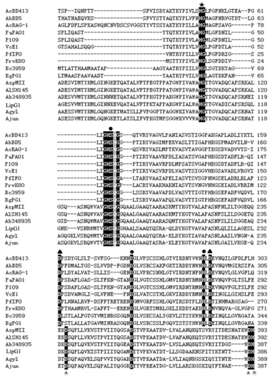
Bioinfomatics analysis showed that thelipG1gene (KM925083) contained an open reading frame with 1,227 nucleotides, encoding a protein of 406 amino acids (Fig A inS2 File). A 20 amino acid residue signal peptide was found at the N-terminal of the lipase with a cleavage position between Cys20 and His21 as predicted by Signal P (Fig A inS2 File). BLASTp analysis indicated that LipG1 was 99% identical to hypothetical protein fromAcinetobacter tandoii
(WP_016167430.1), and was 70%, 68%, 68%, 62% and 61% identical to lipases fromA. bau-mannii348935 (WP_034704719.1),Acinetobactersp. MII (KGH48978.1),A.lwoffiiSH145 (WP_004279025),A.gyllenbergii(WP_032860322.1) andA.junii(WP_005402474.1), respec-tively. Alignment of LipG1 and its relatives with representatives of subfamily I.1 and I.2 of bac-terial true lipases revealed the typical lipase semi-conserved pentapeptide, GXSXG, where the catalytic residue Ser is located, as well as the catalytic residue Asp, at homologous positions in all sequences (Fig 1). The last catalytic residue constituting the lipase consensus catalytic triad, His, was not found at homologous position in LipG1. Nevertheless, a conserved His residue was located in LipG1 and its relatives at the position close to the catalytic His for I.1 and I.2 lipases (Fig 1). Therefore, the consensus lipase catalytic triad in LipG1putatively comprises of Ser 190 (in the motif GHSQG), Asp 354 and His 386, which requires further experimental con-firmation. An HG sequence, which putatively constitutes an oxyanion hole in the three-dimen-sional protein structure, was also located at homologous position for all sequences (Fig 1).
Enzyme expression, purification, and mass spectrometry analysis
The recombinant LipG1 was expressed inE.coliBL21 in the presence of 0.01mM IPTG at 20°C. After Ni-NTA affinity chromatography, the enzymatic activity of recombinant LipG1 was 8908 U mg−1toward C8pNP. The purified enzyme presented as a single band of about
Fig 1. Alignment of amino acid sequences of representative lipases from subfamilies I.1 and I.2 of bacterial lipases and the group of lipases represented by LipG1.Abbreviations and sequence accession numbers: AcBD413 (Acinetobacter calcoaceticusBD413), CAA56780.1; AbBD5 (A.baumanniiBD5), ABW70205.1; AcRAG-1 (A.calcoaceticusRAG-1), AAD29441.1; PaPAO1 (Ps.aeruginosaPAO1), CAA44997.1; P109 (Pseudomonassp. 109), P26877.1; VcE1 (V.choleraeE1), P15493.2; PfIFO (P.fragi IFO-12049), P08658.2; PrvK80 (Proteus vulgarisK80), AAB01071.1; Bc3959 (Burkholderia cepacia DSM3959), P22088.2; BgPG1 (Burkholderia glumaePG1), Q05489.1; AspMII (Acinetobactersp. MII), KGH48978; AlSH145 (A.lwoffiiSH145), WP_004279025; Ab348935 (A.baumannii348935), WP_034704719.1; LipG1, this study; Agyl (A.gyllenbergii), WP_032860322.1; Ajun (A.junii),
WP_005402474.1. Symbols:●Catalytic triad residues,▲Asp residues involved in calcium binding,$HG dipeptide of the oxyanion loop,Putative triad residue,4Putative calcium binding Asp residues.
Effects of pH and temperature on LipG1 activity
LipG1showed maximum activity at pH 8.0, and retained at least 70% of its maximum activity between pH 7.0 and 8.5 (Fig 2a). LipG1 was stable over a wide pH range, remaining at least 60% of its maximum activity after incubation at pH from 3.0 to 12.0 (Fig 2c). The enzyme activity of LipG1 peaked at 40°C, and retained over 40% of its maximum activity at tempera-tures from 20°C to 35°C (Fig 2b). The recombinant lipase showed high stability at 20°C and 30°C. However, the activity decreased quickly when incubated at 50°C (Fig 2d).
Effects of metal ions, organic solvents, detergents and proteases on
LipG1 activity
The effects of different metal ions, organic solvents, and detergents on the activity of LipG1 are shown inTable 2, Tables A and B inS1 File, respectively. At the concentration of 1mM, lipase activity was strongly inhibited byCo2+, Cr3+, Fe3+,Pb+, Ag+and EDTA, and enhanced by Ca2+, Mg2+. At the higher concentration of 10 mM, Cu2+, EDTA,Co2+, Zn2+and Mn2+drastically Fig 2. Characterization of purified recombinant LipG1.a. The effect of pH on lipase activity. The activity assay was performed at 37°C in buffers of pH 3.0–12.0 for 10 min. b. The effect of temperature on lipase activity measured in 0.1 M PBS buffer (pH 8.0) for 10 min. c. pH stability of LipG1. After pre-incubating the enzyme at 37°C for 1 h at various pHs, the residual activity was measured in 0.1 M PBS buffer (pH 8.0), 40°C. d. Thermostability of purified LipG1. The enzyme was preincubated at 20, 30, 40, or 50°C in 0.1 M PBS buffer (pH 8.0). Aliquots were removed at specific time points for measurement of residual activity in the same buffer at 40°C. e. Substrate specificity of LipG1 determined with various chain length fatty acid esters. f. Effects of proteases on the activity of LipG1. Each value in the panel represents the mean±SD (n = 3).
inhibited its activity; Na+, K+, Cr3+, Ni2+, Pb+and Ag+strongly inhibited the activity. Neverthe-less, a stimulating effect was still observed for Ca2+and Mg2+(Table 2). The enzyme was stable in various organic solvents at the concentration of 10%, with extended stability at the concen-tration of 30% for methanol, ethanol, n-heptane, glycerol and DMSO (>75% activity
remained). However, isopropanol and capryl alcohol drastically reduced its activity at the con-centration of 30% (Table A inS1 File). At the concentration of 0.1%, Tween 80 and CTAB didn’t significantly inhibit the activity of LipG1, and the non-ionic detergent Triton X-100 slightly enhanced its activity. Tween 20, Tween 40, SDS reduced its activity by 37.5%, 28.3%, 57.7%, respectively. At the concentration of 1%, all the detergents except Triton X-100 drasti-cally inhibited the lipase activity, with over 50% activity loss observed (Table B inS1 File).
We also investigated the resistance of recombinant LipG1against proteases (Fig 2f). The enzyme retained over 60% activity after treatment with all proteases at 37°C for 1 h, with over 80% activity retained for subtilisin and pepsin. Also, longer incubation time of 120 min didn’t lead to considerable further activity loss for all the tested proteases.
Substrate specificity of LipG1
The substrate specificity of LipG1 was examined usingpNP esters with different chain lengths. Results showed that the lipase showed activity on a wide range of substrates. LipG1 preferred medium chain fatty acid substrates, and the highest activity was registered forpNP octanoate (Fig 2e,S2 Table).
Effect of LipG1 on growth and feed utilization of common carp
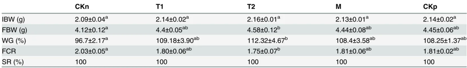
Dietary supplementation of LipG1 at 6 U/g significantly increased the final body weight (FBW) and weight gain (WG) of common carp compared with negative control after feeding for 4 weeks. LipG1 at 3 U/g and commercial lipase at 6 U/g marginally increased the growth of carp compared with the negative control. Overall, all the lipase supplementation groups showed similar growth performance (P>0.05), which is comparable to the group with soybean oil
(Table 3). Also, significantly lower FCR (feed conversion ratio) was observed in 6 U/g LipG1 Table 2. Effects of various metal ions on LipG1 activity.
Metal ion Relative activity (%)
1 mM 10 mM
Na+ 101.6±2.3 88.7±1.7
K+ 107.4±1.9 81.6±2.3
Ca2+ 115.5±±4.4 213.9±3.5
Zn2+ 97.5±±2.1 37.7±2.1
Li+ 104.2±2.5 97.5±2.5
Co2+ 50.6±1.2 30.3±0.9
Cr3+ 79.6±2.2 70.4±1.7
Ni2+ 81.1±3.1 49.2±1.2
Cu2+ 84.9±±2.2 0
Mg2+ 119.4±2.7 171.0±1.4
Fe3+ 66.9±1.8 18.2±0.6
Mn2+ 93.7±2.0 15.1±0.9
Pb+ 76.6±1.5 56.8±1.3
Ag+ 60.6±1.2 43.4±1.5
EDTA 75.5±1.3 11.1±0.4
group compared with the negative control (P<0.05), and marginally decreased FCR was
recorded in 3 U/g LipG1 and the commercial lipase groups, with no significant difference among the lipase addition groups, which were comparable to the soybean oil group(Table 3).
Effect of LipG1 on digestive enzyme activities of common carp
Heptaopancreas trypsin activity was not significantly different among groups, while the gut trypsin activity was similar among the palm oil groups regardless of lipase supplementation, and the activity in soybean oil group was significantly higher (Table 4). Dietary exogenous lipases (T1, T2 and M) significantly increased gut lipase activity compared with the negative control (P<0.05), with an activity comparable to the positive control. The heptaopancreas
lipase activity was significantly increased in groups with lipase supplementation at 6 U/g (T2 and M) (Table 4). For both gut and heptaopancreas lipase activity, the maximal value was observed in the 6 U/g LipG1 group.
Effect of LipG1 on common carp gut microbiota
The 16S rRNA gene V3 region PCR-DGGE fingerprints of the adhesive intestinal microbiota of different groups are shown inS1 FigMost of the OTUs of the intestinal microbiota of com-mon carp were assigned to theProteobacteria(17 OTU) andFirmicutes(4 OTU) (S3 Table). The gut microbiota was affected by dietary lipid sources, as the microbiota of palm oil diet associated groups were clearly distinguishable from the microbiota of the soybean oil group (Fig 3). Lipase supplementation didn’t exert significant change to the intestinal microbiota, and the intestinal microbial profiles of all the lipase supplement groups were similar (Fig 3). Table 3. Effects of dietary lipase on weight gain, feed conversion ratio and survival rate of common carp (Cyprinus carpio) (n = 3).
CKn T1 T2 M CKp
IBW (g) 2.09±0.04a 2.14±0.02a 2.16±0.01a 2.13±0.01a 2.14±0.02a
FBW (g) 4.12±0.12a 4.4±0.05ab 4.58±0.12b 4.44±0.08ab 4.45±0.06ab
WG (%) 96.7±2.17a 109.18±3.90ab 112.32±4.67b 108.4±3.58ab 108.25±1.37ab
FCR 2.03±0.05a 1.80±0.06ab 1.75±0.07b 1.81±0.06ab 1.81±0.02ab
SR (%) 100 100 100 100 100
Values in the same row with different superscript letters (a, b, ab) have significant difference (P<0.05). FBW,final body weight (g); Weight gain (%), WG (%) = 100 × (Final body weight−Initial body weight)/Initial body weight; Feed conversion ratio, FCR = feed fed (g)/weight gained (g).
doi:10.1371/journal.pone.0132049.t003
Table 4. Trypsin and lipase activities in the gut and heptaopancreas of common carp (Cyprinus carpio) fed with different diets (n = 3).
Treatments CKn T1 T2 M CKp
Trypsin activity (U/mg)
Heptaopancreas 127.7±2.4a 125.3±,3.8a 128.7±4.3a 125.3±2.3a 134.3±1.5a
Gut 77.0±1.5a 78.0±2.3a 79.7±3.0a 79.3±2.4a 86.7±1.5b
Lipase activity (U/g)
Heptaopancreas 62.0±0.6a 65.7±1.2ab 68.7±2.9b 68.7±2.0b 67.3±2.0ab
Gut 41.3±2.0a 49.7±1.8b 58.3±1.5c 54.0±1.7bc 54.0±1.7bc
Values in the same row with common superscript (a, b, ab, c, bc) are not significantly different (P>0.05).
Discussion
Bacterial true lipases comprise the family I of bacterial lipolyitc enzymes, and were further clas-sified into six subfamilies [25]. True lipases from Gram-negative bacteria were assigned into the first three subfamilies. Subfamily I.1 were lipases fromPseudomonas aeruginosa,Vibrio cholerae,Acinetobacter calcoaceticus,etc. Subfamily I.2 mainly comprises lipases produced by
Burkholderia, while I.3 contains enzymes from two distinct species:Ps.fluorescensandSerratia marcescens[25]. Many lipase genes have been cloned fromAcinetobacter[14,15,17,18,19]. TheAcinetobacterlipases were assigned to subfamily I.1 of bacterial true lipases [25], and were proposed as a separate‘Acinetobacter’clade based on similarity of amino acid sequences [15, 26]. However, amino acid sequence alignment showed that LipG1 isn’t homologous to any of the above mentionedAcinetobacterlipases and doesn’t belong to the typical‘Acinotebacter’ lipase clade. Based on the phylogenetic tree predicted from multiple sequence alignment of the lipases (Fig 4), and considering the deviation in the position of the catalytic residue His (Fig 1), Fig 3. Cluster analysis of the adhesive gut bacterial communities of common carp (Cyprinus carpio).
doi:10.1371/journal.pone.0132049.g003
Fig 4. Phylogenetic tree of true lipases produced by Gram-negative bacteria, including LipG1 and its relatives.Abbreviations and sequence accession numbers: PaPAO1 (P.aeruginosaPAO1), CAA44997.1; P109 (Pseudomonassp. 109), P26877.1; AcBD413 (Acinetobacter calcoaceticusBD413), CAA56780.1; Bc3959 (Burkholderia cepaciaDSM3959), P22088.2; BgPG1 (Burkholderia glumaePG1), Q05489.1; LipG1, this study; Ab348935 (A.baumannii348935), WP_034704719.1; AspMII (Acinetobactersp. MII), KGH48978; AlSH145 (A.lwoffiiSH145), WP_004279025; Pflu (P.fluorescensSIK W1), BAA02012.1; Smar (Serratia marcescens), BAA02519.1.
we propose that LipG1 and its relatives comprise another subfamily of true lipases from Gram-negative bacteria. The lipases homologous with LipG1 in the BLASTp list all originated from genomic sequence analysis rather than experimental characterization. Also, around half of the hits with over 60% identity in the BLASTp list were designated as hypothetical proteins from
Acinetobacterstrains. Therefore, LipG1 represents a novel group of lipases that deserves further characterization.
LipG1 is most active at 40°C, and retained over 40% of its optimal activity at temperatures from 20°C to 35°C, the temperature range for warm-water aquaculture. Activity of LipG1 peaked at pH 8.0, and showed strong activity in a narrow alkaline pH range, which is similar with otherAcinetobacterlipases [14,16,17]. Remarkably, LipG1 exhibited stability over a wide range of pH from 3.0–12.0. In contrast, incubation of many lipases at acidic pH lower than 5.6 resulted in inactivation [16,26]. The stability of LipG1 at acidic pH is a useful characteristic as feed additive, as the enzyme has to pass through the acidic gastric environment of the host. In line with other lipases fromAcinetobacter, the activity of LipG1 was activated by Ca2+and Mg2+, while inhibited by Zn2+and Cu2+[16,18,19,27]. Particularly, the positive effect of Ca2+ on enzyme stabilization and activity was common inAcinetobacterlipases, most probably due to a function of the Ca2+-binding pocket, leading to correct active-site configuration [26]. The Ca2+-binding Asp residues were conserved in subfamily I.1 and I.2 lipases [15,25]. Alignment didn’t show putative Ca2+-binding Asp residues at the conserved positions in LipG1 (Fig 1). However, in both regions, an Asp residue was located in the close position, which probably constitutes the Ca2+-binding pocket (Fig 1). Another notable characteristic of LipG1 is the strong resistance to proteases, which is rarely reported forAcinetobacterlipases [28]. Collec-tively, the ion, protease resistance and strong pH stability of LipG1 supported it as a candidate aquafeed additive enzyme. Moreover, the stability of lipase in the presence of surfactants and organic solvent suggests its potential application in industry.
metal ions and proteases of LipG1 make it a suitable exogenous lipase as aquafeed additive. Study is underway to further improve the lipolytic efficiency of LipG1 on palm oil.
In this study, the lipase LipG1 exhibited efficient lipolytic activity at environmental factors generally encountered in the warm water aquaculture, and resistance against acidic pH, metal ions and proteases. Amino acid sequence analysis showed that LipG1 and its relatives represent a novel group of uncharacterized lipases. Supplementation of LipG1significantly improved the gut lipase activity, feed conversion ratio, and growth performance of common carp fed diet with high amount of palm oil, to a level comparable to the group of fish fed with soybean-oil-based diet. Collectively, these results paved the way for further application of LipG1 as aqua-feed additive enzyme.
Supporting Information
S1 Fig. Fingerprints of 16S rRNA gene V3 DGGE of the adhesive gut bacterial communities in common carp (Cyprinus carpio).
(DOCX)
S1 File. Effects of various organic solvents (Table A) and detergents (Table B) on LipG1 activity.
(DOCX)
S2 File. Characterization of LipG1. (Fig A):The nucleotide sequence and deduced amino acid sequence oflipG1. The amino acid sequence of LipG1 is below the nucleotide sequence. The putative signal peptide sequence is underlined. The stop codon is marked by an asterisk. (Fig B):SDS-PAGE of purified LipG1. M, protein molecular mass markers (kDa); lane 1, puri-fied LipG1 protein.(Fig C):Peptide mass fingerprint generated by MALDI-TOF mass spec-trometry of the products produced by trypsinisation of the LipG1.
(DOCX)
S1 Table. The basal diet formulation and its calculated chemical compositions. (DOCX)
S2 Table. Kinetic parameters for LipG1. (DOCX)
S3 Table. Representative of intestinal adhesive bacteria from common carp (Cyprinus car-pio).
(DOCX)
Acknowledgments
The authors would like to thank Yang Deng for the fish work.
Author Contributions
Conceived and designed the experiments: ZZ SH. Performed the experiments: SH YY LH. Ana-lyzed the data: CR. Wrote the paper: CR SH ZZ.
References
1. FAO. Fishery statistical collections. 2014. Rome Retrieved from:http://www.fao.org/fishery/statistics/ global-aquaculture-production/en.
3. Ng WK, Campbell PJ, Dick JR, Bell JG. Interactive effects of dietary palm oil concentration and water temperature on lipid digestibility in rainbow trout,Oncorhynchus mykiss. Lipids. 2003; 38: 1031–1038. PMID:14669967
4. Ng WK, Sigholt T, Bell JG. The influence of environmental temperature on the apparent nutrient and fatty acid digestibility in Atlantic salmon (SalmosalarL.) fed finishing diets containing different blends of fish oil, rapeseed oil and palm oil. Aquacult Res. 2004; 35:1228–1237.
5. Johnsen RI, Grahl-Nielsen O, Roem A. Relative absorption of fatty acids by Atlantic salmonSalmosalar from different diets, as evaluated by multivariate statistics. Aquacult Nutr. 2000; 6: 255–261.
6. Caballero MJ, Obach A, Rosenlund G, Montero D, Gisvold M, Izquierdo MS. Impact of different dietary lipid sources on growth, lipid digestibility, tissue fatty acid composition and histology of rainbow trout, Oncorhynchus mykiss. Aquaculture. 2002; 214: 253–271.
7. Ng WK, Tee MC, Boey PL. Evaluation of crude palm oil and refined palm olein as dietary lipids in pel-leted feeds for a tropical bagrid catfishMystusne murus(Cuvier and Valenciennes). Aquacult Res. 2000; 31: 337–347.
8. Torstensen BE, Lie O, Froyland L. Lipid metabolism and tissue composition in Atlantic salmon ( Salmo-salarL.)–Effects of capelin oil, palm oil, and oleic acid-enriched sunflower oil as dietary lipid sources. Lipids. 2000; 35: 653–664. PMID:10901428
9. Varghese S, Oommen OV. Long-term feeding of dietary oils alters lipid metabolism, lipid peroxidation, and antioxidant enzyme activities in a teleost (AnabastestudineusBloch). Lipids. 2000; 35: 757–762. PMID:10941876
10. Bell JG, Henderson RJ, Tocher DR, McGhee F, Dick JR, Porter A, et al. Substituting fish oil with crude palm oil in the diet of Atlantic salmon (Salmosalar) affects muscle fatty acid composition and hepatic fatty acid metabolism. J Nutr. 2002; 132: 222–230. PMID:11823582
11. Samuelsen T, Isaksen M, McLean E. Influence of dietary recombinant microbial lipase on performance and quality characteristics of rainbow trout,Oncorhynchus mykiss. Aquaculture. 2001; 194:161–171.
12. Jaeger KE, Dijkstra BW, Reetz MT. Bacterial biocatalysts:molecular biology, three-dimensional struc-tures, and biotechnological applications of lipases. Annu Rev Microbiol. 1999; 53:315–351 PMID: 10547694
13. Kademi A, Fakhreddine L, Baratti J. Purification and characterization of a thermostable esterase from the moderate thermophileBacillus circulans. Appl Microbiol Biotechnol. 2000; 54:173–179 PMID: 10968629
14. Kok RG, van Thor JJ, Nugteren-Roodzant IM, Brouwer MBW, Egmond MR, Nudel CB, et al. Characteri-zation of the extracellular lipase, LipA, ofAcinetobacter calcoaceticusBD413 and sequence analysis of the cloned structural gene. Mol Microbiol. 1995; 15: 803–818. PMID:7596283
15. Sullivan ER, Leahy JG, Colwell RR. Cloning and sequence analysis of the lipase and lipase chaper-one-encoding genes fromAcinetobacter calcoaceticusRAG-1, and redefinition of a Proteobacterial lipase family and an analogous lipase chaperone family. Gene. 1999; 230: 277–285. PMID:10216267
16. Snellman EA, Sullivan ER, Colwell RR. Purification and properties of the extracellular lipase, LipA, of Acinetobactersp. RAG-1. Eur J Biochem. 2002; 269: 5771–5779. PMID:12444965
17. Han SJ, Back JH, Yoon MY, Shin PK, Cheong CS, Sung MH, et al. Expression and characterization of a novel enantioselective lipase fromAcinetobacterspecies SY-01.Biochimie. 2003; 85:501–510. PMID:12763309
18. Park IH, Kim SH, Lee YS, Lee SC, Zhou Y, Kim CM, et al. Gene cloning, purification, and characteriza-tion of a cold-adapted lipase produced byAcinetobacter baumanniiBD5. J Microbiol Biotechnol. 2009; 19:128–135. PMID:19307760
19. Zheng X, Chu X, Zhang W, Wu N, Fan Y. A novel cold-adapted lipase from Acinetobacter sp. XMZ-26: gene cloning and characterisation. Appl Microbiol Biotechnol. 2011; 90:971–980. doi:10.1007/ s00253-011-3154-1PMID:21336927
20. Timothy M. CODEHOP-mediated PCR—A powerful technique for the identification and characteriza-tion of viral genomes. Virol J. 2005; 2: 1–24. PMID:15769292
21. Nielsen H, Engelbrecht J, Brunak S, von Heijne G. A neural network method for identification of pro-karyotic and eupro-karyotic signal peptides and prediction of their cleavage sites. Int J Neural Syst. 1997; 8:581–599 PMID:10065837
22. Tamura K, Peterson D, Peterson N, Stecher G, Nei M, Kumar S. MEGA5: molecular evolutionary genet-ics analysis using maximum likelihood, evolutionary distance, and maximum parsimony methods. Mol Biol Evol. 2011; 28(10): 2731–2739. doi:10.1093/molbev/msr121PMID:21546353
24. Zhou Z, He S, Liu Y, Shi P, Huang G, Yao B. The effects of dietary yeast culture or short-chain fructo-oli-gosaccharides on the intestinal autochthonous bacterial communities in juvenile hybrid tilapia Oreo-chromis niloticus. J World Aquacult Soc. 2009; 40:450–459.
25. Arpigny JL, Jaeger KE. Bacterial lipolytic enzymes: classification and properties. Biochem J. 1999; 343: 177–183. PMID:10493927
26. Snellman EA, Colwell RR. Acinetobacter lipases: molecular biology, biochemical properties and bio-technological potential. J Ind Microbiol Biotechnol. 2004; 31(9): 391–400. PMID:15378387
27. Ahmed EH, Raghavendra T, Madamwar D. An alkaline lipase from organic solvent tolerant Acinetobac-ter sp. EH28: Application for ethyl caprylate synthesis. Bioresour Technol. 2010; 101(10): 3628–3634. doi:10.1016/j.biortech.2009.12.107PMID:20096565
28. Wang H K, Shao J, Wei YJ, Zhang J, Qi W. A Novel low-temperature alkaline lipase fromAcinetobacter johnsoniiLP28 suitable for detergent formulation. Food Technol Biotechnol. 2011; 49(1): 96–102
29. Koven WM, Kolkovski S, Tandler A, Kissil G, Sklan D. The effect of dietary lecithin and lipase, as a func-tion of age, onn−9 fatty acid incorporation in the tissue lipids ofSparusauratalarvae. Fish Physiol