RESEARCH ARTICLE
Discovery of piRNAs Pathway Associated with
Early-Stage Spermatogenesis in Chicken
Lu Xu1☯, Lingling Qiu1☯, Guobin Chang1
*, Qixin Guo1, Xiangping Liu2, Yulin Bi1,
Yu Zhang1, Hongzhi Wang2, Zhiteng Li1, Xiaoming Guo1, Fang Wan1, Yang Zhang1, Qi Xu1, Guohong Chen1*
1College of Animal Science & Technology, Yangzhou University, Yangzhou, Jiangsu, 225009, China, 2Poultry Institute, Chinese Academy of Agricultural Sciences, Yangzhou, Jiangsu, 225003, China
☯These authors contributed equally to this work.
*[email protected](GBC);[email protected](GHC)
Abstract
Piwi-interacting RNAs (piRNAs) play a key role in spermatogenesis. Here, we describe the piRNAs profiling of primordial germ cells (PGCs), spermatogonial stem cells (SSCs), and the spermatogonium (Sp) during early-stage spermatogenesis in chicken. We obtained
31,361,989 reads from PGCs, 31,757,666 reads from SSCs, and 46,448,327 reads from Sp cells. The length distribution of piRNAs in the three samples showed peaks at 33 nt. The resulting genes were subsequently annotated against the Gene Ontology (GO) database. Five genes (RPL7A,HSPA8,Pum1,CPXM2, andPRKCA) were found to be involved in cel-lular processes. Interactive pathway analysis (IPA) further revealed three important path-ways in early-stage spermatogenesis including the FGF, Wnt, and EGF receptor signaling pathways. The genePum1was found to promote germline stem cell proliferation, but it also plays a role in spermatogenesis. In conclusion, we revealed characteristics of piRNAs during early spermatogonial development in chicken and provided the basis for future research.
Introduction
Great losses have been reported for farms due to the large number of chickens that suffer from azoospermia; however, it has so far been difficult to elucidate the mechanism of spermatogene-sis in chicken. Recently, researchers have suggested that Piwi-interacting RNAs (piRNAs) play a key role in this process. PiRNAs are non-coding RNAs of 24–35 nucleotides (nt) in length that were first discovered in 2006 and are enriched in animal gonads, where they repress trans-posons to maintain genome integrity [1,2]. In the porcine testis, the Piwi/piRNA-mediated post-transcriptional silencing pathway plays a conserved role in mammalian spermatogenesis [3,4]. In mice, Lim et al. defined a critical role for HEN methyltransferase 1 and piRNAs in the maintenance of transposable element repression in adult germ cells and in setting the sper-matogenic program [5]. In developingDrosophilaovaries, secondary piRNA-guided target slic-ing is the predominant mechanism that specifies transcripts, includslic-ing those from piRNAs clusters, as primary piRNAs precursors and defines the spectrum of Piwi-bound piRNAs in
a11111
OPEN ACCESS
Citation:Xu L, Qiu L, Chang G, Guo Q, Liu X, Bi Y, et al. (2016) Discovery of piRNAs Pathway Associated with Early-Stage Spermatogenesis in Chicken. PLoS ONE 11(4): e0151780. doi:10.1371/ journal.pone.0151780
Editor:Wei Shen, Qingdao Agricultural University, CHINA
Received:February 1, 2016
Accepted:March 3, 2016
Published:April 5, 2016
Copyright:© 2016 Xu et al. This is an open access article distributed under the terms of theCreative Commons Attribution License, which permits unrestricted use, distribution, and reproduction in any medium, provided the original author and source are credited.
Data Availability Statement:All relevant data are within the paper and its Supporting Information files.
germline cells during oogenesis. Additionally, target slicing defines the nuclear piRNAs pool during spermatogenesis in mice [6]. Moreover, during early meiosis, the transcription factor
A-MYBinitiates pachytene piRNAs production and regulation of piRNAs pathway proteins and piRNAs genes create a coherent feed-forward loop that ensures the robust accumulation of pachytene piRNAs in mouse testes [7]. In mice, pachytene piRNAs are the end-products of RNA processing during spermiogenesis by RNA-seq [8]. During meiosis, piRNAs populations are selected to enable successful spermatogenesis, both driving the response away from essen-tial genes and directing the pathway toward mRNA targets that are regulated by small RNAs in meiotic cells [9]. Furthermore, in the mouse elongating spermatid phase, pachytene piRNAs are responsible for massive mRNA elimination and inactivating vast cellular programs in prep-aration for sperm production [10]. Therefore, a large number of reports have suggested that piRNAs are involved in spermatogenesis, but few reports can be found in poultry. We used chicken as a model to find the relationship between piRNAs and spermatogenesis. Here, we analyzed the piRNAs profile in three types of cells–PGCs), SSCs, and Sp during the early sper-matogenesis stage. We aimed to find several key piRNAs that are involved in germ cell and spermatogonial development via small RNA sequencing and to understand these processes from an epigenetic perspective.
Materials and Methods
Samples
All animal experimental procedures were approved and guided by the Institutional Animal Care and Use Committee of the School of Animal Science and Technology, Yangzhou Univer-sity (Permit Number: 45, Government of Jiangsu Province, China) and the U.S. National Insti-tute of Health guidelines (NIH Pub. No. 85–23, revised 1996).
We purchased 25 male Langshan chickens at 28 weeks of age and 2000 freshly fertilized eggs from the Poultry Institute, Chinese Academy of Agricultural Sciences (Yangzhou, China). Half of the eggs were used to isolate PGCs from the gonads of chicks hatched for 5.5 days (Stage 28) and SSCs from the left testis of chicks hatched for 18 days. PGCs and SSCs were sep-arated by density gradient equilibrium centrifugation and the differential adhesion method [11,12]. PGC colonies were identified using the mouse anti-chicken c-Kit antibody stain (Santa Cruz Biotechnology, Santa Cruz, CA, USA; 1:50); the second antibody was a goat-anti-mouse IgM-FITC antibody (Santa Cruz Biotechnology, Santa Cruz, CA, USA; 1:50). SSCs were identi-fied using a rabbit polyclonal antibody to Integrin alpha 6 conjugated to FITC (Biorbyt Bio-technology, Biorbyt, United Kindom; 1:50) according to each manual provided with each reagent. Sp were isolated from the testis of cocks and were sorted by flow cytometry [13].
PGCs and SSCs were screened with the c-Kit antibody and Integrin alpha 6 antibody, respectively, using a FACSAria flow cytometer (FACSAria, BD Biosciences, San Jose, CA, USA). According to a method by Chang et al., the Sp single-cell suspensions from testicular tis-sues were prepared and isolated by flow cytometer [14]. Sp were dyed with 2-(4-amidinophe-nyl)-6-indolecarbamidine dihydrochloride (DAPI) (Beyotime Biotechnology, Shanghai, China) during flow cytometry.
According to the viewpoint of animal welfare, 25 male Langshan chickens were stay in 25°C (average temperature) environment. Each chicken feed in one cage and volume of cage was 35
38
42 centimeters. They feed with complete diet three times a day. The treatment of 25 chickens were as followed: firstly, we use Xylazine Hydrochloride Injection to anesthetize the chicken with the volume of medical was 0.4ml/kg. After 3 minutes, two wings and tail feathers were fall down. 8 minutes later, the eyes were closed and fall into the sleep. Secondly, cut off the right abdomen feathers with surgical scissors and clean the skin with alcohol. Then used Competing Interests:The authors have declared
scalpel to gash skin tissues and find the testis. Finally, we performed wound closure to chicken. It would take 30 minutes to finish this surgery and the chicken kept sleeping during surgery. Thirdly we used brachial vein injection to treat with chicken euthanasia. Find brachial vein under the wing and clean the skin with alcohol to make the vein expand, then we inject air into the vein with injector. According to Animal Management Rules of Yangzhou University, we transfer these chickens to that office. All the treatment were performed in aseptic environment and we covered with protection suit.
Small RNA library construction and sequencing
According to the mirVana™miRNA Isolation Kit (Ambion, Austin, TX, USA) manual, we iso-lated all samples of total RNA. Approximately 3μg total RNA from each test sample (PGCs,
SSCs, and Sp) was used for small RNA sequencing. The quality of RNA samples was measured by an Agilent 2100 Bioanalyzer (Agilent technologies, Santa Clara, CA, USA). Small RNA frac-tions were ligated to 50and 30adaptors. After amplification, the three cell libraries (140
–150 bp) underwent quantification and quality assessment by Qubit12.0 Fluorometer and Agilent 2100 Bioanalyzer (Agilent technologies, Santa Clara, CA, USA). Small RNA sequencing was performed on the HiSeq 2500 platform at Shanghai Biotechnology Co., Ltd (Illumina, Shanghai Biotechnology Co., Ltd, China). The initial sequencing results were converted into sequence data by base calling to generate raw data.
Bioinformatics analysis
Raw data were processed to obtain clean reads by filtering adaptor-ligated contaminants, low-quality reads (Q-value<20), and short read tags (<18 nt) using Fastx (fastx_toolkit-0.0.13.2,
FastQC:http://www.bioinformatics.babraham.ac.uk/projects/fastqc/). Briefly, to identify miR-NAs, the reads were aligned with the miRbase, ncRNA, and Rfam databases using CLC geno-mics workbench. We then set three capture premises to investigate the piRNAs by Excel: (a) inclusion of the phrase“-binding small RNA”, (b) length of 26–39 nt, and (c) count30. We used DEGseq R and perl to analyze differentially expressed piRNAs. We analyzed these data from two sides. First, we performed a Venn diagram analysis of common piRNAs in the three types of cells. We verified these piRNAs by piRNAspredictor (http://122.228.158.106/piRNAs/ analysis.php) after searching in the database [4]. We mapped these sequences onto the refer-ence genome from the NCBI (Gallus gallusv.4) using Bowtie 2.0. Gene expression levels were calculated by the TPM (transcripts per million) method. Gene Ontology (GO) and Kyoto Encyclopedia of Genes and Genomes (KEGG) pathway analyses were performed on these genes. Gene Ontology (GO) analyses of differentially expressed genes (DEGs) were based on DAVID (https://david.ncifcrf.gov/) and WEGO (http://wego.genomics.org.cn/cgi-bin/wego/ index.pl). KEGG pathways were based on KOBAS 2.0 (http://www.kobas.cbi.pku.edu.cn/ home.do) and SBC Analysis System (SAS, Shanghai Biotechnology Co., Ltd, Shanghai, China). An interactive pathway analysis (IPA) was performed using the Ingenuity software (http:// www.ingenuity.com). Secondly, we defined two groups (PGCs vs. SSCs and SSCs vs. Sp). We performed a Venn diagram analysis to identify common unique piRNAs in the two groups. We verified these piRNAs with piRNAspredictor after searching in the database. We mapped these sequences onto the reference genome from NCBI (Gallus gallusv.4) using Bowtie 2.0. GO and KEGG pathway analyses were also performed on these genes, as above.
Quantitative RT-PCR
Quantitative RT-PCR (RT-qPCR) using SYBR green (Takara) was performed according to the manual. RT-qPCR was performed on three samples for each candidate gene. GAPDH, a
housekeeping gene, was used as a control. Reverse transcriptase reactions included 1μg of total
RNA per sample. Quantitative PCR reactions were used to calculate the relative fold-change in accordance with theΔΔCT method. Where appropriate, comparisons of gene expression levels were analyzed by ANOVA using SPSS19.0 and visualized with SigmaPlot 12.5.
Results
Identification of cells
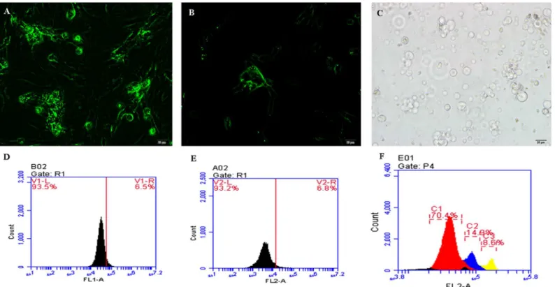
We used flow cytometry to identify the three types of cells (Fig 1). The PGCs were large cells and became a mulberry-like shape after being cultured for 48 h, while the SSCs became bird nest-like after 48 h. The Sp cells were isolated from the testes of sexually mature cocks and were larger than the other two types of cells. Three peaks can be seen inFig 1F, reflecting hap-loid, diphap-loid, and tetraploid cells. We collected the diploid cells (second peak) for sequencing.
Sequencing results
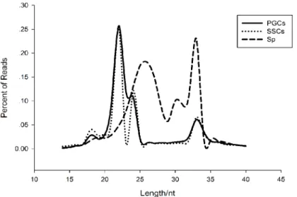
We obtained 31,361,989 reads from PGCs, 31,757,666 reads from SSCs, and 46,448,327 reads from Sp cells, representing an effective ratio of>95% (Table 1). Examining the length
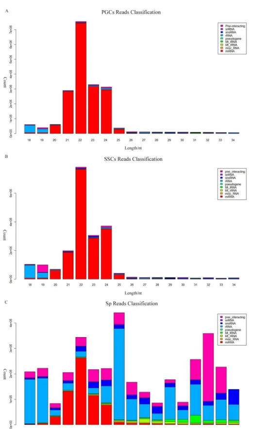
distribu-tion of the reads, we found that all the samples have two peaks, both PGCs and SSCs showed one peak at 22 nt and one at 33nt, while the Sp distribution showed one peak at 26 nt and one at 33 nt (Fig 2). According to Chen et al. and Juliano et al., the 22-nt peak represents mainly microRNAs, whereas from 26-nt to 33-nt peaks represent piRNA clusters [15,16]. We were able to classify and annotate 9 groups of small RNAs (Fig 3), with different distributions appearing in different cell types. The 9 groups represent miRNAs, misc_RNAs, mt_rRNAs, mt_tRNAs, pseudogenes, rRNAs, snoRNAs, snRNAs, and piRNAs. The number small RNAs found in each group is shown inS1 Table.
Common piRNAs in the three cell types
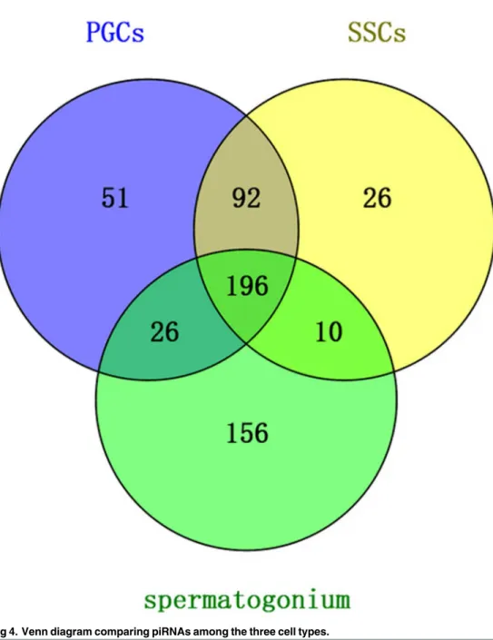
Venn diagram analysis. Venn diagram analysis revealed that 196 unique piRNAs appeared in all three cell types. Of these, the PGCs contained 365 total reads, the SSCs con-tained 324 reads, and the Sp cells concon-tained 388 reads. Only 10 piRNAs were present in SSCs and Sp but not PGCs, while 92 reads were present in PGCs and SSCs only and 26 reads were present in PGCs and Sp only (Fig 4). The uploading of the 196 common piRNAs to piRNA-spredictor software showed that 153 of these piRNAs could be verified (S2 Table).
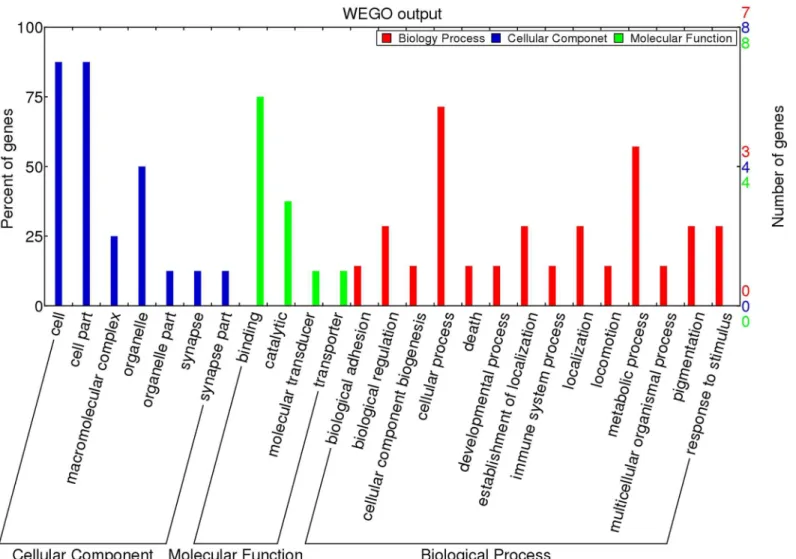
GO and KEGG pathway analyses. We mapped the 153 validated piRNAs sequences onto the chicken reference genome (Gallus gallusv.4) using Bowtie 2.0. After filtering for repeats, 22 genes were annotated (S3 Table). Gene Ontology (GO) analysis of these genes showed that nearly 75% of the genes are related to cellular processes (includingRPL7A,PRKCA,PUM1,
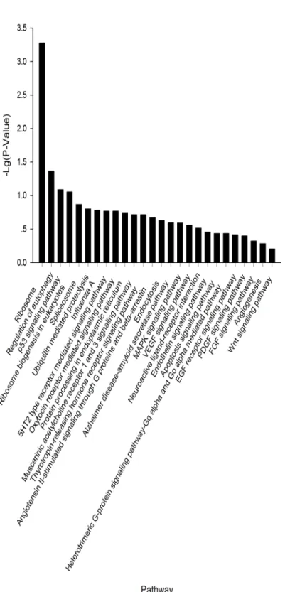
HSPA8, andCPXM2) while 20% of the genes are involved in developmental processes within the Biological Process category. Within the Cellular Component category, nearly 80% of the genes contained the ontology“cell part”. For the Molecular Function category, 75% of the genes were associated with binding, including ATP binding, nucleotide binding, or metal ion binding (Fig 5). In the KEGG pathway analysis, 26 pathways were identified. Of these, 9 path-ways were found to be involved in cellular processes, including migration, proliferation, devel-opment, and signal transduction (Fig 6).
Different expressed piRNAs in two groups
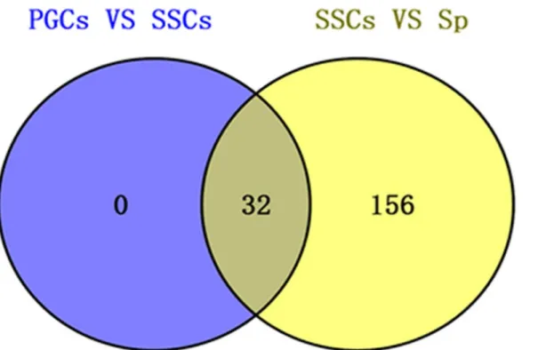
no unique common different piRNAs, while the SSCs vs. Sp group contained 156 common dif-ferent piRNAs (Fig 7). Of these, 32 piRNAs overlapped with the former group of 153 validated piRNAs that were common to all three groups (S4 Table). The number of differnet expressed piRNAs in two groups showed inFig 8. In the PGCs vs. SSCs group, 8 piRNAs were up-regu-lated and 24 were down-reguup-regu-lated. In the SSCs vs. Sp group, 155 piRNAs were up-reguup-regu-lated and 33 were down-regulated.
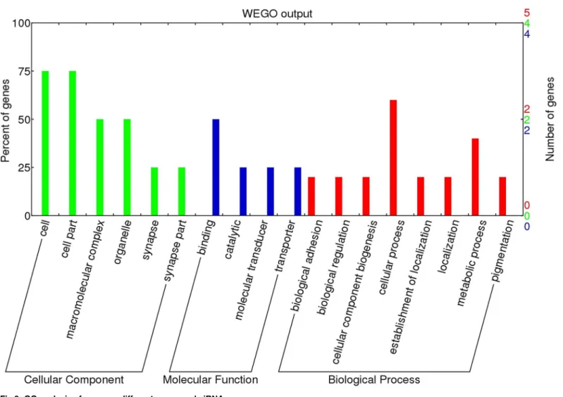
GO and KEGG pathway analyses. We mapped 32 piRNAs sequences onto the chicken reference genome (Gallus gallusv.4) using Bowtie 2.0. After filtering for repeats, we were able to annotate 9 (marked in red inS3 Table). GO analysis of these genes showed that within the Biological Process category, nearly 60% of genes were related to cellular processes (including
RPL7A,PUM1,CPXM2), while 20% of genes were involved in cellular component biogenesis. Nearly 75% of genes are present in the cell part within the Cellular Component category, and 55% of genes were associated with binding, including ATP binding, nucleotide binding, or metal ion binding among the Molecular Function category (Fig 9). In the KEGG analysis,
Fig 1. Cell sorting via flow cytometry.(A) The primordial germ cells (PGCs) were identified with a c-Kit antibody marked with FITC (200×). (B) The spermatogonial stem cells (SSCs) were marked with an Integrin alpha 6 antibody with FITC (200×). Both PGCs and SSCs were cultured for 48 h. (C) The spermatogonial (Sp) cells were directly isolated from testes and identified under light microscopy (200×). Scale bar = 20μm. (D-F) Flow cytometry sorting for
PGCs (D), SSCs (E), and Sp cells (F). The PGC, SSC, and Sp sorting ratios were 6.5%, 6.8%, and 14.9% respectively. Sp cells were stained with DAPI.
doi:10.1371/journal.pone.0151780.g001
Table 1. Number of reads obtained for the three sample libraries.
Sample Clean Reads Effective reads Effective ratio
PGCs 31361989 30851348 98.37%
SSCs 31757666 31461002 99.07%
Sp 46448327 46273957 99.62%
doi:10.1371/journal.pone.0151780.t001
genes were mainly identified in two pathways: the ribosome pathway and the neuroactive ligand-receptor interaction pathway.
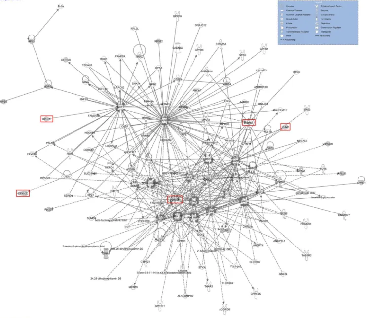
Potential network of 22 piRNAs genes
In order to identify a common network among the 22 genes, including the 5 genes, we per-formed an Ingenuity pathway analysis (IPA) (Fig 10). The results revealed a potential network including the 22 genes. Focusing on the 5 genes we acquired from the GO analysis (PRKCA,
EGFR,GNB2L1,APP, andMAPK), we found that several key elements combined these 5 genes (Fig 10).
Screening of candidate genes involved in germline stem cell proliferation
and spermatogenesis
We searched for candidate genes and pathways with two separate approaches. First, we identi-fied genes with piRNAs common to all three cell types. Using this approach, we identiidenti-fied 22 genes that are involved in processes such as immune system processes, death, pigmentation, and biological regulation. We also acquired 5 genes involved in cellular processes (RPL7A,
HSPA8,Pum1,CPXM2, andPRKCA). Protein kinase C alpha (PRKCA) is a key element with many functions, including the ability to regulate catalytic activity, inactivate MAPK activity, and regulate transferase activity. We speculate that this gene could occupy a central role, involved in inactivating or activating several pathways. Pumilio RNA-binding family member 1 (Pum1) is mainly involved in the regulation of cellular protein metabolic processes and trans-lation. We infer this gene performs the same function in germline stem cell proliferation or spermatogenesis. Heat Shock 70kDa Protein 8 (HSPA8) is involved in protein folding, and ribosomal Protein L7a (RPL7A) mainly participates in ribosome biogenesis according to the GO analysis. Carboxypeptidase X (M14 family) member 2 (CPXM2) is related to cell adhesion according to the GO analysis. Using the second approach of identifying the common different piRNAs, results showed thatRPL7A,Pum1, andCPXM2were involved in this process. These genes overlap with the genes from the former approach.
According to KEGG analysis, 3 out of 26 pathways were related to embryonic development and stem cell proliferation or spermatogenesis: the Wnt, FGF, and EGF receptor signaling
Fig 2. Length distribution of small RNAs in the three types of cells.
Fig 3. Classification and annotation of small RNAs from different cell types.Small RNAs from (A) PGCs, (B) SSCs, and (C) Sp cells were shown. Different small RNA groups were represented by different colors.
doi:10.1371/journal.pone.0151780.g003
pathways. Furthermore, five pathways were associated with cell differentiation, migration, and programmed cell death, namely the apoptosis, angiogenesis, MAPK, p53, and PDGF pathways.
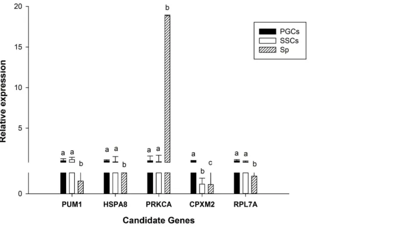
RT-qPCR of candidate genes
We assessed the relative expression of the above 5 candidate genes (Fig 11). The SYBR green primers used are provided inS5 Table. From this analysis, we found that there was no differ-ence betweenPum1,HSPA8,PRKCA, andRPL7Agenes in PGCs and SSCs but in Sp there was significant difference in these genes. ThePRKCAgene were expressed highest in Sp and lowest in PGCs. Furthermore, except PRKCA, the other three genes expressed higher in PGCs and SSCs than in Sp, indicating that they play a role in the three cell types. On the other hand,
CPXM2expression showed significant differences in the three cell types. Notably, the
Fig 4. Venn diagram comparing piRNAs among the three cell types.
expression ofPRKCAin Sp was much higher than in the other two cell types, indicating that it may act as a key element during spermatogenesis.
Discussion
Spermatogenesis is a complicated process that requires the coordinated efforts of germ cells and several somatic cells within the tubular structure of the testis [17]. We have examined three cell types in chicken through a small RNA sequencing approach. Through comparison of the results, we revealed that germline stem cell differentiation is a complex regulatory process associated with several genes and signaling pathways.
Germline stem cells are of interest in that they are involved in the passing of genetic infor-mation from one generation to the next via sexual reproduction [18]. Following the process of stem cell development and formation of sperm, primordial germ cells (PGCs) and spermatogo-nial stem cells (SSCs) are the precursors of germ cells and are specified during the early days of embryonic development in birds [19–22]. The first cells involved in spermatogenesis are called spermatogonia, which yield primary spermatocytes via mitosis [23]. GO analysis revealed that thePum1gene is involved in many processes. Several reports have identified that Pum1 plays a
Fig 5. Gene Ontology classification of piRNAs target genes which were found in all three cell types (PGCs, SSCs, and Sp cells).
doi:10.1371/journal.pone.0151780.g005
Fig 6. KEGG analysis of piRNAs common to all three cell types (PGCs, SSCs, and Sp cells).
Fig 7. Venn diagram analysis of common different piRNAs in two groups (PGCs vs. SSCs and SSCs vs. Sp).
doi:10.1371/journal.pone.0151780.g007
Fig 8. Number of piRNAs up- or down-regulated among two groups (PGCs vs. SSCs and SSCs vs. Sp).The x-axis represents the number of piRNAs, and the y-axis reflects the groups.
doi:10.1371/journal.pone.0151780.g008
role in embryogenesis or spermatogenesis. In haploid ESCs, it was found that Pum1 disruption prompted self-renewal when cells were cultured in conditions allowing for differentiation in mice [24]; however, in mammals,Pum1and the p53-mediated pathway act as a guard to main-tain germline homeostasis. After removal ofPum1, males have a significantly reduced sperm count and reduced fertility, and they display elevated rates of apoptosis in spermatocytes [25]. Furthermore, in mice, the lack ofPum1gene trap homozygotes suggested that an early loss of homozygous premplantation embryos because of 96-hr in vitro culture of 1-cell embryos either by natural mating or in vitro fertilization between heterozygotes failed to uncover any homozy-gous blastocysts [26]. Thus, we infer thatPum1plays a similar role in chickens during sper-matogenesis. In early spermatogenesis,Pum1acts as a marker of non-differentiation in germline stem cells and promotes cell proliferation.
The result of IPA revealed a potential network of 22 genes that is similar to the results of the KEGG analysis implicating the FGF, Wnt, and EGF receptor signaling pathways. Some reports have suggested that these pathways are involved in germline stem cell development. For exam-ple, the role of Wnt in embryonic development was discovered when genetic mutations in pro-teins in the Wnt pathway produced abnormal fruit fly embryos. Later research found that the genes responsible for these abnormalities also influenced breast cancer development in mice [27]. Saito-Diaz et al. found that the Wnt pathway was required for stem cell self-renewal in
Fig 9. GO analysis of common different expressed piRNAs.
developing organisms [28]. Therefore, we infer that the Wnt pathway promotes the develop-ment and proliferation of germline stem cells (PGCs, SSCs, Sp) in chicken. Fibroblast growth factor (FGF) acts on a variety of different cell types, functioning as both a direct and an indirect stimulator of angiogenesis. It has been reported that members of the FGF family function in the earliest stages of embryonic development and during organogenesis to maintain progenitor cells and mediate their growth, differentiation, survival, and patterning [29]. Choi et al. revealed that basic fibroblast growth factor (bFGF) is one of the key factors that enable prolifer-ation of chicken PGCs via MEK/ERK signaling, regulating downstream genes that may be important for PGC proliferation and survival [30].
piRNAs are a new kind of non-coding RNAs that are important in spermatogenesis. In this study, we found several piRNAs that are associated with early-stage spermatogenesis, and we
Fig 10. Potential network of 22 piRNAs genes.Different shapes indicate different functions in different pathways. Solid lines and dashed lines indicate direct interactions and indirect interactions, respectively. The red box indicates the 5 genes that were acquired from the GO analysis.
doi:10.1371/journal.pone.0151780.g010
identified additional piRNAs that could be further studied in the future. In order to further illustrate the role of piRNAs in spermatogenesis, additional research should focus on the next two stages of development: from spermatogonium to primary spermatocyte during the first meiotic division and from primary spermatocyte to secondary spermatocyte during the second meiotic division.
Conclusions
This study revealed a key gene,Pum1, which promotes both spermatogenesis and germline stem cell development. Additionally, we found that the Wnt, FGF, and EGF receptor signaling pathways are important in germ cell development.
Supporting Information
S1 Table. Number of piRNAs classified into 9 groups in three cell types. (DOCX)
S2 Table. Sequences of 153 piRNAs. (DOCX)
S3 Table. List of 22 genes acquired from 153 sequences.The 9 genes marked in red were acquired by mapping 32 piRNAs sequences.
(XLSX)
Fig 11. RT-qPCR of candidate genes.Lowercase letters indicated p0.01.
S4 Table. Sequences of 32 piRNAs. (DOCX)
S5 Table. qRT-PCR primers of five candidate genes. (DOCX)
Author Contributions
Conceived and designed the experiments: G. Chang G. Chen. Performed the experiments: LX LQ QG. Analyzed the data: LX XL QX Yang Zhang. Contributed reagents/materials/analysis tools: YB Yu Zhang HW ZL XG FW. Wrote the paper: LX GC.
References
1. Aravin A, Gaidatzis D, Pfeffer S, Lagos-Quintana M, Landgraf P, Iovino N, et al. A novel class of small RNAs bind to MILI protein in mouse testes. Nature. 2006; 442: 203–207. PMID:16751777
2. Hirakata S, Siomi MC. piRNA biogenesis in the germline: From transcription of piRNA genomic sources to piRNA maturation. Biochim Biophys Acta. 2015.
3. Gebert D, Ketting RF, Zischler H, Rosenkranz D. piRNAs from pig testis provide evidence for a con-served role of the Piwi pathway in post-transcriptional gene regulation in mammals. PloS One. 2015; 10: e0124860. doi:10.1371/journal.pone.0124860PMID:25950437
4. Liu G, Lei B, Li Y, Tong K, Ding Y, Luo L, et al. Discovery of potential piRNAs from next generation sequences of the sexually mature porcine testes. PloS One. 2012; 7.
5. Lim SL, Qu ZP, Kortschak RD, Lawrence DM, Geoghegan J, Hempfling AL, et al. HENMT1 and piRNA stability are required for adult male germ cell transposon repression and to define the spermatogenic program in the mouse. PLoS Genet. 2015; 11: e1005620. doi:10.1371/journal.pgen.1005620PMID: 26496356
6. Senti KA, Jurczak D, Sachidanandam R, Brennecke J. piRNA-guided slicing of transposon transcripts enforces their transcriptional silencing via specifying the nuclear piRNA repertoire. Genes Dev. 2015; 29: 1747–1762. doi:10.1101/gad.267252.115PMID:26302790
7. Li XZ, Roy CK, Dong X, Bolcun-Filas E, Wang J, Han BW, et al. An ancient transcription factor initiates the burst of piRNA production during early meiosis in mouse testes. Mol Cell. 2013; 50: 67–81. doi:10. 1016/j.molcel.2013.02.016PMID:23523368
8. Vourekas A, Zheng Q, Alexiou P, Maragkakis M, Kirino Y, Gregory BD, et al. Mili and Miwi target RNA repertoire reveals piRNA biogenesis and function of Miwi in spermiogenesis. Nat Struct Mol Bio. 2012; 19: 773–781.
9. Goh WS, Falciatori I, Tam OH, Burgess R, Meikar O, Kotaja N, et al. piRNA-directed cleavage of mei-otic transcripts regulates spermatogenesis. Genes Dev. 2015; 29: 1032–1044. doi:10.1101/gad. 260455.115PMID:25995188
10. Gou L-T, Dai P, Yang J-H, Xue Y, Hu Y-P, Zhou Y, et al. Pachytene piRNAs instruct massive mRNA elimination during late spermiogenesis. Cell Res. 2014; 24: 680–700. doi:10.1038/cr.2014.41PMID: 24787618
11. Li BC, Tian ZQ, Sun M, Xu Q, Wang XY, Qin YR, et al. Directional differentiation of chicken primordial germ cells into adipocytes, neuron-like cells, and osteoblasts. Mol Reprod Dev. 2010; 77: 795–801. doi:10.1002/mrd.21224PMID:20722070
12. Yu F, Ding LJ, Sun GB, Sun PX, He XH, Ni LG, et al. Transgenic sperm produced by electrotransfection and allogeneic transplantation of chicken fetal spermatogonial stem cells. Mol Reprod Dev. 2010; 77:340–347. doi:10.1002/mrd.21147PMID:20063420
13. Mozdziak PE, Angerman-Stewart J, Rushton B, Pardue SL, Petitte JN. Isolation of chicken primordial germ cells using fluorescence-activated cell sorting. Poult Sci. 2005; 84: 594–600. PMID:15844816 14. Chang G, Chen R, Xu L, Ma T, Wang H, Chen J, et al. DNA methylation and NF-Y regulate Piwil1
expression during chicken spermatogenesis. Anim Reprod Sci. 2015; 162: 95–103. doi:10.1016/j. anireprosci.2015.09.016PMID:26471838
15. Chen R, Chang G, Zhang Y, Dai A, Ma T, Li J, et al. Cloning of the quail PIWI gene and characterization of PIWI binding to small RNAs. PloS One. 2012; 7: e51724. doi:10.1371/journal.pone.0051724PMID: 23284755
16. Juliano C, Wang J, Lin H. Uniting germline and stem cells: the function of Piwi proteins and the piRNA pathway in diverse organisms. Ann Rev Genet. 2011; 45: 447–469. doi: 10.1146/annurev-genet-110410-132541PMID:21942366
17. Song H-W, Wilkinson MF. In vitro spermatogenesis: A long journey to get tails. Spermatogenesis. 2012; 2: 238–244. PMID:23248764
18. Zhang Z, Elsayed AK, Shi Q, Zhang Y, Zuo Q, Li D, et al. Crucial genes and pathways in chicken germ stem cell differentiation. J Biol Chem. 2015; 290: 13605–13621. doi:10.1074/jbc.M114.601401PMID: 25847247
19. Park TS, Han JY. Conservation of migration and differentiation circuits in primordial germ cells between avian species. J Reprod Dev. 2013; 59: 252–257. PMID:23386102
20. Pramod RK, Mitra A. In vitro culture and characterization of spermatogonial stem cells on Sertoli cell feeder layer in goat (Capra hircus). J Assist Reprod Genet. 2014; 31: 993–1001. doi: 10.1007/s10815-014-0277-1PMID:24958548
21. Caires K, Broady J, McLean D. Maintaining the male germline: regulation of spermatogonial stem cells. J Endocrinol. 2010; 205: 133–145. doi:10.1677/JOE-09-0275PMID:20147357
22. Phillips BT, Gassei K, Orwig KE. Spermatogonial stem cell regulation and spermatogenesis. Phil Trans R Soc B. 2010; 365: 1663–1678. doi:10.1098/rstb.2010.0026PMID:20403877
23. Song N, Liu J, An S, Nishino T, Hishikawa Y, Koji T. Immunohistochemical analysis of histone H3 modi-fications in germ cells during mouse spermatogenesis. Acta Histochem Cytochem. 2011; 44: 183–190. doi:10.1267/ahc.11027PMID:21927517
24. Leeb M, Dietmann S, Paramor M, Niwa H, Smith A. Genetic exploration of the exit from self-renewal using haploid embryonic stem cells. Cell Stem Cell. 2014; 14: 385–393. doi:10.1016/j.stem.2013.12. 008PMID:24412312
25. Chen D, Zheng W, Lin A, Uyhazi K, Zhao H, Lin H. Pumilio 1 suppresses multiple activators of p53 to safeguard spermatogenesis. Curr Biol. 2012; 22: 420–425. doi:10.1016/j.cub.2012.01.039PMID: 22342750
26. Zhang C, Zhu T, Chen Y, Xu EY. Loss of preimplantation embryo resulting from a Pum1 gene trap mutation. Biochem Biophys Res Comm. 2015; 462: 8–13. doi:10.1016/j.bbrc.2015.04.019PMID: 25896760
27. van Amerongen R, Fuerer C, Mizutani M, Nusse R. Wnt5a can both activate and repress Wnt/beta-catenin signaling during mouse embryonic development. Dev Biol. 2012; 369: 101–114. doi:10.1016/j. ydbio.2012.06.020PMID:22771246
28. Saito-Diaz K, Chen TW, Wang X, Thorne CA, Wallace HA, Page-Mccaw A, et al. The way Wnt works: Components and mechanism. Growth Factors. 2013; 31: 1–31. doi:10.3109/08977194.2012.752737 PMID:23256519
29. Ornitz DM, Itoh N. The fibroblast growth factor signaling pathway. Wiley Interdiscip Rev Dev Biol. 2015; 4: 215–266. doi:10.1002/wdev.176PMID:25772309