Advanced Pharmaceutical Bulletin, 2013, 3(2), 333-337 doi: http://dx.doi.org/10.5681/apb.2013.054
http://apb.tbzmed.ac.ir/
*Corresponding author: Jafar Majidi, Immunology Research Center, Tabriz University of Medical Sciences, Tabriz, Iran. Tel: +98 (411) 3364665,
A Unique Report: Development of Super Anti-Human IgG Monoclone
with Optical Density Over Than 3
Leili Aghebati Maleki1,2, Behzad Baradaran1,2, Jalal Abdolalizadeh1,2, Fatemeh Ezzatifar1, Jafar Majidi1,2*
1Immunology Research Center, Tabriz University of Medical Sciences, Tabriz, Iran. 2
Tabriz Pharmaceutical Technology Incubator(TPTI).
Introduction
Monoclonal antibodies can be produced in specialized cells through a method now popularly known as hybridoma technology.1 Hybridoma technology was
first invented by two scientists, Georges Kohler and Cesar Milstein. From 1975, Köhler and Milstein successfully fused antibody- producing mouse spleen cells with mouse myeloma cells, the fusion of somatic cells has been carried out for many years with a variety of different aims and this technique quickly became
one of immunology΄s key technologies.2 Using
hybridoma technology, monoclonal antibodies have been prepared against a wide range of antigens including growth factors, growth factor receptors, viruses, bacterial products, hormones and differentiated antigens.3
In fact, monoclonal antibodies (mAbs) have been widely applied in various fields such as diagnosis applications, purification, disease monitoring, identifying prognostic markers and therapy.4 For most
research, diagnostic and therapeutic purposes, monoclonal antibodies derived from a single clone and thus specific for a single epitope, are preferable.5
In most diagnostic kits of the infectious diseases, for instance, Rubella, H.pylor Toxoplasmosis and etc, the
conjugated monoclonal antibodies against human IgG carry out as a key role. Due to in this need, generation of mAb seems invaluable. To approach these goals, generation and characterization of a highly specific mAb against human IgG was investigated.
The prim aim of this study including: production and application of mabs against human IgG for development of diagnostic kits and large scale, semi-industrial production and standardization of this product towards self-sufficiency of the country.
Materials and Methods
Immunization Procedure and Screening of
Immunized Animals
Four female Balb/c (6-8 weeks old) mice were used for purified human IgG (Affinity Purified Human IgG, Sigma) immunization. Each mouse was immunized 4 times with an interval of two-three weeks subcutaneously. The first Immunization was performed using Freund's complete adjuvant(Sigma-Aldrich Co. Louis, USA). Incomplete Freund's adjuvant (Sigma-Aldrich Co. Louis, USA) was used for the 2nd, 3rd and
4th immunization.For the first immunization, 50 µg of
purified human IgG was mixed with an equal volume A R T I C L E I N F O A B S T R A C T
Article Type: Research Article
Article History: Received: 13 March 2013 Revised: 18 March 2013 Accepted: 6 April 2013 ePublished: 20 August 2013
Keywords:
Monoclonal antibody Human IgG Balb/c mice ELISA
Aghebati Maleki et al.
of Freund's complete adjuvant. For the subsequent immunizations 50 µg purified human IgG were injected with Freund's incomplete adjuvant. Three days before the cell fusion, 50 µg of antigen (without any adjuvant) was injected intravenously.
A week after the second injection, blood was taken from each mouse by a vertical incision of the tail vein and the antibody response was measured by ELISA. The mouse with the highest serum antibody titre was selected as the spleen donor. Sera collected from non-immunized and non-immunized mice served as negative and positive controls.
Cell Fusion and Hybridoma Production
Three days after final immunization, spleen of the immunized mouse was aseptically removed and fused with SP2/0 myeloma cell line at a ratio of 1:5 (1 SP2/0 and 5 spleen cells) by PEG (polyethyleneglycol, MW 1450, Sigma) as fusogen. Selective HAT medium (Gibco) was added to the fused cells and cells were seeded into five 96-well microtitre plates (Nunc) containing feeder layer. The cells were incubated at 37 °C with 5% CO2 for 2-3 days. Cell growth and colony formations were examined daily. Colonies were appeared after 5-7 days. Once the colony diameter reached to 1 mm the presence of antibody against the immunized antigen was determined by ELISA method. Then, positive hybridomas were selected for further studies.
Cloning of Hybridoma Cells by Limiting Dilution
Assay
After screening, the clones with high absorbance were selected for cloning by Limiting Dilution (L.D) method. The cells were diluted so that contained only
one cell in each 10 μl. Twelve days after L.D, the
supernatants of monoclones were screened for production of antibody. Suitable monoclones possessing high absorbance were selected for further characterization and considered for mass production.
Assessment of Anti-IgG Cross-Reactivity with other Classes of Immunoglobulins by a Sandwich ELISA In order to determine the cross reactivity with IgM & IgA, the micro titer plates were coated with each class
of the purified immunoglobulins (5μg/ml in coating buffer, 100μl/well) and incubated for 1.5 hour at 37 °c. Plates were saturated with 2% BSA at 37 °C for 1.5 hr. Wells were then washed 3 times with PBS containing 0.05% Tween 20 (PBS-T) for 5 min.At the next step, Hybridoma culture supernatants with distinct dilution was added to the wells and incubated as described
above. Then, 100 μl of 1:4000 dilution of H RP-conjugated rabbit anti-mouse IgG (Sigma-Aldrich Co. Louis, USA ) was added to the wells and incubation was continued for 1.5 hrs at 37 °C. After washing, 100 µl of Tetramethylbenzidine (TMB) substrate was added to each well and the plate was incubated at room temperature in a dark place. After 15 min, the reaction
was stopped by adding 100 µl of stopping solution (0.16 M H2SO4) to each well. The Optical Density
(OD) of the reactions was measured at 450 nm by an ELISA reader (STAT FAX 303+). In the end of the reaction and by reading their absorbance, cross reactivity of the monoclonal antibodies with IgM & IgA was determined.
Isotype Determination
The class and subclass of the mAbs were determined by an ELISA with a mouse monoclonal sub isotyping kit containing rabbit anti-mouse IgG1, IgG2a, IgG2b, IgG3, IgM and IgA, following the procedure provided by the manufacturer (Thermo, USA).
Production of Ascitic Fluids
Hybridoma cells producing IgG mAbs were grown in RPMI1640 supplemented with 10% fetal bovine serum,
harvested and washed twice in PBS (pH 7.2).Ten days after pristane injection, 6-8 weeks old Balb/c mice were injected intraperitoneally with 2-3×10⁶ hybridoma cells
suspended in 0.5 ml PBS (pH 7.2). Fluid was collected from the peritoneal cavity 8 to 10 days after the injection of the cells, by using of needle 19. Ascitic fluid was kept at 4 °C for 1 h and centrifuged at 5000 g for 15 min. Supernatant was collected and stored at -20 °C until used.
Antibody Purification
The monoclonal antibodies were purified from ascitic fluid by Ion exchange chromatography based on its isotype. Briefly, ascitic fluids were filtered through 45
μm filter and were diluted 1 to 2 with PBS and
fractionated with 40% saturated ammonium sulfate. After several times of washing with 40% ammonium sulfate, the fraction was centrifuged for 15minutes in 5000g. The precipitated fraction was dialyzed against 0.05 M phosphate buffer pH 7.4 containing 0.05 M Nacl. The final dialyze was exchanged against the column washing buffer (Tris 40 mM). Purification of ascitic fluid was done by Ion exchange chromatography (DEAE-Sepharose 6B) which is a simple and economical method. At first, the column was eluted with washing buffer (Tris 40 mM, pH 8.1) in order that the pH of the external buffer to be the same as the pH of internal buffer. Then the dialyzed sample in 60 mg/3ml concentration was run to the column with dimensions of 1.6×15cm. Distinct antibody was eluted from the column through washing buffer containing 50 mM Nacl, and the fractions were collected in 5 ml/20 ml. Finally, the purified fractions were kept for conjugation with Peroxidase.
SDS-PAGE Gel Electrophoresis
A Unique Report: Development of Super Anti-Human IgG
were mixed with sample buffer, then boiled for 2 - 5 min and cooled on ice. Electrophoresis was done in a 12.5% SDS-PAGE gel with a mini- PROTEAN electrophoresis instrument (Bio- Rad Laboratories, Hercules, CA, USA) 100 mA for 1 hr. The gel was stained with Coomassie Brilliant Blue R-250 (Sigma).
Western Blotting
Western blotting technique was used for confirming the result of ELISA and to see pattern of specificity and cross -reactivity of anti-IgG monoclonal antibody. Briefly, the nitrocellulose membrane and several thicknesses of Whatman chromatography papers were soaked in the transfer buffer (25 mM Tris, 192 mM glycine, 20% V/V methanol, pH 8.3). The wet nitrocellulose membrane was overlaid on the wet Whatman sheets by taking precaution to avoid bubbles. Then, the gel of SDS-PAGE was placed on the wet nitrocellulose membrane and then several wet Whatman papers were placed on it. Transfer of the proteins from gel to nitrocellulose membrane was done in 100V for 3 hours. Then, non- specific sites were blocked with 2% BSA solution. After three times of washing, the membrane was cut into strips and incubated for 2 hours at 37 °C with the supernatants of suitable clones. Again, after five times of washing, the strips were incubated for 2 hours at 37 °C with Rabbit Anti- Mouse IgG conjugate (1/2000 dilution). The strips were washed and detected by ECL (Amersham Phamacia Biotech Inc, USA) hyperfilm after exposure for 5 min.
Labelling of Monoclonal Antibody with Horseradish Peroxidase Enzyme (HRP)
For conjugation, the Nakan and Periodate methods were used. Briefly, 4 mg of HRP was dissolved in 1 ml of distillated water. Then 0.2 ml freshly prepared sodium Periodate solution (0.1M) was added to the enzymatic solution and incubated on shaker for, 20 minutes at room temperature. The solution was dialyzed against acetate buffer (pH 4.4), overnight at 4 °c. 8 mg of the purified monoclonal antibody was dissolved in 1 ml sodium carbonate (10 mM, pH 9.5).The pH of the dialyzed enzyme was reached to 9 and immediately the solution containing monoclonal antibody was added to it and shaked for 2 hours at room temperature. Then 0.1 ml of the freshly prepared sodium brohydrate was added and incubated for 30 minutes at room temperature. The final solution was precipitated with ammonium sulfate and then was dialyzed against PBS buffer.
Results
Among the five immunized Balb/c mice against human IgG, the serum of the immune mouse at 1/32000 dilution, indicated the highest absorbance in reaction with human IgG using ELISA method. So the immune mouse was selected for the fusion. The final result of the successful fusion of the immune mouse spleen cells
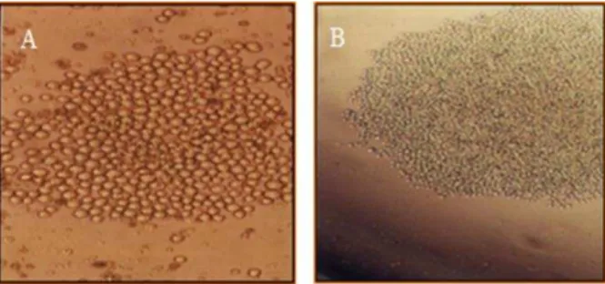
with myeloma SP2/0 cells were about 50 wells containing positive clones with high absorbance in reaction with IgG that 1 clone had optical density over than 3. We named this clone as supermonoclone (Figure 1) and was selected for cloning by limiting dilution (L.D) method. The yield of limiting dilution was many clones with absorbance over than 3 and about 2 at 0.01 dilutions which did not show any cross-reactivity with IgM and IgA by ELISA method (Table l).
Figure 1. Proliferated suitable mono clone (supermonoclone)
selected for injection into the peritoneum of mice. Supermonoclone in the growing form (Mag.10X) (A), Supermonoclone in the highly proliferated form (Mag.4X) (B).
Table 1. Comparison of the mean absorbance of supernatant
of the supermonoclone at 450 nm.
IgG IgM IgA
SP2/0 (N.C) 0.05 0.07 0.06
Mouse Serum (N.C)* 0.1 0.08 0.09
Immune mouse serum (P.C)** 1.1 0.2 0.2
Super monoclone >3 0.21 0.18
Supermonoclone(1/100 dilution) 2 0.11 0.1
*negative control with 1/32000 dilution, **positive control with 1/32000 dilution
Aghebati Maleki et al.
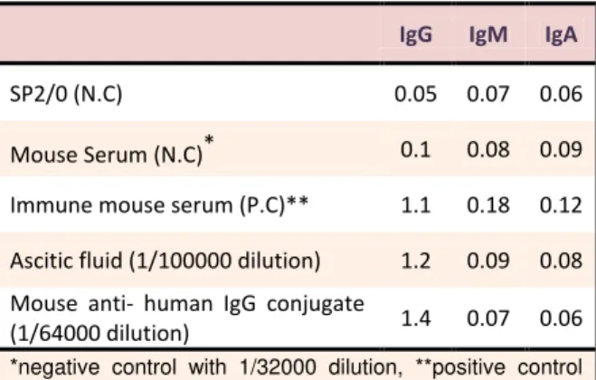
1/100,000 dilution has high absorbance with IgG (above 1) but has no absorbance with IgM & IgA (Table 3).Also, the results of conjugation indicated that
1/64000dilution of conjugate had high absorbance with
IgG (above 1) and didn’t show any cross reactivity with
IgM & IgA(Table 3).
Table 2. Results of subclass isotyping of mouse mAb at 450 nm.
IgG1 IgG2a IgG2b IgG3 IgA IgM Kappa Lambda
Mouse anti-human IgG 1.101 0.131 0.154 0.168 0.117 0.136 1.692 0.138
Figure 2. SDS- PAGE of purified monoclonal antibody. In
reduced form (A), two bands were seen in 50 & 25 kDa but in non-reduced condition (B), only one band was seen in about 150 kDa.
Table 3. Comparison of the mean absorbance and cross
reactivity of ascitic fluid and anti- human IgG conjugate at 450 nm.
IgG IgM IgA
SP2/0 (N.C) 0.05 0.07 0.06
Mouse Serum (N.C)* 0.1 0.08 0.09
Immune mouse serum (P.C)** 1.1 0.18 0.12
Ascitic fluid (1/100000 dilution) 1.2 0.09 0.08
Mouse anti- human IgG conjugate
(1/64000 dilution) 1.4 0.07 0.06
*negative control with 1/32000 dilution, **positive control with 1/32000 dilution
Discussion
Hybridoma technology has been rapidly and successfully applied to a wide variety of biological problems of both theoretical and practical importance.6
MAbs are therefore of enormous utility in applications such as experimental biology, medicine, biomedical research, diagnostic testing, and therapy.7 More
importantly, each antibody is highly specific for a particular antigen; this characteristic feature of antibodies has led to their routine usage in diagnostic kits.8
IgG antibodies are involved in predominantly the secondary immune response.9 The presence of specific
IgG, in general, corresponds to maturation of the antibody response. In addition, in chronic condition of infectious diseases; the class of the produced antibody against pathogen is IgG.10 Therefore monoclonal anti- human IgG is very important, significant and as a key reagent for its recognition. Accordingly, production of monoclonal antibody without any cross reactivity with homologous molecules such as other classes of immunoglobulins can be used in diagnostic kits of infectious diseases.11
When mAbs are produced, it is important to consider antigen features, which include the quality and quantity of the antigen and the antigen preparation. The specificity of the immune response obtained depends on the purity of the antigen applied.12 Therefore in this study, purifiedhuman IgG immunoglobulins were used to immunize Balb/c mice. Upon such procedure, we observed high immunologic response in the immunized mouse, whose serum resulted in absorbance above 1 with 1/32000 dilution and the most immune mouse was selected for the fusion.
In similar previous study, Majidi et al produced monoclonal antibody against human IgE which could be used in designing ELISA kits. Purified IgE was used for immunization, in order to produce mAb.13
In a study, YÜCEL et al produced hybridomas against Hepatitis B virus (HBV) by fusing spleen cells from hyper immunized mice with SP2/O mouse myeloma cell. HBsAg proteins were used for immunization then; they concluded that these mAbs were valuable for diagnosis of hepatitis B surface antigen in human serum.1 In all of these studies, native proteins purified
from the natural source were preferred for its most likely to produce useful antibodies and high concentrations of mAbs produced.
In the present study, the best clone designated as supermonoclone, was injected intraperitoneally to pristane-injected mice. The production of monoclonal antibody in the ascitic fluid is commercially cost effective also; a rapid, reproducible method for large-scale production in comparison with expensive and time-consuming culture methods.14 Anti-IgG mAb was
A Unique Report: Development of Super Anti-Human IgG
recognizes human IgG and there was no cross-reactivity with other classes of immunoglobulins. Furthermore, anti-IgG mAb was interacted with human IgG with a very high specificity and affinity. These mAbs with high affinity are useful tools for quantitation of human IgG subclass levels in various diseases. So, this mAb could be a useful tool for use in the development of diagnostic kits based on sandwich ELISA. Taking all together, the conjugated monoclonal antibody could have application in diagnosis of infectious diseases like Toxoplasmosis, Rubella, H.Pylori and IgG class of other infectious and non- infectious diseases. Importantly, production of purified monoclonal antibody and HRP conjugated IgG could be consider another step toward Iran self-sufficiency.
Acknowledgements
We would like to thank for Tabriz Pharmaceutical Technology Incubator (TPTI) and Immunology Research Center (IRC) for kind assistance, respectively. This work was supported by a grant from Tabriz Pharmaceutical Technology Incubator (TPTI).
Conflict of Interest
The authors report no conflicts of interest.
References
1. Yucel F, Manav A, Basalp A. Production and characterization of monoclonal antibodies against hepatitis B viruses and application of a quick sandwich ELISA. Hybrid Hybridomics
2003;22(3):173-7.
2. Modjtahedi H. Monoclonal Antibodies as Therapeutic Agents: Advances and Challenges. Iran
J Immunol 2005;2(1):3-21.
3. Kohler G, Milstein C. Continuous cultures of fused cells secreting antibody of predefined specificity.
Nature 1975;256(5517):495-7.
4. Guan M, Su B, Ye C, Lu Y. Production of extracellular domain of human tissue factor using maltose-binding protein fusion system. Protein
Expr Purif 2002;26(2):229-34.
5. Daginakatte GC, Chard-Bergstrom C, Andrews GA, Kapil S. Production, characterization, and uses of monoclonal antibodies against recombinant
nucleoprotein of elk coronavirus. Clin Diagn Lab
Immunol 1999;6(3):341-4.
6. Sangdokmai A, Pimpitak U, Buakeaw A, Palaga T, Komolphis K. Production and Characterization of Monoclonal Antibodies Against Aflatoxin M1. International Conference on Environmental, Biomedical and Biotechnology; Singapoore: IACSIT Press; 2011.
7. Fraser PD, Misawa N, Sandmann G, Johnson J, Schuch W, Bramley PM. Production and characterisation of monoclonal antibodies to phytoene synthase of Lycopersicon esculentum.
Phytochemistry 1998;49(4):971-8.
8. Lang AB, Schuerch U, Cryz SJ Jr. Optimization of growth and secretion of human monoclonal antibodies by hybridomas cultured in serum-free media. Hybridoma 1991;10(3):401-9.
9. Chevrier MC, Chateauneuf I, Guerin M, Lemieux R. Sensitive detection of human IgG in ELISA using a monoclonal anti-IgG-peroxidase conjugate. Hybrid
Hybridomics 2004;23(6):362-7.
10. Hajighasemi F, Khoshnoodi J, Shokri F. Development of two murine monoclonal antibodies recognizing human nG1m(a)-like isoallotypic markers. Hybridoma (Larchmt) 2008;27(6):473-9.
11. Hussain A, Pankhurst T, Goodall M, Colman R, Jefferis R, Savage CO, et al. Chimeric IgG4 PR3-ANCA induces selective inflammatory responses from neutrophils through engagement of Fcgamma receptors. Immunology 2009;128(2):236-44. 12. Baradaran B, Hosseini AZ, Majidi J, Farajnia S,
Barar J, Saraf ZH, et al. Development and characterization of monoclonal antibodies against human epidermal growth factor receptor in Balb/c mice. Hum Antibodies 2009;18(1-2):11-6.
13. Majidi J, Zavaran Hosseini A, Hassan ZM, Alimohamadian MH. Production of monoclonal antibody against human Immunoglobulin E. Iran J
Allergy Asthma Immunol 2000;1(2):81-7.
14. Aghebati Maleki L, Majidi J, Baradaran B, Abdolalizadeh J, Kazemi T, Aghebati Maleki A, et al. Large Scale Generation and Characterization of Anti-Human CD34 Monoclonal Antibody in Ascetic Fluid of Balb/c Mice. Adv Pharm Bull