Associated with Protection against Human Visceral
Leishmaniasis
Daniel R. Aba´nades1, Leonardo V. Arruda1,2, Elaine S. Arruda1, Jose´ Roberto A. S. Pinto3, Mario S. Palma3, Dorlene Aquino4, Arlene J. Caldas4, Manuel Soto5, Aldina Barral1,2,6, Manoel Barral-Netto1,2,6*
1Centro de Pesquisas Gonc¸alo Moniz (CPqGM), Fundac¸a˜o Oswaldo Cruz (FIOCRUZ), Salvador, Bahia, Brazil,2Faculdade de Medicina da Bahia, Universidade Federal da Bahia, Salvador, Brazil,3Center of the Study of Social Insects, Institute of Biosciences of Rio Claro, Department of Biology, University of Sa˜o Paulo State (UNESP), Rio Claro, Sa˜o Paulo, Brazil,4Departamento de Enfermagem, Universidade Federal do Maranha˜o, Sa˜o Luis, Maranha˜o, Brazil,5Centro de Biologı´a Molecular Severo Ochoa (CSIC-UAM), Departamento de Biologı´a Molecular, Universidad Auto´noma de Madrid, Madrid, Spain,6Instituto Nacional de Cieˆncia e Tecnologia de Investigac¸a˜o em Imunologia (iii-INCT), Salvador, Bahia, Brazil
Abstract
Background:Protection and recovery from visceral leishmaniasis (VL) have been associated with cell-mediated immune (CMI) responses, whereas no protective role has been attributed to humoral responses against specific parasitic antigens. In this report, we compared carefully selected groups of individuals with distinct responses toLeishmania chagasito explore antigen-recognizing IgG present in resistant individuals.
Methodology and Principal Findings:VL patients with negative delayed-type hypersensitivity (DTH) were classified into the susceptible group. Individuals who had recovered from VL and converted to a DTH+response, as well as asymptomatic infected individuals (DTH+), were categorized into the resistant group. Sera from these groups were used to detect antigens fromL. chagasiby conventional and 2D Western blot assays. Despite an overall reduction in the reactivity of several proteins after DTH conversion, a specific group of proteins (approximately 110–130 kDa) consistently reacted with sera from DTH converters. Other antigens that specifically reacted with sera from DTH+ individuals were isolated and tandem mass
spectrometry followed by database query with the protein search engine MASCO were used to identify antigens. The serological properties of recombinant version of the selected antigens were tested by ELISA. Sera from asymptomatic infected people (DTH+) reacted more strongly with a mixture of selected recombinant antigens than with total soluble Leishmaniaantigen (SLA), with less cross-reactivity against Chagas disease patients’ sera.
Significance: Our results are the first evidence of leishmania proteins that are specifically recognized by sera from individuals who are putatively resistant to VL. In addition, these data highlight the possibility of using specific proteins in serological tests for the identification of asymptomatic infected individuals.
Citation:Aba´nades DR, Arruda LV, Arruda ES, Pinto JRAS, Palma MS, et al. (2012) Immunodominant Antigens ofLeishmania chagasiAssociated with Protection against Human Visceral Leishmaniasis. PLoS Negl Trop Dis 6(6): e1687. doi:10.1371/journal.pntd.0001687
Editor:Edgar M. Carvalho, Hospital Universita´rio, Brazil
ReceivedJanuary 4, 2012;AcceptedApril 30, 2012;PublishedJune 19, 2012
Copyright:ß2012 Aba´nades et al. This is an open-access article distributed under the terms of the Creative Commons Attribution License, which permits unrestricted use, distribution, and reproduction in any medium, provided the original author and source are credited.
Funding:The research and publication process was supported by grants from FAPESB: PES 0074/2008 Projetos Estrate´gicos, from whom DRA and EA received fellowships, and from Pronex: CNPq/FAPESB. MSP, AB, JC and MB-N are senior investigators from CNPq. The funders had no role in study design, data collection and analysis, decision to publish, or preparation of the manuscript.
Competing Interests:The authors have declared that no competing interests exist. * E-mail: mbarral@bahia.fiocruz.br
Introduction
Visceral Leishmaniasis (VL) is a potentially fatal disease caused by infection with Leishmania chagasi in the New World and
Leishmania donovani or Leishmania infantum in the Old World [1]. Infection leads to a spectrum of clinical outcomes ranging from asymptomatic infection to active disease. The anti-Leishmania
immune response during asymptomatic infection is characterized by a low serological and positive cellular response, which is demonstrated by a positive delayed-type hypersensitivity skin response (DTH+) [2]. Patients with active VL, on the other hand, present a strong positive serological and a negative cell-mediated immune (CMI) response with low IFN-cproduction [3]. Treated patients that recover from illness (accounts for 90%) only exhibit a
positive DTH response long after treatment [4]. Epidemiological studies in Brazil showed that a positive DTH response is a marker of protection against VL [5].
Serological diagnosis ofL. chagasiduring asymptomatic infection is complicated by low antibody titers and frequent cross-reactivity with other diseases [6]. In addition, serologic markers of recovery or resistance to infection have not been characterized. There is no effective and safe vaccine approved for human use against any form of visceral leishmaniasis despite the obvious need and considerable effort that has been made.
DTH positivity was observed in both asymptomatic individuals and in recovered patients. In addition, the recombinant version of select antigens appeared to be a valuable tool for the serological identification of asymptomatic patients.
Materials and Methods
Ethics statement
This study was approved by the Research Ethics Committee of the Federal University of Maranha˜o University Hospital, Brazil (localities where the field study was performed). All clinical investigations were conducted according to the Declaration of Helsinki. Written, informed consent was obtained from all participants or legal guardians.
Parasites
Leishmania chagasi(MHOM/BR00/MER/STRAIN2) promasti-gotes were cultured in Schneider’s medium supplemented with 10% inactive FBS, 2 mM L-glutamine, 100 U/mL penicillin, and 100mL/mL streptomycin.
Patients, sera and delayed-type hypersensitivity reaction Sera were obtained at two distinct settings as described below: The patients were classified as VL (pre-treatment, samples obtained during active disease previous to treatment) and post-treatment (samples from treated and cured patients). All patients were from the Maranha˜o Federal University Hospital. Diagnosis was confirmed by identification ofLeishmania sp.in Giemsa-stained smears of bone marrow aspirates (parasitological test). The study was conducted from August 2000 to July 2002 and information on the individuals has been previously reported [4,7].
Sera were stored at 220uC without thawing. All patients received adequate treatment. DTH skin reactivity assays (Mon-tenegro test) were performed with SLA prepared as described elsewhere [8].
Sample preparation
Parasites in logarithmic growth phase were washed twice with PBS, lysed with Laemmli’s Buffer [9], sonicated, heated at 95uC
for 5 min and centrifuged for 15 min at 12,0006g at 4uC. Extracts
from 106parasites were loaded by line into an acrylamide gel. For 2D electrophoresis, parasites were lysed at 4uC with 200mL of Buffer A (0.5% Nonidet P40, 0.1 mM PMSF, 1 mM DTT, 10 mM Tris-HCl, pH 7.4) followed by addition of 200mL of phenol and vortex. The samples were centrifuged at 6,0006g for
5 min, and the aqueous phase was discarded. The proteins were precipitated by adding 1 mL of 0.1 M ammonium acetate in absolute methanol and centrifuged at 6,0006g for 10 min at 4uC.
The resultant pellet was washed with 80% acetone and dried. The proteins were solubilized for 3 h at 30uC in RP3 Buffer (7 M urea, 2 M thiourea, 4% CHAPS, 40 mM Tris-HCl pH 8.8, 0.5% ampholytes, pH 4–7), followed by 15 min of centrifugation at 12,0006g at 4uC. The amount of proteins in the supernatants was
quantified using the Quick Start Bradford Protein Assay (BioRad. USA), and the proteins were then stocked at 20uC.
2D gel electrophoresis
Samples containing 250mgL. chagasiprotein extract in 200mL RP3 Buffer supplemented with DTT (50 mM) were applied by rehydration to 11 cm IPG strips (pH 4–7). Isoelectric focusing (IEF) was performed using a Multiphor II electrophoresis unit (GE Healthcare, UK) at 3,500 V for 15,000 Vh. Subsequently, the IPG strips were reduced (130 mM DTT) and alkylated (135 mM iodoacetamide) for 15 min in equilibration buffer (0.375 M Tris-HCl, pH 8.8, 6 M urea, 20% vol/vol glycerol, 2% wt/vol SDS). The second dimension was run on home-casted SDS-PAGE gels (10% or 8% wt/vol polyacrylamide) at 50 V for 30 min and then at 160 V until the dye front reached the bottom of the gel. With the separation in the second dimension, the proteins were visualized by staining with PlusOneTM Silver Staining Kit or Colloidal Coomassie staining (GE Healthcare, UK).
Western blot
The electrophoresed proteins were transferred to nitrocellulose membranes (GE Healthcare, UK) and were stained with Ponceau S. Membranes were blocked with 5% non-fat dried milk powder in wash solution (PBS and 0.05% Tween 20). The membranes were probed with sera (1:1000 or 1:500), and an anti-human-IgG Phosphatase Alkaline (PA) immunoconjugate (Sigma-Aldrich. Germany) was used as a secondary antibody (1:2000). To measure the recognition of sera by IgG subclasses after incubation, mouse anti-human-IgG1, IgG2, IgG3 and IgG4 were employed. After three washes with wash solution, an anti-mouse-IgG PA immunoconjugate was used (1:2000). Specific IgG-PA binding was measured with Western BlueH Stabilized Substrate for Alkaline Phosphatase (Promega, USA).
Matching antigens and protein spots
To match antigen spots in Western blots with the corresponding protein spot in the Coomassie gels, the blot coordinates were defined after the Ponceau S staining pattern of the blot filter was aligned with the spot pattern of the Coomassie gel. Only perfect overlap (position and form) between blot-spot and Ponceau S staining-spot was accepted (mapped spot). Spots in Western blots without overlap with Ponceau S or Coomassie spot were defined as unmapped spots.
In-gel digestion
The protocol that was used for in-gel digestion was based on that in a previous publication [10]. Briefly, gel pieces were distained twice for 30 min at 25uC with 25 mM ammonium bicarbonate/50% (w/v) acetonitrile, dehydrated in acetonitrile,
Author Summary
dried, and treated with trypsin (20mg/mL, Promega, USA) in 25 mM ammonium bicarbonate pH 7.9 at 37uC for 16 h. Digests were extracted from gel pieces with 50% (v/v) acetonitrile/water and 0.1% (v/v) formic acid and subsequently combined and vacuum-dried. The concentrated digests were mixed with 0.5mL of a-cyano-4-hydroxycinnamic acid matrix (10 mg/mL) in 50% (v/v) acetonitrile/0.1% (v/v) trifluoroacetic acid and were spotted onto a MALDI target plate.
MALDI-ToF/ToF mass spectrometry data
Mass spectrometric analysis was performed using MALDI ToF/ ToF-MS/MS (matrix-assisted laser desorption ionization time of flight/time of flight-mass spectrometry) on a Shimadzu instrument (model Axima Performance). MS data were acquired in the m/z range of 700 to 4,000, with an accelerating voltage of 20 kV, delayed extraction, a peak density of maximum 50 peaks per 200 Da, a minimal S/N ratio of 10 and a maximum peak at 60. MS/MS data were acquired in the mass range of 60 Da to each precursor’s mass, with a minimum S/N ratio of 10, a maximum number of peaks set at 65 and a peak density maximum of 50 peaks per 200 Da.
Protein identification
LaunchPad 2.8.4 (Shimadzu Biotech) was used to submit the combined MS and MS/MS data to the MASCOT protein search engine version 2.2 using the National Center for Biotechnology Information (NCBI) protein database. The search parameters were as follows: no restrictions on protein molecular weight; one tryptic missed cleavage allowed; peptide mass tolerance in the searches was 0.2 Da for MS spectra and 0.8 Da for MS/MS spectra. Carbamide-methylation due to treatment of sulfhydryl with iodoacetamide and oxidation of methionine and tryptophan were specified in MASCOT as fixed and variable modifications, respectively.
Cloning and recombinant protein purification
For expression of antigens identified by MALDI ToF/ToF-MS/MS, coding regions were amplified by polymerase chain reaction (PCR) and were subcloned into the pQE30 expression vector (Qiagen, Germany). The following primers were employed for amplification: Enolase, forward: 59 -CGGGATCCATGCC-GATCCAAAAGGTTTAC-39 and reverse: 59 -CCAAGCTTT-TACGCCCAGCCGGGGTAG-39; S-adenosylmethionine syn-thetase, forward: CGGGATCCATGTCTGTCCACAGCATCC TC, and reverse: 59-CCCAAGCTTTTACTCGACCATCTT CTTGG-39; Alpha tubulin, forward: 59 -CGGGATCCATGCA-CACAGACACGCACGC-39, reverse: 59-GGGGTACCCCTTC GCTTCACTATTTTTG -39; Heat shock protein 70, forward: 59 -CGGGATCCATGTCGTCTACCAACGCCATC-39, reverse: 59-CCCAAGCTTTTAGTCAACGTCTTCGGCG-39; Heat shock 70, mitochondrial precursor, forward: 59 -CGGGATC-CATGTTCGCTCGTCGTGTG-39, reverse: 59-GGGGTACC TCAACTATTACCTGAGTAGG-39 and heat shock protein 83-1, forward: 59-CGGGATCCATGACGGAGACGTTCGCG TT-39, reverse: 59 -CCCAAGCTTTCAGTCCACCTGCTC-CATGC-39. Underlined sequences in primers indicate restriction sites for cloning.
Recombinant antigens were over-expressed inE. colicultures, transformed with serial pQE30s by the addition of 2 mM isopropyl b-D-1-thiogalactopyranoside (IPTG), followed by 3 h at 37uC incubation. Non-native bacterial lysates were subjected to Ni-nitrilotriacetic acid agarose columns chromatography (Qiagen, Germany). Purification was performed according to the manufac-turer’s instructions.
ELISA
Soluble Leishmaniaantigen (SLA) fromL. chagasiand recombi-nant purified proteins were diluted in PBS buffer to 1mg/100mL, and then 100mL of each sample were placed into wells of 96-well
microtiter plates (Probind; Falcon, Becton Dickinson, USA) and were incubated overnight at 4uC. Wash solution (PBS 16with 0.5% Tween 20) was used three times for 10 min at room temperature. To block wells, a blocking solution (PBS plus 0.5% Tween 20 and 5% non-fat milk) was used for 1 h at room temperature. Serum samples diluted at 1:100 in blocking solution were added at 100mL/well, and plates were then incubated for
2 h at room temperature. A new round of washes was performed as indicated previously, followed by an incubation of 1 h at room temperature with a 1:2000 dilution of alkaline phosphatase-conjugated anti-human IgG antibody (Sigma-Aldrich, Germany). Antibody excess was removed by four rounds of 10 min washes using wash solution. The plates were developed using a chromogenic solution of p-nitrophenylphosphate in sodium carbonate buffer (pH 9.6) with 1 mg/mL MgCl2. The absorbance
was recorded at 405 nm.
Results
IgG reaction with totalL. chagasiprotein in patients during DTH conversion
After treatment, VL patients develop anti-LeishmaniaCMI, as evidenced by positive Montenegro reaction followed by decreases in titers of IgG againstLeishmaniatotal protein [2]. However, even a year after treatment and curing disease, anti-Leishmania
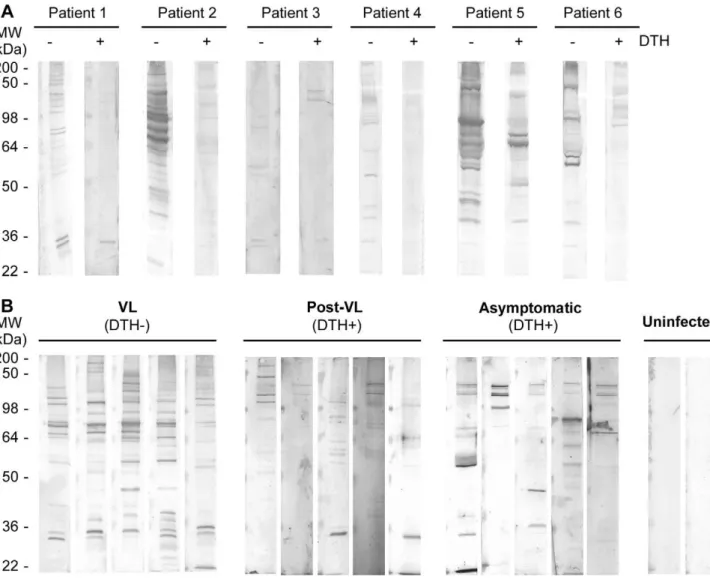
antibodies are still present in the sera [4]. To understand if this reduction in IgG recognition is associated with changes in antigen specificity, we screened sera from patients before and after DTH conversion for reactivity withL. chagasi total protein by Western blot. A decrease in the number of proteins with reactivity was observed in sera from VL patients after their recovery, and new reactivity patterns were observed after DTH conversion (Figure 1A). Next, we compared the reactivity pattern of sera from symptomatic VL patients (DTH2), recovered patients (DTH+), asymptomatic donors (DTH+) and uninfected volunteers from the same endemic area (Figure 1B). Sera from post-treatment and asymptomatic patients both of which were DTH+, showed weak or no reactivity to a majority of the bands. A high mass proteins group (approximately 110–130 kDa) was detected in all naturally resistant patient samples and in 60% of post-treatment DTH+ sera tested (Figure 1; Supplementary Figure S1; Baseline characteristics of the study population used in Table S1). To determine if any IgG subclass was involved in the differential reactivity of sera between groups, IgG1, IgG2, IgG3 and IgG4 binding was tested. Only IgG1-antibody showed significant reactivity that was similar to that of the total IgG pattern in all cases (Table S1).
Characterization of 2D IgG recognizing by sera of VL and DTH+patients
To characterize the reactivity patterns associated with protec-tion against VL, 200mg of total L. chagasi protein was 2D
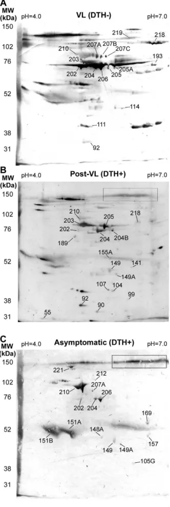
Table 1. In VL, post-treatment VL and asymptomatic patients, 58, 62 and 33 spots were detected respectively (Table 1). It is remarkable that the ratio between mapped spots versus total spots reactions were higher when we used sera from asymptomatic (14/ 33) than those found we used sera from VL (15/58) or Post-VL (17/62) groups. Each group showed specific mapped spots, whereas two mapped spots (202 and 204) were recognized by all groups. In asymptomatic individuals, spots mapped were not detected in the 110–130 kDa range, but a specific non-mapped signal was detected (delineated in a quadrant in Figure 2). This signal was also recognized by sera from a post-treatment VL sample; however it was not observed when we used VL patient sera.
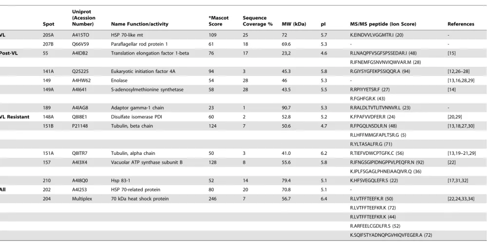
MALDI ToF/ToF-MS/MS analysis of the 24 identified spots (of which 6 spots were specifically recognized by VL patient sera, 8 spots by post-treatment VL sera, 8 spots by asymptomatic patient sera and 2 spots recognized by all groups) resulted in the identification of 14 (58.3%) proteins. These results, combined with MASCOT protein identification, are summarized in Table 2.
We identified the following two proteins that specifically reacted with sera from VL patients (DTH2): Mitochondrial 70 kDa heat shock protein (MPT70, spot 205A) and paraflagellar rod protein 1 (PFR1, spot 207B). Additionally, eleven proteins specifically reacting with sera from DTH+patients were identified. Five proteins reacting with sera from post-VL treatment patients (translation elongation factor 1-beta [LieEF1B, spot 55], eukaryotic initiation factor 4A [LieIF4A, spot 141A], enolase [spot 149], S-adenosylmethionine synthetase [MAT2, spot 149A] and adaptor gamma-1 chain [spot 189]) and 6 proteins with specific reactivity with sera from asymptomatic infected patients (disulfate isomerase [PDI, spot 148A], alpha- and beta-tubulin [spots 151A and 151B, respectively], vacuolar ATP synthase subunit B [spot 157] and 83 kDa heat shock protein [HSP83, spot 210]) were identified. Finally, the two proteins recognized by all groups (spots 202 and 204) were both identified as 70 kDa heat shock protein (HSP70). The putative functions and immunological properties of the identified proteins were retrieved from published literature and also from the L. infantum Genome Project database (www.genedb.org).
Figure 1. IgG reaction pattern associated to DTH conversion. Total protein extract from L. chagasi was SDS-PAGE resolved (10% polyacrilamide) and electrotransferred to nitrocellulose filter. In all blots, sera were used at 1:1000, and secondary anti-human IgG was used at 1:2000. A) Sera from six VL patients before and after DTH+conversion. B) Western blot results using sera from active VL and recovered VL patients, asymptomatic infected and non-infected individuals. Molecular weight markers are shown on the left panel.
Characterizing the humoral response against recombinant proteins fromL. chagasiidentified by proteomic approaches
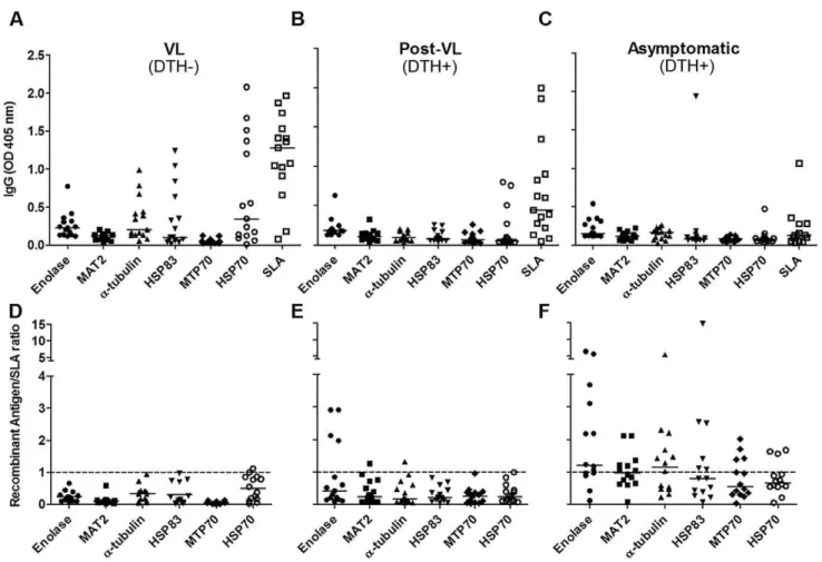
To analyze whether the identified proteins were indeed reactive with sera from the respective groups and could be used in a serological test for potentially asymptomatic L. chagasi infected individuals, we measured reactivity of the following recombinant proteins (Figure S3 for supporting information) by ELISA: alpha-tubulin, enolase, MAT2, and HSP83, which were reactive with DTH+individuals sera; MPT70, which reacted with VL patients’ sera and HSP70, which reacted with sera from both groups (Figure 3). Recombinants antigens reacted with some of the serum samples from VL (DTH2), post-treatment (DTH+) and asymp-tomatic patients (DTH+) (Figures 3 A–C). However, when we normalized the titers of IgG reactive with antigens from SLA, we observed that asymptomatic patients presented higher specific reactivity with DTH+ antigens (Figure 3D–F). We then used a Figure 2. 2D Western blot using sera from VL (DTH2), post-VL (DTH+) and naturally resistant individuals (DTH+).Total protein extract fromL. chagasiwere added to 11 cm strips (pH 4–7), followed by SDS-PAGE (8% polyacrylamide). After Ponceau staining, the filters were incubated with a mixture of sera at 1:1000 from VL (A) and 1:500 from post-VL (B) and naturally resistant donors (C). Only mapped spots are indicated. Numbers corresponding with spots from 2D maps (supplementary Figure S2). A specific DTH+signal (none mapped) is shown in the designated box. MW, Molecular weight markers. doi:10.1371/journal.pntd.0001687.g002
Table 1.Summary of 2D Western blot results.
VL Post-VL VL-Resistant
Total Spot Reactions 58 62 33
Specific Mapped Spots 219 189 210
207B 155A 212
205A 141 151B
193 107 151A
114 104 148A
111 99 169
90 157
55 105G
Non-Specific Mapped Spots 202 202 202
204 204 204
207A 207A
206 206
218 218 210* 210*
203A/B 203A/B
205 205
92 92
149 149
149A 149A
Total Mapped Spots 15 18 14
Total Unmapped Spots 43 44 19
Reproducible spots reacting with each group are indicated. Mapped and non-mapped spots are defined in Materials and Methods. Asterisks indicate low recognition.
Table 2.Summary of MALDI ToF/ToF-MS/MS results.
Spot
Uniprot (Acession
Number) Name Function/activity
*Mascot Score
Sequence
Coverage % MW (kDa) pI MS/MS peptide (Ion Score) References
VL 205A A415TO HSP 70-like mt 109 25 72 5.7 K.EINDVVLVGGMTR.I (20)
-207B Q66V59 Paraflagellar rod protein 1 61 18 69.6 5.3 -
-Post-VL 55 A4IDB2 Translation elongation factor 1-beta 76 17 23,2 4.6 R.LNAQPFVSGFSPSSEDAR.I (48) [15]
R.IFNEMFGSNVNVIQWVAR.M (28)
141A Q25225 Eukaryotic initiation factor 4A 94 3 45.3 5.8 R.GIYSYGFEKPSSIQQR.A (94) [12,26–28]
149 A4HW62 Enolase 54 28 46 5.3 - [13,16,28,29]
149A A4I641 S-adenosylmethionine synthetase 58 28 43.5 5.5 R.RPIYYETSR.F (27) [14]
R.FGHFGR.K (43)
189 A4IAG8 Adaptor gamma-1 chain 23 1 90.7 5.3 R.RALDLTVTLITVNNVR.L (23)
-VL Resistant 148A Q8I8E1 Disulfate isomerase PDI 60 2 52.8 5.2 K.FPAFVVDFER.R (24) [20,29]
151B P21148 Tubulin, beta chain 124 7 50.6 4.7 R.FPGQLNSDLR.N (48) [13,18,27,30]
R.LHFFMMGFAPLTSR.G (5)
R.YLTASALFR.G (71)
151A Q8ITR7 Tubulin, alpha chain 50 3 41.0 6.2 R.TIEFVDWCPTGFK.C (56) [13,19–21,29]
157 A4I3X4 Vacuolar ATP synthase subunit B 128 8 55.6 5.8 R.IFNGSGIPIDNGPPVLPEQFR.N (92) [22]
K.IPLFSGAGLPHNEIAAQIVR.Q (36)
210 A4I8Q0 Hsp 83-1 52 14 79.4 5.1 K.HFSVEGQLEFR.S (22) [17,31,32]
All 202 A4I253 HSP 70-related protein 80 20 70.8 5.1
-204 Multiplex 70 kDa heat shock protein 246 7 56.7 6.4 R.LVTFFTEEFK.R (50) [22,24,33,34]
R.LVTFFTEEFKR.K (72)
R.LVTFFTEEFKR.K (44)
R.ARFEELCGDLFR.S (52)
K.SQIFSTYADNQPGVHIQVFEGER.A (72)
Note.References are related to the significance of these antigens with regard to drug resistance, host parasite survival or immunological properties. *Mascot Score, protein overall scores greater than 20 are significant (P,0,05). MW, molecular weight; Ip, isoelectric point.
doi:10.1371/journal.pntd.0001687.t002
Immunodom
inant
Leishmani
a
Antigens
ntds.org
6
June
2012
|
Volume
6
|
Issue
6
|
combination of recombinants proteins composed of enolase, MAT2, alpha-tubulin and HSP83 (MIX) to test the sensitivity in the sera from asymptomatic patients and their specificity against sera from patients with Chagas disease, which presents high
cross-reactivity with Leishmania antigens [11]. We verified that asymptomatic individuals had a higher reaction with MIX than SLA, and sera from patients with Chagas disease showed lower cross-reactivity (Figure 4).
Figure 3. ELISA with individual recombinant proteins.SLA and obtained recombinants proteins (10mg/mL) were tested for reactivity with sera (diluted 1:100) from those groups shown above the panels. Absolute OD obtained (A–C) and the ratio between antigen-OD versus SLA-OD for each serum sample (D–F)) are shown. The dotted line shows the OD ratio at 1. Medians are shown as horizontal lines.
doi:10.1371/journal.pntd.0001687.g003
Figure 4. Mixture of DTH+antigens in serological diagnosis of asymptomatic infected individuals.ELISA results were obtained using 10mg/mL SLA or DTH+recombinant antigen mixture (MIX, contained 2.5mg/mL of each of the following proteins: enolase, MAT2, alpha tubulin and HSP83). Sera from asymptomaticL. chagasiinfected patients, Chagas-infected patients were used (1:100).
Discussion
The decrease in anti-LeishmaniaIgG titers during conversion to DTH+ in effectively treated VL patients has been previously associated with the development of cell-mediated immune responses [4]. Herein, we show that decreased antibody levels in VL-related states and positive DTH reactions are associated with a reduction in the reactivity against most of the parasite proteins and, more relevant, the recognition of previously unrecognized proteins. These observations indicate that impor-tant changes in the humoral response occurs during DTH conversion and reveals a reactivity pattern associated with recovery and natural resistance to VL that is characterized by high-mass group proteins (approximately 110–130 kDa). These high-mass proteins did not react with sera from VL patients, indicating that some specific immunodominant antigens are specifically associated with DTH conversion and putatively with protection against VL. Unfortunately, high-mass group proteins were not 2D-resolved, and their identification was not possible; however, 2D-recognizing map analyses of sera from VL patients and DTH+ individuals (post-VL or naturally resistant) showed other immunodominant antigens whose recognition was DTH status-dependent. A lower number of reactive spots were observed in sera from DTH+ individuals living in the endemic area who are presumably naturally resistant. It is noteworthy that sera from post-VL patients (DTH+) reacted with a larger number of proteins, which suggests that post-VL patients likely share antigenic recognition patterns with both VL patients and naturally resistant individuals which reinforces the idea that some antigens are specifically associated with positive DTH response and therefore protection against VL.
Proteomic analysis revealed the identification of 14 antigens, including proteins not previously reported as antigenic, such as adaptor gamma-1 chain, MPT70 and PFR1. Among the identified antigens associated with the post-VL subset, four of them (LieIF4A, LieEF1B, enolase and MAT2) have been reported as highly expressed in drug-resistant strains [12–14] and have been implicated in the survival of the parasite inside the host [15,16]. For the six identified antigens that reacted with sera from the VL naturally resistant people (PDI, alpha- and beta-tubulin, vacuolar ATP synthase subunit B and HSP83), drug resistance implications and host parasite survival properties have been previously described too [13,17–21]. The stress state imposed on the parasite in a DTH+ environment (resulting either from drug treatment or natural resistance) can induce the overexpression of these proteins, and this fact may explain their immunodominant antigenicity in these individuals. It is notable that LieIF4A protein was described as antigen reactive with sera from VL patients infected byL. donovanifrom India [25] and we found a clear reactivity against this protein using sera from post-VL patients. It could be interpreted as yet another indication that IgG pattern recognition in post-VL patients preserved reactivity against some antigens of active illness. In addition, it was already observed that a Th1 activation response, associated with healing, was induced in cutaneous leishmaniasis patients’ PBMC by recombinant LieIF4A [26]. We consider the hypothesis that some correlation may exist between the antigenicity of DTH+ recognized proteins and its capacity to induce anti-Leishmania
cellular responses. On the other side, it is possible that the highly immunogenic proteins, such as HSP70 [22–24], are implicated in its recognition by all groups.
To determine if the immunodominant antigens identified could serve as serological markers, some of them were expressed as recombinant proteins, purified and then employed as antigen in
ELISA (Figure 3). When we normalized to SLA, enolase exhibited a remarkable increase in reactivity against sera from post-VL group respect to other antigens. Moreover, an increased enolase recognition was observed using sera from asymptomatic people, strengthening the idea that antigens recognized by post-VL sera could also be reactive in VL patients and resistant people sera. In the other hand, alpha tubulin and HSP83, showed a remark recognition increase from asymptomatic donors’ sera. Meanwhile, MTP70 that was identified as a VL-specific protein did not react with any groups. There are four not identical copies of MPT70 in
L. infantumgenome (www.genedb.org), and only one of them was cloned to obtain MPT70 recombinant protein. Future studies with the other 3 variants will shed light about the real serological properties of these proteins. Finally, and as expected, HSP70 reacted with several serum samples from all groups.
Lastly, we proved that a mixture of DTH+ antigens (enolase, MAT2, alpha-tubulin and HSP83) had higher reactivity with sera from asymptomatic individuals than SLA and lower cross-reactivity with sera from patients with Chagas disease. Our results are the first description of several antigens that are immunodo-minant in DTH+ individuals (post-VL and naturally resistant people). Future studies using these antigens may help to identify potent serological tools that could be useful for determining patient disease status as well as new anti-Leishmaniavaccine candidates.
Supporting Information
Figure S1 Characterization of IgG reaction pattern associated to DTH conversion. Total protein extract from
L. chagasi was SDS-PAGE resolved (10% polyacrilamide) and electrotransferred to nitrocellulose filter. In all blots, sera were used at 1:1000 (1:100 in B - line 5) and secondary anti-human IgG (or subclasses IgG1, IgG2, IgG3 and IgG4 in E and F) was used at 1:2000. A) Sera from VL patients. B) Sera from uninfected people. C) Sera from post-VL patients (DTH+). D) Sera from asymptom-atic infected people. E) VL sera mix (VL0678, VL0669, VL0660, VL0671, VL0666). F) Asymptomatic sera mix (576, 421, 689, 011, 417). Under panels are show the serum code. Asterisk over panel indicate filters with DTH+patter (110–130 KDa proteins). MW, Molecular weight markers. Baseline characteristics of the study population are shown in supplementary Table S1.
(TIF)
Figure S2 2D proteomic map ofL. chagasi.Approximately 200mg of total protein fromL. chagasiwas 2D resolved on 11 cm strips with a pH range of 4–7 (first dimension) and SDS-PAGE. The numbers of mapped spots are indicated. A) Gel after electrotransfer to a nitrocellulose filter and Ponceau Red staining (8% polyacrylamide in second dimension). B) Gel with 10% polyacrylamide and Colloidal Coomassie staining. C) Gel with 10% polyacrylamide and Silver staining.
(TIF)
Figure S3 Purified recombinant antigens of L. chagasi identified by proteomic approaches.IdentifiedE. coli(M15) overexpressing recombinant antigens (fused to a 6xHis tag) were used for Ni-affinity chromatography. Obtained purified proteins resolved in 10% polyacrylamide gel stained with Comassie are shown. MW, Molecular weight marker.
(TIF)
Acknowledgments
The´o de Arau´jo-Santos provided assistance with statistical analyses and manuscript preparation.
Author Contributions
Conceived and designed the experiments: DRA MS AB MBN. Performed the experiments: DRA LVA ESA JRASP DA AJC. Analyzed the data: DRA MSP AB MBN. Contributed reagents/materials/analysis tools: MSP DA AJC. Wrote the paper: DRA LVA MBN.
References
1. Wilson ME, Jeronimo SM, Pearson RD (2005) Immunopathogenesis of infection with the visceralizing Leishmania species. Microb Pathog 38: 147– 160.
2. Ribeiro-de-Jesus A, Almeida RP, Lessa H, Bacellar O, Carvalho EM (1998) Cytokine profile and pathology in human leishmaniasis. Braz J Med Biol Res 31: 143–148.
3. Oliveira JM, Fernandes AC, Dorval ME, Alves TP, Fernandes TD, et al. (2010) [Mortality due to visceral leishmaniasis: clinical and laboratory characteristics]. Rev Soc Bras Med Trop 43: 188–193.
4. Caldas A, Favali C, Aquino D, Vinhas V, van Weyenbergh J, et al. (2005) Balance of IL-10 and interferon-gamma plasma levels in human visceral leishmaniasis: implications in the pathogenesis. BMC Infect Dis 5: 113. 5. Jeronimo SM, Teixeira MJ, Sousa A, Thielking P, Pearson RD, et al. (2000)
Natural history of Leishmania (Leishmania) chagasi infection in Northeastern Brazil: long-term follow-up. Clin Infect Dis 30: 608–609.
6. Davies CR, Kaye P, Croft SL, Sundar S (2003) Leishmaniasis: new approaches to disease control. BMJ 326: 377–382.
7. Aquino DMC, Caldas AJM, Miranda JC, Silva AAM, Barral-Netto M, et al. (2010) Epidemiological study of the association between anti-Lutzomyia longipalpis saliva antibodies and development of delayedtype hypersensitivity to Leishmania antigen. Am J Trop Med Hyg 83: 825–827.
8. Reed SG, Badaro´ R, Masur H, Carvalho EM, Lorenco R, et al. (1986) Selection of a skin test antigen for American visceral leishmaniasis. Am J Trop Med Hyg 35: 79–85.
9. Laemmli UK (1970) Cleavage of structural proteins during the assembly of the head of bacteriophage T4. Nature 227: 680–685.
10. Shevchenko A, Wilm M, Vorm O, Mann M (1996) Mass spectrometric sequencing of proteins silver-stained polyacrylamide gels. Anal Chem 68: 850– 858.
11. Vexenat Ade C, Santana JM, Teixeira AR (1996) Cross-reactivity of antibodies in human infections by the kinetoplastid protozoa Trypanosoma cruzi, Leishmania chagasi and Leishmania (viannia) braziliensis. Rev Inst Med Trop Sao Paulo 38: 177–185.
12. Singh G, Chavan HD, Dey CS (2008) Proteomic analysis of miltefosine-resistant Leishmania reveals the possible involvement of eukaryotic initiation factor 4A (eIF4A). Int J Antimicrob Agents 31: 584–586.
13. Kumar A, Sisodia B, Misra P, Sundar S, Shasany AK, et al. (2010) Proteome mapping of overexpressed membrane-enriched and cytosolic proteins in sodium antimony gluconate (SAG) resistant clinical isolate of Leishmania donovani. Br J Clin Pharmacol 70: 609–617.
14. Drummelsmith J, Girard I, Trudel N, Ouellette M (2004) Differential protein expression analysis of Leishmania major reveals novel roles for methionine adenosyltransferase and S-adenosylmethionine in methotrexate resistance. J Biol Chem 279: 33273–33280.
15. Vickers TJ, Wyllie S, Fairlamb AH (2004) Leishmania major elongation factor 1B complex has trypanothione S-transferase and peroxidase activity. J Biol Chem 279: 49003–49009.
16. Vanegas G, Quinones W, Carrasco-Lopez C, Concepcion JL, Albericio F, et al. (2007) Enolase as a plasminogen binding protein in Leishmania mexicana. Parasitol Res 101: 1511–1516.
17. Vergnes B, Gourbal B, Girard I, Sundar S, Drummelsmith J, et al. (2007) A proteomics screen implicates HSP83 and a small kinetoplastid calpain-related protein in drug resistance in Leishmania donovani clinical field isolates by modulating drug-induced programmed cell death. Mol Cell Proteomics 6: 88– 101.
18. Prasad V, Kumar SS, Dey CS (2000) Resistance to arsenite modulates levels of alpha-tubulin and sensitivity to paclitaxel in Leishmania donovani. Parasitol Res 86: 838–842.
19. Mojtahedi Z, Clos J, Kamali-Sarvestani E (2008) Leishmania major: identification of developmentally regulated proteins in procyclic and metacyclic promastigotes. Exp Parasitol 119: 422–429.
20. Ben Achour Y, Chenik M, Louzir H, Dellagi K (2002) Identification of a disulfide isomerase protein of Leishmania major as a putative virulence factor. Infect Immun 70: 3576–3585.
21. Bakker-Grunwald T (1992) Ion transport in parasitic protozoa. J Exp Biol 172: 311–322.
22. Skeiky YA, Benson DR, Guderian JA, Whittle JA, Bacelar O, et al. (1995) Immune responses of leishmaniasis patients to heat shock proteins of Leishmania species and humans. Infect Immun 63: 4105–4114.
23. Rafati S, Gholami E, Hassani N, Ghaemimanesh F, Taslimi Y, et al. (2007) Leishmania major heat shock protein 70 (HSP70) is not protective in murine models of cutaneous leishmaniasis and stimulates strong humoral responses in cutaneous and visceral leishmaniasis patients. Vaccine 25: 4159–4169. 24. Quijada L, Requena JM, Soto M, Alonso C (1998) Analysis of the antigenic
properties of the L. infantum Hsp70: design of synthetic peptides for specific serodiagnosis of human leishmaniasis. Immunol Lett 63: 169–174.
25. Forgber M, Basu R, Roychoudhury K, Theinert S, Roy S, et al. (2006) Mapping the antigenicity of the parasites in Leishmania donovani infection by proteome serology. PLoS One 1: e40.
26. Skeiky YA, Guderian JA, Benson DR, Bacelar O, Carvalho EM, et al. (1995) A recombinant Leishmania antigen that stimulates human peripheral blood mononuclear cells to express a Th1-type cytokine profile and to produce interleukin 12. J Exp Med 181:1527–37.
27. Probst P, Skeiky YA, Steeves M, Gervassi A, Grabstein KH, et al. (1997) A Leishmania protein that modulates interleukin (IL)-12, IL-10 and tumor necrosis factor-alpha production and expression of B7-1 in human monocyte-derived antigen-presenting cells. Eur J Immunol 27: 2634–2642.
28. Gupta SK, Sisodia BS, Sinha S, Hajela K, Naik S, et al. (2007) Proteomic approach for identification and characterization of novel immunostimulatory proteins from soluble antigens of Leishmania donovani promastigotes. Proteomics 7: 816–823.
29. Kumari S, Samant M, Misra P, Khare P, Sisodia B, et al. (2008) Th1-stimulatory polyproteins of soluble Leishmania donovani promastigotes ranging from 89.9 to 97.1 kDa offers long-lasting protection against experimental visceral leishmaniasis. Vaccine 26: 5700–5711.
30. Bhowmick S, Ali N (2009) Identification of novel Leishmania donovani antigens that help define correlates of vaccine-mediated protection in visceral leishmaniasis. PLoS One 4: e5820.
31. Rico AI, Girones N, Fresno M, Alonso C, Requena JM (2002) The heat shock proteins, Hsp70 and Hsp83, of Leishmania infantum are mitogens for mouse B cells. Cell Stress Chaperones 7: 339–346.
32. Echeverria P, Dran G, Pereda G, Rico AI, Requena JM, et al. (2001) Analysis of the adjuvant effect of recombinant Leishmania infantum Hsp83 protein as a tool for vaccination. Immunol Lett 76: 107–110.
33. Rasouli M, Zavaran Hoseini A, Kazemi B, Alborzi A, Kiany S (2009) Expression of recombinant heat-shock protein 70 of MCAN/IR/96/LON-49, a tool for diagnosis and future vaccine research. Iran J Immunol 6: 75–86.