Serum levels of soluble TNF-
α
receptors
but not BDNF are associated with apathy
symptoms in mild Alzheimer’s disease and
amnestic mild cognitive impairment
Henrique Cerqueira Guimarães1, Paulo Caramelli1, Patricia Paes Araujo Fialho1,
Elisa de Paula França1, Marcelo Pelizzaro Dias Afonso1, Antonio Lucio Teixeira1,2
ABSTRACT. Apathy is intimately associated with dementia. Unfortunately, its pathophysiology remains poorly understood. The motivational impairment that characterizes this disorder might share the same inflammatory mechanisms, as suggested by the sickness behavior theory. Objective: The primary aim of this study was to investigate the association between apathy symptoms and serum levels of tumor necrosis factor alpha (TNF-α) and its soluble receptors. Brain-derived neurotrophic factor (BDNF) levels were also analyzed since these have been associated with depression, a condition which shares abulic features with apathy. Methods: The sample consisted of 27 subjects with mild Alzheimer’s disease or amnestic mild cognitive impairment, who were submitted to specific apathy evaluation using the Apathy Scale (AS) and provided blood samples for biomarker analysis. Participants were categorized into two groups according to median AS scores (17 points). Results: Subjects with higher apathy symptoms (n=13) displayed higher levels of TNF-α soluble receptors (type 1: p=0.03; type 2: p=0.04). No other difference was found between groups. Conclusion: These findings point to the involvement of inflammatory mediators in the genesis of apathy symptoms, as suggested by the sickness behavior theory.
Key words: apathy, dementia, Alzheimer’s disease, mild cognitive impairment, TNF-α, sTNFR1, sTNFR2, BDNF.
NÍVEIS SÉRICOS DE RECEPTORES SOLÚVEIS DO TNF-α MAS NÃO DE BDNF ESTÃO ASSOCIADOS A SINTOMAS DE APATIA NA DOENÇA DE ALZHEIMER LEVE E NO COMPROMETIMENTO COGNITIVO LEVE AMNÉSTICO
RESUMO. Apatia está intimamente associada à demência. Lamentavelmente, sua fisiopatologia ainda é pouco compreendida. O comprometimento motivacional que caracteriza este transtorno poderia compartilhar mecanismos inflamatórios como sugere a teoria do comportamento associado à doença. Objetivo: O principal objetivo deste estudo foi investigar a associação entre apatia e os níveis séricos do fator de necrose tumoral alfa (TNF-α) e de seus receptores solúveis. Os níveis de fator neurotrófico derivado do cérebro também foram analisados já que estes foram associados à depressão, que compartilha aspectos abúlicos com a apatia. Métodos: A amostra consistiu de 27 indivíduos com doença de Alzheimer leve ou com comprometimento cognitivo leve amnéstico, que foram submetidos à avaliação de apatia pela Escala de Apatia (EA), e proveram amostra de sangue para análise de biomarcadores. De acordo com a mediana de escores na EA (17 pontos), a amostra foi divida em dois grupos. Resultados: O grupo com mais sintomas de apatia apresentou maiores níveis séricos de receptores solúveis de TNF-α (tipo 1: p=0,03 ; tipo 2: p=0,04). Nenhuma outra diferença foi encontrada entre os grupos. Conclusão: Estes achados sugerem o envolvimento de mediadores inflamatórios na gênese de sintomas de apatia, assim como sugere a teoria do comportamento associado à doença.
Palavras-chave: apatia, demência, doença de Alzheimer, comprometimento cognitivo leve, TNF-α, sTNFR1, sTNFR2,BDNF.
INTRODUCTION
A
pathy is a pervasive feature in several neu-ropsychiatric disorders.1 Most of ourcur-rent understanding regarding this behavioral syndrome was built upon research on
neuro-1Behavioral and Cognitive Neurology Research Group and 2Translational Psychoneuroimmunology Group, Department of Internal Medicine, Faculty of Medicine of
the Federal University of Minas Gerais, Belo Horizonte (MG), Brazil.
Henrique Cerqueira Guimarães. Av. Contorno, 4747 / sala 1710 – 30110-921 Belo Horizonte MG – Brasil. E-mail: [email protected]
Disclosure: The authors report no conflicts of interest.
degenerative conditions.2 Generally speaking, apathy has been shown to be the most prevalent behavioral disorder in dementia.3 Once identiied, apathetic symp-toms follow a prolonged and mostly deinitive course throughout cognitive decline,4 leading to a sharp in-crease in apathy prevalence and severity as dementia reaches its moderate to advanced stages.3
here is fairly good agreement in the literature that apathy should be considered an independent syndrome in dementia, with speciic clinical implications and probable worse outcomes.5,6 Another emerging consen-sus is that apathy can be recognized in a signiicant pro-portion of mild cognitive impairment (MCI) subjects, especially those regarded as amnestic.7,8 Longitudinal studies have suggested a substantially increased risk of dementia conversion in these MCI apathetic subjects.9 Unfortunately, scant therapeutic options are available to improve this devastating disorder, a fact which, at least partially, can be ascribed to a poor understanding of the neurobiological underpinnings of apathy.
Marin10 deined apathy as “lack of motivation, rela-tive to the patient’s previous level of functioning or the standards of his or her age and culture, not attribut-able to intellectual impairment, emotional distress or diminished level of consciousness”. he motivational feature has remained the core diagnostic criteria in a consensus proposition to identify apathy in Alzheimer disease (AD) and other neuropsychiatric disorders.3 Lack of motivation is also the core feature of sickness behavior. his is a coordinated set of behavioral adap-tations that also includes a neurovegetative dimen-sion (fatigue, loss of appetite and sleep disorders), and a psychological dimension (depressed mood, anxiety and cognitive dysfunction), that are supposed to reor-ganize the organism’s priorities to cope with infectious pathogens.11 TNF-α, a proinlammatory cytokine, plays a pivotal role in triggering sickness behavior.12 he levels of this cytokine have also been shown to be increased in a sample of MCI and AD patients.13 Since inlam-matory mechanisms play a putative role in the patho-physiology of several types of neurodegeneration,14 and apathy is a pervasive feature in dementia, it is reason-able to investigate whether these two phenomena are associated.
he primary aim with this study was to investigate whether blood TNF-α, and its soluble receptors, are associated with apathy symptoms. Soluble forms of TNF-α receptors represent reliable markers of this cy-tokine activity, binding to and protecting TNF-α from proteolytic degradation, therefore, extending its efects systemically.15 We also investigated the possible
associa-tion between BDNF and apathy, since there has been a previous report of reduced levels of this neurotrophic factor in subjects with late-life depression,16 a condition that shares abulic features with apathy. To accomplish this objective we evaluated only subjects with cogni-tive impairment in its very early stages, namely mild AD and amnestic MCI (aMCI), since inlammatory sta-tus has been related to frailty,17,1, an almost inexorable outcome in moderate and advanced stages of late-onset dementia.
METHODS
Participants and procedures. his study was a retrospec-tive analysis of the available clinical and laboratory data for a small subset of participants from the Pietà Study, a community-based survey of successful aging, carried out in Caeté, Southeast Brazil, in the summer of 2008. Detailed methodology has been described previously.19 Briely, the study invited all of the city’s inhabitants aged 75 years or older to participate, and those who agreed gave written informed consent. he study was approved by the University’s research ethics committee. A total of 639 subjects were submitted to a thorough functional, clinical, psychiatric and neurological evalu-ation, including the Functional Activities Questionnaire (FAQ), the motor section of the Uniied Parkinson’s disease rating scale (UPDRSm), the M.I.N.I. structured psychiatric interview, Geriatric Depression Scale (GDS), and a Brief Cognitive Screening Battery (BCSB), consist-ing of the Mini-Mental State Examination (MMSE), ani-mal category semantic luency test, and picture draw-ings memory test (PDMT). Individuals suspected of having cognitive impairment, together with a subset of putative cognitively healthy control subjects, were fur-ther submitted to a comprehensive neuropsychological evaluation with the following instruments: Rey Audi-tory Verbal Learning Test, naming and praxis tests from the CERAD (Consortium To Establish A Registry For Alzheimer’s Disease) protocol, phonemic verbal luency tasks (FAS), Frontal Assessment Battery, and the De-mentia Rating Scale. A subset of 358 subjects provided blood samples for routine and inlammatory biomarker analyses.
in Alzheimer Disease (FAST).21 Only those categorized as FAST 4 or below were included. None of the AD sub-ject were taking cholinesterase inhibitors at the time of evaluations. Vascular dementia was ruled out accord-ing to NINDS-AIREN criteria.22 he second group con-sisted of 26 subjects with aMCI, diagnosed according to Petersen’s criteria.23 Amnestic impairment was estab-lished by comparing performances on the Rey Auditory Verbal Learning Test (RAVLT) by cognitively healthy subjects from the same population. Subjects with a major depressive episode, diagnosed according to the Diagnostic and Statistical Manual of Mental Disorders fourth edition, were excluded.24 Of the 52 subjects with either mild AD or amnestic MCI who were submitted to apathy and behavioral evaluation, 27 underwent blood analysis, giving the inal study sample reported.
Apathy and behavioral assessment. Since there were no pub-lished consensual diagnostic criteria for apathy at the time of data collection, this disorder was evaluated di-mensionally through the Apathy Scale (AS),25 which was administered to caregivers or to a very close member of the household. he AS has 14 questions with a score ranging from zero to 42 points, where higher scores rep-resent more severe apathetic symptoms. Despite a lack of consensus regarding this issue,3 previous studies have suggested a cut-of of 14 points in order to identify clini-cally relevant apathy.25,26 Subjects were also submitted to the Neuropsychiatric Inventory (NPI).27
Laboratory assessment. Blood was drawn during fasting and in the early morning for all the participants. Serum levels of BDNF, TNF-α, sTNF-R1, and sTNF-R2 were
Table 1. Comparison of higher vs lower apathy symptoms groups, according to demographics.
Apathy scale
p - value
Lower apathy symptoms (n=14) Higher apathy symptoms (n=13)
% mean SD % mean SD
Age, years -- 82.9 3.9 -- 80.5 4.6 0.08
Gender (male, %) 28.6 -- -- 61,5 -- -- n.s.*
Education, years -- 3.6 3.8 -- 3.3 2 n.s.
Weight, Kg -- 62.9 15.9 -- 67.6 13.9 n.s.
Body mass index -- 26.6 5.1 -- 28.1 4.5 n.s.
Alzheimer’s disease (%) 35.7 -- -- 61.5 -- -- n.s.*
Cognitive and functional measures
Functional Assessment Questionnaire -- 3.5 5 -- 6.1 6.2 n.s.
Mini-Mental State Examination -- 20.3 4.4 -- 20.4 4.8 n.s.
Dementia Rating Scale -- 100.3 19.8 -- 105.1 11.7 n.s.
Frontal Assessment Battery -- 6.2 2.7 -- 6.7 2.3 n.s.
Behavioral evaluation
Apathy Scale -- 10.4 4.5 -- 26.1 3.9 <0.0001
NPI - apathy -- 0 0 -- 2.75 3.3 0.003
NPI - total -- 7.3 14.8 -- 10.3 9.8 0.06
Geriatric Depression Scale -- 4 2.1 -- 4 3.4 n.s.
Motor exam
UPDRSm -- 6,5 6 -- 9 8.3 n.s.
Biomarkers
BDNF (pg/ml) -- 1418.3 353.9 -- 1668.9 398.6 0.10
TNF-α (pg/ml) -- 227.9 546.8 -- 260.5 814.8 n.s.
sTNF-R type1(pg/ml) -- 273.9 86.3 -- 360.5 111.9 0.03
sTNF-R type2 (pg/ml) -- 291.6 93.8 -- 377.2 138.6 0.04
measured according to the procedures supplied by the manufacturer, and using ELISA kits for TNF-α (Quan-tikine, R&D Systems, Minneapolis, MN, USA), soluble TNF-α receptors and BDNF (DuoSet, R&D Systems, Minneapolis, MN, USA). All samples were assayed in du-plicate, yielding concentrations expressed as pg/ml. he detection limits were 0.1 pg/ml for TNF-α, 10 pg/ml for both soluble receptors, and 5 pg/ml for BDNF.
Statistical analysis. An exploratory analysis with the D’Agostino-Pearson test showed that most of the data had non-normal distributions. he 27 eligible subjects were split into two groups according to median AS scores (17 points), forming one group with higher (AS >17) and another with lower apathy symptoms (AS ≤17). Diferences between groups were investigated using the Mann-Whitney test for continuous variables and Chi-square test for categorical data. Correlation analysis was performed using Spearman’s rho coeicient. Statistical
signiicance was set as a p-value ≤0.05.
RESULTS
he target sample consisted of 27 subjects (15 wom-en, 12 men), with a mean age of 81.5±4.6 years, and 3.2±2.7 mean years of formal education. According to cognitive status, 14 subjects presented mild AD and 13 had aMCI. he median score on the AS was 17 points, providing two groups with diferent intensity of apathy symptoms.
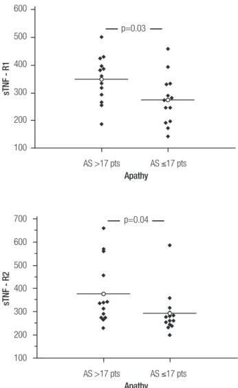
Table 1 displays the comparison between the groups, regarding demographics, anthropometrics, cognitive status; cognitive, motor, functional and behavioral mea-sures, and, lastly, BDNF and inlammatory biomarker levels. As expected, apathy measures difered between the groups. he higher apathy symptoms group had higher levels of both types of TNF-α soluble receptors (sTNF-R1: p=0.03; sTNF-R2: p=0.04). hese results are illustrated in Figure 1. Soluble TNF-α receptors levels also correlated with AS scores (sTNF-R1: rho=0.49; p=0.01; sTNF-R2: rho=0.40; p=0.037), considering the whole sample. BDNF and TNF-α levels did not dif-fer signiicantly between groups. No other variables reached statistical signiicance, except for age and NPI-total scores which displayed a tendency towards signii-cance. Nevertheless, NPI-total scores did not correlate with levels of any of the TNF-α soluble receptor types (sTNF–R1: rho=0.05, p=0.82; sTNF-R2: rho=0.22, p=0.29). Additionally, although there was a marked dif-ference between groups regarding proportions of gen-der and AD subjects, levels of TNF-α soluble receptors were not diferent in pairwise comparison
(Mann-Whit-600
500
400
300
200
100
sTNF - R1
AS >17 pts AS ≤17 pts Apathy
p=0.03
700
600
500
400
300
200
100
sTNF - R2
AS >17 pts AS ≤17 pts Apathy
p=0.04
Figure 1. Comparison of sTNF-R1 and sTNF-R2 levels (pg/ml) between
higher and lower apathy symptoms groups. AS: Apathy Scale.
ney test), either between genders (sTNF-R1: p=0.09; sTNF-R2: p=0.3) or between diferent cognitive status groups (sTNF-R1: p=0.7; sTNF-R2: p=0.29), suggesting that these variables were not driving the results.
DISCUSSION
In this retrospective investigation of a small subset of subjects from the Pietà study involving participants who had provided a blood sample for inlammatory biomarker analysis and were also submitted to apathy evaluation, we found elevated levels of both soluble TNF-α receptors in subjects with greater apathy symp-toms. he negative results found for TNF-α levels are unsurprising. his cytokine has a very short half-life and remains below the assay threshold in a signiicant proportion of subjects. As stated earlier, soluble recep-tors constitute more reliable markers of blood TNF-α
study that has used a speciic apathy scale to investigate association of the condition with TNF-α and its soluble receptors. A previous report found an association be-tween GDS items, assumed to mirror apathy, and higher TNF-α levels in male patients with AD.28 he indings herein reported are in accordance with the sickness behavior motivational impairment theory, and might constitute a reasonable explanation for apathy ubiqui-ty and its worsening in progressive neurodegenerative disorders. Surprisingly, very few studies have explored this association. A previous report found no relation-ship between a GDS surrogate apathy measure and C-reactive protein (CRP) levels.29 However, this acute phase reaction protein may not be the most suitable inlammatory biomarker to investigate this issue, since apathy and cognitive impairment are supposed to result from a chronic phenomenon. In line with the theoretical framework underlying this report, Ferretti et al report-ed, for a cohort of schizophrenic patients, a signiicant improvement in negative symptoms, including apathy, by treating them with minocycline,30 a drug with puta-tive anti-inlammatory properties.31
None of the other investigated variables were found to be associated with AS scores. he two groups dis-played similar depression scores, and also had similar mean performances on motor, global cognitive and executive function measures. he apparent diference regarding gender and dementia proportions between groups did not reach statistical signiicance, and these
features were not associated with soluble TNF-α re-ceptors. Although there was a clinically signiicant dif-ference between higher and lower apathy symptoms groups regarding mean performance on the FAQ, cor-roborating results of a previous report,32 these indings did not reach statistical signiicance. It is important to note that BDNF levels were higher in the more apathetic group, in contrast to previous indings from a late-life depression cohort.16 his inding adds to a large body of evidence regarding clinical and neuroimaging data that dissociates apathy from depression.2
he small sample size can be considered a serious limitation of our study. Additionally, this is an emerg-ing research ield, precludemerg-ing a robust hypothesis that might explain the neuroimmunology of these indings. Another important point is that the concept of MCI has proven to be, in recent years, a highly heterogeneous condition.33 hus, it is reasonable to question the va-lidity of pooling clinically deined AD and MCI groups together. However, as stated previously, inlammatory mechanisms seem to play a fundamental role in sev-eral neurodegenerative diseases, independently of their etiology, constituting a pervasive feature of dementia, akin to apathy. Lastly, the indings herein reported are preliminary and should be replicated in larger samples.
Acknowledgements. he study was supported by CNPq, FAPEMIG, Instituto Hermes Pardini, and Laboratório Geraldo Lustosa.
REFERENCES
1. van Reekun R, Stuss DT, Ostrander L. Apathy: why care? J Neuropsy-chiatry Clin Neursoci 2005;17:7-19.
2. Guimaraes HC, Levy R, Teixeira AL, Beato RG, Caramelli P. Neurobi-ology of apathy in Alzheimer’s disease. Arq Neuropsiquiatr 2008;66: 436-443.
3. Robert P, Onyike CU, Leentjens AF, et al. Proposed diagnostic criteria for apathy in Alzheimer’s disease and other neuropsychiatric disorders. Eur Psychiatry 2009;24:98-104.
4. Craig D, Mirakhur A, Hart DJ, McIlroy SP, Passmore AP. A cross-sec-tional study of neuropsychiatric symptoms in 435 patients with Alzheim-er’s disease. Am J Geriatr Psychiatry 2005;13:460-468.
5. Starkstein SE, Petraca G, Chemerinski E, Kremer J. Syndromic validity of apathy in Alzheimer’s disease. Am J Psychiatry 2001;158:872-877. 6. Starkstein SE, Jorge R, Mizrahi R, et al. A prospective longitudinal
study of apathy in Alzheimer’s disease. J Neurol Neurosurg Psychiatry 2006;77:8-11.
7. Robert PH, Berr C, Volteau M, et al. Neuropsychological performance in mild cognitive impairment with and without apathy. Dement Geriatr Cogn Disord 2006;21:192-197.
8. Geda YE, Roberts RO, Knopman DS, et al. Prevalence of neuropsychi-atric symptoms in mild cognitive impairment and normal cognitive ag-ing: a population-based study. Arch Gen Psychiatr 2008;65:1193-1198. 9. Robert PH, Berr C, Volteau M, et al. Apathy in patients with mild cognitive impairment and the risk of developing dementia of Alzheimer’s disease: a one-year follow-up study. Clin Neurol Neurosurg 2006;108:733-736. 10. Marin RS. Apathy: a neuropsychiatric syndrome. J Neuropsychiatry Clin
Neurosci 1991;3:243-254.
11. Dantzer R. Cytokine-induced sickness behavior: mechanisms and im-plications. Ann N Y Acad Sci 2001;933:22-34.
12. Palin K, Bluthe RM, McCusker RH, et al. The type 1 TNF receptor and its associated adapter, FAN, are required to TNF-α induced sickness behavior. Psychopharmcaology 2009;201:549-56.
13. Bermejo P, Martín-Aragón S, Benedí J, et al. Differences in peripheral inflammatory markers between mild cognitive impairment and Alzheim-er’s disease. Immunol Lett 2008;117:198-202.
14. Amor S, Puentes F, Baker D, van der Valk P. Inflammation in neurode-generative diseases. Immunology 2010;129:154-169.
15. Wajant H, Pfizenmaier K, Scheurich P. Tumor necrosis factor signaling. Cell Death Differ 2003;10:46-65.
16. Diniz BS, Teixeira AL, Talib LL, Mendonça VA, Gattaz WF, Forlenza OV. Serum brain-derived neurotrophic factor level is reduced in antide-pressant-free patients with late-life depression.World J Biol Psychiatry 2010;11:550-555.
17. Hubbard RE, O’Mahony MS, Sawa GM, Calver BL, Woodhouse KW. Inflammation and frailty measures in older people. J Cell Mol Med 2009;13:3103-3109.
18. Coelho FM, Narciso FM, Oliveira DM, et al. sTNFR-1 is an early inflam-matory marker in community versus institutionalized elderly women. Inflamm Res 2010;59:129-134.
19. Caramelli P, Barbosa MT, Sakurai E, et al. The Pietà Study: epide-miological investigation in successful brain aging in Caeté(MG), Bra-zil. Methods and baseline cohort characteristics. Arq Neuropsiquatr 2011;69:579-584.
EM. Clinical Diagnosis of Alzheimer’s disease: report of the NINCDS-ADRDA work group under the auspices of department of health and human services task force on Alzheimer’s disease. Neurology 1984;34: 939-944.
21. Reisberg B. Functional assessment staging (FAST). Psychopharmacol Bull 1988;24:653-659.
22. Roman GC, Tatemichi TK, Erkinjuntti T, et al. Vascular dementia: diag-nostic criteria for research studies. Report of the NINDS-AIREN Interna-tional Workshop. Neurology 1993;43:250-260.
23. Petersen RC. Mild cognitive impairment as a diagnostic entity. J Intern Med 2004;256:183-194.
24. American Psychiatric Association. Diagnostic and Statistical Manual of Mental Disorders (DSM). 4th ed. Washington, DC: American Psychiatric Association, 1994.
25. Starkstein SE, Mayberg HS, Preziosi TJ, Andrezejewski P, Leiguarda R, Robinson RG. Reliability, validity and clinical correlate of apathy in Parkinsons’s disease. J Neuropsychiatry Cli Neurosci 1992;4;134-139 26. Funkiewiez A, Bertoux M, de Souza LC, Levy R, Dubois B. The SEA
(Social Cognition and Emotional Assessment): A clinical neuropsycho-logical tool for early diagnosis of frontal variant frontotemporal lobar de-generation. Neuropsychology 2012;26:81-90.
27. Cummings JL, Mega M, Gray K, Rosenberg-Thompson S, Carusi DA, Gornbein J. The Neuropsychiatric Inventory: comprehensive
as-sessments of psychopathology in dementia. Neurology 1994;44: 2308-2313.
28. Hall JR, Vo HT, Johnson LA, Winter S, Barber RC, O’Bryant SE. Bio-markers and depressive symptoms in a sample of cognitively intact and Alzheimer’s disease elderly males. Neuroscience Med 2011;2:306-312. 29. Maas DW, van der Mast RC, de Craen AJ. Increased C-reactive protein
is not associated with apathy: the Leiden 85-Plus Study. Int J Geriatr Psychiatry 2009;24:1177-1184.
30. Chaudhry IB, Hallak J, Husain N, et al. Minocycline benefits negative symptoms in early schizophrenia: a randomised double-blind placebo-controlled clinical trial in patients on standard treatment. J Psychophar-macol 2012;26:1185-93.
31. Ferretti MT, Allard S, Partridge V, Ducatenzeiler A, Cuello AC. Minocy-cline corrects early, pre-plaque neuroinflammation and inhibits BACE-1 in a transgenic model of Alzheimer’s disease-like amyloid pathology. J Neuroinflammation 2012;9:62.