Ablation of Prion Protein in Wild Type Human
Amyloid Precursor Protein (APP) Transgenic
Mice Does Not Alter The Proteolysis of APP,
Levels of Amyloid-β
or Pathologic Phenotype
Isobel J. Whitehouse1☯, Deborah Brown2☯, Herbert Baybutt2, Abigail B. Diack2, Katherine
A. B. Kellett3, Pedro Piccardo2, Jean C. Manson2*, Nigel M. Hooper3*
1School of Molecular and Cellular Biology, Faculty of Biological Sciences, University of Leeds, Leeds, United Kingdom,2The Roslin Institute and Royal (Dick) School of Veterinary Studies, University of Edinburgh, Roslin, Midlothian, United Kingdom,3Institute of Brain, Behaviour and Mental Health, Faculty of Medical and Human Sciences, University of Manchester, Manchester, United Kingdom
☯These authors contributed equally to this work.
*jean.manson@roslin.ed.ac.uk(JM);nigel.hooper@manchester.ac.uk(NH)
Abstract
The cellular prion protein (PrPC) has been proposed to play an important role in the patho-genesis of Alzheimer’s disease. In cellular models PrPCinhibited the action of theβ -secre-tase BACE1 on wild type amyloid precursor protein resulting in a reduction in amyloid-β
(Aβ) peptides. Here we have assessed the effect of genetic ablation of PrPCin transgenic mice expressing human wild type amyloid precursor protein (line I5). Deletion of PrPChad no effect on theα- andβ-secretase proteolysis of the amyloid precursor protein (APP) nor on the amount of Aβ38, Aβ40 or Aβ42 in the brains of the mice. In addition, ablation of PrPC did not alter Aβdeposition or histopathology phenotype in this transgenic model. Thus using this transgenic model we could not provide evidence to support the hypothesis that PrPCregulates Aβproduction.
Introduction
Alzheimer’s disease (AD) is the most common form of dementia affecting 30 million individu-als world-wide [1,2]. Age is the greatest risk factor for AD, with the incidence doubling every 5 years after age 65. Therefore, with our ageing population, AD is placing immense financial and social pressure on society. Currently there are no treatments that either cure or halt the pro-gression of this neurodegenerative disease [3]. The majority (>95%) of AD cases have no
underlying genetic mutation and are referred to as sporadic or late-onset AD [4]. In a small proportion of cases, mutations in the genes encoding the amyloid precursor protein (APP) or presenilin (PS) 1 or PS2 give rise to early onset, familial AD [4]. The disease is characterised by the deposition in the brain of extracellular plaques of amyloid-β(Aβ) which is derived from the proteolytic processing of APP [5], along with intracellular neurofibrillary tangles of
a11111
OPEN ACCESS
Citation:Whitehouse IJ, Brown D, Baybutt H, Diack AB, Kellett KAB, Piccardo P, et al. (2016) Ablation of Prion Protein in Wild Type Human Amyloid Precursor Protein (APP) Transgenic Mice Does Not Alter The Proteolysis of APP, Levels of Amyloid-βor Pathologic Phenotype. PLoS ONE 11(7): e0159119. doi:10.1371/ journal.pone.0159119
Editor:Roberto Chiesa, IRCCS - Mario Negri Institute for Pharmacological Research, ITALY
Received:May 12, 2016
Accepted:June 27, 2016
Published:July 22, 2016
Copyright:© 2016 Whitehouse et al. This is an open access article distributed under the terms of the
Creative Commons Attribution License, which permits unrestricted use, distribution, and reproduction in any medium, provided the original author and source are credited.
Data Availability Statement:All relevant data are within the paper.
hyperphosphorylated tau protein [6]. APP is cleaved first by theβ-secretase,β-site APP cleav-ing enzyme-1 (BACE1), and then by the PS-containcleav-ingγ-secretase complex to release Aβ, the predominant forms of which are 40 or 42 amino acids in length (Aβ40and Aβ42, respectively)
[5].
Cleavage of APP by BACE1 is the rate-limiting step in Aβproduction [7] and various cellu-lar proteins have been reported to influence this step, including the cellucellu-lar form of the prion protein (PrPC) [8]. PrPCinhibited the action of BACE1 on wild type APP (APPWT) in various
cellular models, in part through glycosaminoglycan-mediated interaction at the cell surface and in part through retaining the pro-domain containing form of BACE1 in the early secretory pathway [8,9]. In the brains of PrPCnull mice there was a significant increase in the amount of endogenous murine Aβ[8], consistent with PrPChaving a role in regulating the production of Aβfrom APPin vivo. Together with the observation that PrPCwas decreased in the brains from sporadic AD individuals and that the amount of PrPCinversely correlated with BACE1 activity, soluble and insoluble Aβand Braak stage in the human brain [10,11], led us to propose that PrPCmay function to protect against AD and that loss of PrPCwould lead to the earlier onset of AD [12,13].
The inhibitory effect of PrPCon the BACE1 cleavage of APP was only apparent on APPWT
and was lost on APP with the Swedish double point mutation adjacent to the BACE1 cleavage site (APPSwe) [9]. APPSweis subject to BACE1 cleavage in the secretory pathway [14,15] rather
than in the endosomal pathway as for APPWT[16,17]. As PrPCinteracted directly with the
pro-domain of the immature Golgi-localised form of BACE1, decreasing the amount of BACE1 at the cell surface and in endosomes, this provided a mechanism to explain the differential inhibi-tory effect of PrPCtowards APPWTand APPSwe[9]. In transgenic mice expressing human
APPSwe.Indwe [9] and others [18–20] have reported that upon genetic ablation of PrPCthere is
no alteration in APP processing, Aβlevels or plaque pathology, consistent with this differential inhibitory mechanism. Therefore, in this study we aimed to determine whether ablation of PrPCin transgenic mice expressing human APPWTresults in increased Aβand subsequently
causes the premature appearance of plaque pathology.
Materials and Methods
Transgenic Animals
Transgenic APPWTmice over expressing human wild-type APP (line I5) or APPSwe,Indmice
over expressing human APP with the Swedish (K670N/M671L) and Indiana (V717F) familial AD mutations (line J20) [21] were obtained from The Jackson Laboratory, (Line B6.Cg-Tg (PDGFB-APP)5Lms/J, stock number 004662 and B6.Cg-Tg(PDGFB-APPSwe,Ind)20Lms/2J,
stock number 006293, respectively) and The J. David Gladstone Institutes, San Francisco, CA 94158) and crossed with inbred PrP knockout mice (129Ola PrP-/-) [22]. The genetic back-ground of all mice used in this study was mixed B6/129Ola and only female mice were used. Operators were blinded to genotype and animals were randomly assigned to cages of n = 4 and given access to food and water ad libitum. During housing, animals were monitored daily for health status and no adverse effects were noted. All the transgenic mice used in this study were genotyped. DNA was prepared from ear punch tissue using a DNeasy Kit (Qiagen). PCR was performed using the protocol specific for these mice from The Jackson Laboratory. At end point, animals were culled by cervical dislocation and brain hemispheres were either frozen at -80°C for biochemical analysis or fixed in 10% formol saline for histopathological analysis. These experiments were approved by The Roslin Institute’s Animal Welfare and Ethical Review Board and were conducted according to the regulations of the UK Home Office Ani-mals (Scientific Procedures) Act 1986. All efforts were made to minimise suffering.
Competing Interests:The authors have declared that no competing interests exist.
Homogenisation
Brain hemispheres were homogenised using a two-step extraction protocol [23]. Briefly, initial homogenisation (120 mg/ml wet weight) was carried out using an electrical homogeniser in 2% (w/v) SDS containing protease inhibitor cocktail (Roche Diagnostics GMbH, Germany) and PhosSTOP phosphatase inhibitor (Roche Diagnostics GMbH, Germany), followed by centrifu-gation at 100,000gfor 1 h at 4°C. The resultant supernatant (containing‘soluble’Aβ) was col-lected and analysed as described below. The pellet was extracted in 70% (v/v) formic acid in dH2O followed by centrifugation at 100,000gfor 1 h at 4°C. The supernatant (containing ‘insoluble’Aβ) was collected and analysed as described below.
SDS-PAGE and Immunoblot Analysis
Samples were mixed with an equal volume of SDS dissociation buffer (125 mM Tris/HCl, pH 6.8, 2% (w/v) SDS, 20% (v/v) glycerol, 100 mM dithiothreitol, 0.002% (w/v) bromophenol blue) and boiled for 5 min. Proteins from mouse brain homogenate (30μg) were resolved by
SDS-PAGE using 7–17% polyacrylamide gradient gels. Resolved proteins were then transferred to Immobilon P polyvinylidene difluoride membrane (Amersham, Little Chalfont, UK). The membrane was blocked by incubation for 1 h with PBS containing 0.1% (v/v) Tween-20 and 5% (w/v) dried milk powder. Antibody incubations were performed in PBS-Tween containing 2% (w/v) BSA. Antibody 6D11 (Eurogentec Ltd., Liege, Belgium) recognises PrPC, antibody Y188 (Abcam, Cambridge, UK) was used to detect full length APP and antibody AC15 (Sigma, Dorset, UK) was used to detect actin. Horseradish peroxidase (HRP)-conjugated secondary antibodies were used in the same buffer. Bound antibody was detected using the enhanced chemiluminescence detection method (Amersham Biosciences, Amersham, UK).
Measurement of A
β
and Soluble APP Fragments by Mesoscale
Discovery Analysis
Biochemical analysis was performed on APPWT/PrP+/+(n = 3), APPWT/PrP-/-(n = 6) mice at
32 and 75 weeks of age, and APPSwe,Ind/PrP+/+(n = 5, 7 and 4 at 5, 10 and 40 weeks,
respec-tively) Aβpeptides (Aβ38, Aβ40and Aβ42) and soluble APP fragments (sAPPαand sAPPβ) con-tained in the soluble (SDS extracted) fraction and Aβpeptides in the insoluble (formic acid extracted) fraction were assessed using the Mesoscale Discovery (MSD) platform. Aβ38, Aβ40 and Aβ42were measured using the V-Plex Aβpeptide panel (6E10) kit and sAPPαand sAPPβ
using the sAPPα/sAPPβmultiplex kit according to the manufacturer’s instructions (MSD, Maryland US). SDS samples were diluted 1:40 and formic acid samples 1:500 in diluent 35.
Histology
Pathological analysis was performed on APPWT/PrP+/+(n = 4), APPWT/PrP-/-(n = 4) and
APPSwe,Ind/PrP+/+(n = 4) mice at 32 and 75 weeks of age (the group size dropped to an n = 1 in
the 75 week APPWT/PrP+/+group due to intercurrent losses). Sections from a 129Ola (WT)
mouse at 75 weeks of age were also analysed. Neuropathologic evaluation was performed blind by two operators. Fixed brain tissue was processed and tissue sections were prepared as described [24]. Paraffin sections (6μm) were stained with hematoxylin-eosin and
(1/400) (DAKO Z0334); anti-Iba1 (1/1000) a calcium-binding protein specifically expressed in macrophages and microglia (Wako 019–9741). Primary antibody binding was detected with biotinylated goat anti-species specific IgG (Stratech Scientific Ltd.) and the Vectastain Elite ABC Kit (Vector Laboratories), visualized with diaminobenzidine. For synaptophysin immu-nolabelling antigen retrieval (DAKO S1699) and enhancement visualization system (EnVision™ K5007) were used. Antigen retrieval using 0.1M citrate buffer was performed with the Iba1 labelling.
Statistical Analysis
Densitometric analysis was performed using the advanced image data analyser (AIDA) pro-gramme (Raytest Scientific Ltd). The Kolmogorov-Smirnov test was used to determine that the data in each group was normally distributed. Following this the Levene’s test was used to ensure that the data sets were of equal variance. In samples where the data met the criteria of a normal distribution and equal variance, the parametric independent t-test was used to calculate significance. In the samples which were not normally distributed the non-parametric two-tailed Mann-Whitney U test was used to compare two independent samples. The data were analysed using the Statistical Package for Social Sciences (SPSS 19) program (Chicago, USA).
Results and Discussion
In order to investigate whether ablation of PrPCin transgenic mice expressing human APPWT
results in increased Aβ, mice expressing human APPWT[21] were crossed with PrPC-null
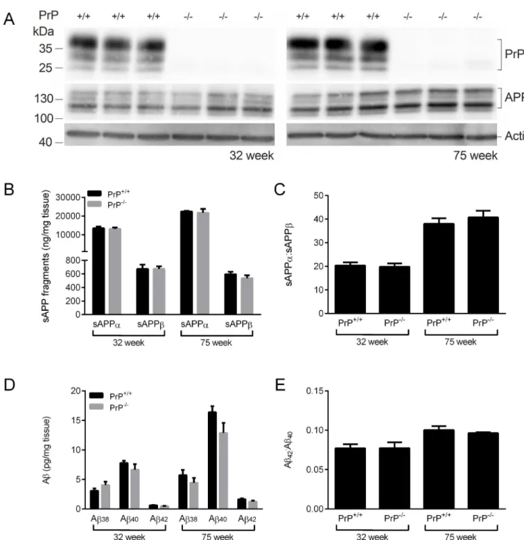
129/Ola mice [22]. Immunoblot analysis of mice at 32 and 75 weeks of age confirmed the absence of PrPCin the APPWT/PrP-/-mice and that the lack of PrPChad no effect on APP
expression (Fig 1A). The amounts of the soluble APP species, sAPPαand sAPPβ, were no dif-ferent between the APPWT/PrP-/-and APPWT/PrP+/+mice at either 32 or 75 weeks of age (Fig
1B), indicating that deletion of PrPChad no effect on either theα- orβ-secretase cleavage of APP. There was a significant increase in sAPPαin both genotypes at 75 weeks of age compared to 32 weeks of age (Fig 1B) which resulted in an increased sAPPα:sAPPβratio (Fig 1C). The levels of soluble (SDS-extracted) Aβ38, Aβ40and Aβ42were investigated. At both 32 and 75
weeks of age there was no difference in the amount of soluble Aβ38, Aβ40or Aβ42in the brain homogenates of the APPWT/PrP-/-mice compared to the APPWT/PrP+/+mice (Fig 1D). The
amount of Aβ40and Aβ42was higher at 75 weeks of age than at 32 weeks of age regardless of Prnpgenotype (Fig 1D), with a proportionately larger increase in Aβ42such that the Aβ42: Aβ40
ratio increased in both the APPWT/PrP-/-and APPWT/PrP+/+mice with age (Fig 1E). The levels
of Aβpeptides in the insoluble (formic acid extracted) fraction from either the APPWT/PrP
-/-or the APPWT/PrP+/+mice were below the limit of detection in the assay. These data indicate
that lack of PrPCin transgenic mice expressing human APPWTdoes not alter the proteolysis of
APP or the levels of Aβ.
To investigate whether lack of PrPCin the APPWT/PrP-/-mice affected the deposition of Aβ
in the brain, histopathological analysis of the hippocampus was carried out at 32 weeks of age and compared to APPWT/PrP+/+mice. APPSwe,Ind/PrP+/+mice, which are known to
accumu-late limited Aβ-positive plaques at this age, were also analysed [9,21]. APPWT/PrP+/+and
APPWT/PrP-/-mice at 32 weeks of age showed no obvious neuropathologic alterations (data
not shown). To explore whether histopathologic alterations were detected in older animals, we also examined mice at 75 weeks of age. Histopathological analysis of WT, APPWT/PrP+/+and
APPWT/PrP-/-mice at 75 weeks of age showed similar histopathologic features; i.e. absence of
Fig 1. Ablation of PrPCdoes not affect APP proteolysis or Aβpeptide levels in APP
WTmice.Brain hemispheres from 32 week and 75 week old
APPWT/PrP+/+and APPWT/PrP-/-mice were subjected to two-step homogenisation, and the soluble proteins (SDS extracted) were subjected to
SDS-PAGE and immunoblotting. (A) representative immunoblots of PrPCand APP, with actin as a loading control. (B) Soluble (SDS extracted) sAPPαand sAPPβdetected by MSD ELISA and (C) the ratio of sAPPαto sAPPβ. (D) Soluble (SDS extracted) Aβ38,Aβ40and Aβ42detected by
MSD ELISA and (E) the ratio of Aβ42to Aβ40. Data shown as mean±SEM (n = 3–6). There was no significant difference between the APPWT/PrP+/+
and APPWT/PrP-/-at the same time point for any of the APP or Aβpeptides.
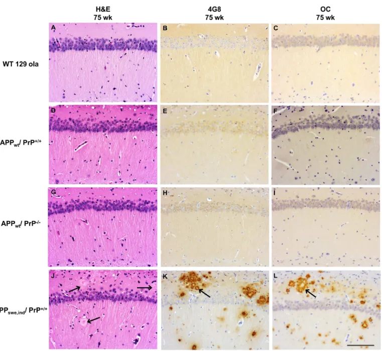
We investigated whether the ablation of PrPCaffected the level of Aβfibrils and fibrillar oligo-mers by using the OC antibody [25]. We observed that WT, APPWT/PrP+/+and APPWT/PrP
-/-mice did not accumulate OC immunopositive deposits as shown by immunohistochemistry (Fig 2C, 2F and 2I). As expected, in the APPSwe,Ind/PrP+/+mice at 75 weeks of age there was
disruption of the hippocampal structure with accumulation of 4G8 and OC positive Aβ deposi-tion forming numerous uni and multicentric plaques (Fig 2J, 2K and 2L), as seen previously [9]. Synapses are particularly vulnerable to the toxic effect of protein oligomers. Thus, we Fig 2. Ablation of PrPCdoes not affect Aβdeposition in APPWTmice.Histopathologic analysis of the hippocampus of hematoxylin-eosin
stained sections of WT, APPWT/PrP++and APPWT/PrP–mice show absence of obvious alterations in the pyramidal cell layer, stratum oriens and
lacunosum-molecular (A, D and G). No Aβdeposits are seen in sections probed with antibody 4G8 (B, E and H) or antibody OC (C, F and I). In contrast, APPSwe,Ind/PrP+/+mice show plaques (arrows) in sections stained with hematoxylin-eosin (J) and abundant Aβ-reactive deposits in
sections probed with antibody 4G8 (K) and OC (L). All images x20 magnification and scale bar = 100μm.
performed immunostaining for the detection of the presynaptic protein synaptophysin and observed disruption of immunoreactivity in 75 week old APPSwe,Ind/PrP+/+mice but not in
APPWT/PrP+/+or APPWT/PrP-/-mice (Fig 3A, 3D and 3G). Glial markers (GFAP and Iba1)
showed similar intensity and pattern of reactivity in APPWT/PrP+/+and APPWT/PrP-/-mice
(Fig 3B, 3C, 3E and 3F). In contrast, gliosis was observed particularly in the vicinity of amyloid deposits in APPSwe,Ind/PrP+/+mice (Fig 3H and 3I). Thus, we did not detect any effect of PrPC
ablation on Aβdeposition using immunohistochemistry.
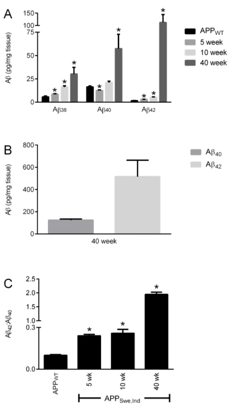
A comparison of the Aβlevels between the APPWT/PrP+/+mice at 75 weeks of age with
APPSwe,Ind/PrP+/+mice at 5 and 10 weeks (before significant Aβdeposition occurs) and at 40
weeks (after Aβdeposition in plaques appears) revealed that at 75 weeks of age the amount of Aβpeptides in the soluble (SDS extracted) fraction of the APPWT/PrP+/+mice was comparable
to the amount of each peptide in the 5 week old APPSwe,Ind/PrP+/+mice (Fig 4A). Although
Aβ40and Aβ42were readily detectable in the insoluble (formic acid extracted) fraction from the
APPSwe,Ind/PrP+/+mice (Fig 4B), no Aβwas detected in the insoluble fraction from the
APPWT/PrP+/+mice. The soluble Aβ42:Aβ40ratio increased with age in both APPWT/PrP+/+
Fig 3. Ablation of PrPCdoes not affect synaptophysin immunoreactivity and does not induce gliosis in APP
WTmice.APPWT/PrP+/+and
APPWT/PrP-/-mice show retention of the pre-synaptic marker synaptophysin (A, D) while APPSwe.Ind/PrP+/+mice reveal disorganization of
synaptophysin immunoreactivity (arrow) (G). Astrogliosis and microgliosis are observed in 75 week old APPSwe.Ind/PrP+/+mice, (arrows) (H, I) but not
in mice expressing APPWT/PrP+/+and APPWT/PrP-/-(B, C, E, F). Section were probed with synaptophysin (A, D, G), GFAP (B, E, H), and
anti-Iba1 (C, F, I) antibodies. Figures A,D,G (synaptophysin) x60 magnification scale bar = 20μm, figures B,E,H (GFAP) and C,F,I (Iba1) x40 magnification scale bar = 50μm.
Fig 4. Comparison of Aβlevels in APPWTand APPSwe.Indmice.Brain hemispheres from 75 week old
APPWT/PrP+/+and from 5, 10 and 40 week old APPSwe,Ind/PrP+/+mice were subjected to two-step
homogenisation. (A) Soluble (SDS extracted) and (B) insoluble (formic acid extracted) Aβ38,Aβ40and Aβ42
detected by MSD ELISA and (C) the ratio of Aβ42:Aβ40. Data shown as mean±SEM (n = 3–6),*p<0.05
compared to the 75 week old APPWT/PrP+/+group.
and APPSwe,Ind/PrP+/+mice (Fig 4C) but was significantly higher in the APPSwe,Ind/PrP+/+mice
than in the APPWT/PrP+/+mice consistent with the presence of the Indiana mutation
increas-ing production of Aβ42as observed previously [21]. Even at 75 weeks of age the amount of
Aβ42in the APPWT/PrP+/+mice is significantly lower than that in the APPSwe,Ind/PrP+/+mice
at 5, 10 and 40 weeks of age (Fig 4A). Thus in the APPWT/PrP+/+mice even at 75 weeks of age
the low level of Aβ42is likely below the threshold required for deposition to occur.
Conclusion
In cellular models PrPCdifferentially affected the activity of theβ-secretase BACE1 towards APPWTand APPSwe, inhibiting the processing of the former but having no effect on the
cleav-age of the latter [8,9]. Thus we undertook this study in order to investigate the effect of genetic ablation of PrPCon human APPWTprocessingin vivo. In contrast to the cellular models, lack
of PrPChad no effect on theα- andβ-secretase proteolysis of APP or on the amount of Aβ pep-tides in the brains of mice expressing human APPWT. Also we did not detect any Aβdeposition
as shown by immunohistochemistry in the mice lacking PrPCas compared to those with a nor-mal level of the protein. In the human brain the amount of PrPCinversely correlated with BACE1 activity, soluble and insoluble Aβand Braak stage [11], consistent with levels of PrPC affecting APP processing and Aβproduction. However, we could find no evidence that loss of PrPCaffected APP proteolysis, Aβlevels or plaque pathologyin vivoin transgenic mice expressing human APPWT. Whether this reflects a difference in the role of PrPCin regulating
APP processing and Aβproduction between this transgenic mouse model and the situation in the human brain will require further study.
Author Contributions
Conceived and designed the experiments: IJW DB HB ABD KABK JCM NMH. Performed the experiments: IJW DB HB ABD KABK PP. Analyzed the data: IJW DB HB ABD KABK PP JCM NMH. Wrote the paper: IJW DB ABD KABK PP JCM NMH.
References
1. Burns A, Iliffe S. Alzheimer's disease. BMJ. 2009; 338: 467–471.
2. Holtzman DM, Morris JC, Goate AM. Alzheimer's disease: the challenge of the second century. Sci-ence translational medicine. 2011; 3: 77sr71.
3. Selkoe D, Mandelkow E, Holtzman D. Deciphering Alzheimer disease. Cold Spring Harb Perspect Med. 2012; 2: a011460. doi:10.1101/cshperspect.a011460PMID:22315723
4. Walsh DM, Teplow DB. Alzheimer's disease and the amyloid beta-protein. Progress in molecular biol-ogy and translational science. 2012; 107: 101–124. doi:10.1016/B978-0-12-385883-2.00012-6PMID: 22482449
5. Vardy ER, Catto AJ, Hooper NM. Proteolytic mechanisms in amyloid-beta metabolism: therapeutic implications for Alzheimer's disease. Trends Mol Med. 2005; 11: 464–472. PMID:16153892 6. Lee VM, Brunden KR, Hutton M, Trojanowski JQ. Developing therapeutic approaches to tau, selected
kinases, and related neuronal protein targets. Cold Spring Harb Perspect Med. 2011; 1: a006437. doi: 10.1101/cshperspect.a006437PMID:22229117
7. Cole SL, Vassar R. The Alzheimer's disease Beta-secretase enzyme, BACE1. Mol Neurodegener. 2007; 2: 22 PMID:18005427
8. Parkin ET, Watt NT, Hussain I, Eckman EA, Eckman CB, Manson JC, et al. Cellular prion protein regu-lates beta-secretase cleavage of the Alzheimer's amyloid precursor protein. Proc Natl Acad Sci USA. 2007; 104: 11062–11067. PMID:17573534
10. Whitehouse IJ, Jackson CD, Turner AJ, Hooper NM. Prion protein is reduced in aging and in sporadic but not in familial Alzheimer's disease. J Alzheimers Dis. 2010; 22: 1023–1031. doi: 10.3233/JAD-2010-101071PMID:20930299
11. Whitehouse IJ, Miners JS, Glennon EB, Kehoe PG, Love S, Kellett KA, et al. Prion protein is decreased in Alzheimer's brain and inversely correlates with BACE1 activity, amyloid-beta levels and Braak stage. PLoS One. 2013; 8: e59554. doi:10.1371/journal.pone.0059554PMID:23577068
12. Hooper NM, Turner AJ. A new take on prions: preventing Alzheimer's disease. Trends Biochem Sci. 2008; 33: 151–155. doi:10.1016/j.tibs.2008.01.004PMID:18343669
13. Griffiths HH, Whitehouse IJ, Hooper NM. Regulation of amyloid-beta production by the prion protein. Prion. 2012; 6: 217–222. doi:10.4161/pri.18988PMID:22449984
14. Haass C, Lemere CA, Capell A, Citron M, Seubert P, Schenk D, et al. The Swedish mutation causes early-onset Alzheimer's disease by b-secretase cleavage within the secretory pathway. Nature Med. 1995; 1: 1291–1296. PMID:7489411
15. Thinakaran G, Teplow DB, Siman R, Greenberg B, Sisodia SS. Metabolism of the 'Swedish' amyloid precursor protein variant in Neuro2a (N2a) cells. Evidence that cleavage at the 'b-secretase' site occurs in the Golgi apparatus. J Biol Chem. 1996; 271: 9390–9397. PMID:8621605
16. Vassar R, Bennett BD, Babu-Khan S, Kahn S, Mendiaz EA, Denis P, et al. b-secretase cleavage of Alz-heimer's amyloid precursor protein by the transmembrane aspartic protease BACE. Science. 1999; 286: 735–741. PMID:10531052
17. Huse JT, Pijak DS, Leslie GJ, Lee VM, Doms RW. Maturation and endosomal targeting of beta -site amyloid precursor protein-cleaving enzyme. THE ALZHEIMER'S DISEASE beta -SECRETASE. J Biol Chem. 2000; 275: 33729–33737. PMID:10924510
18. Calella AM, Farinelli M, Nuvolone M, Mirante O, Moos R, Falsig J, et al. Prion protein and Abeta-related synaptic toxicity impairment. EMBO Mol Med. 2010; 2: 306–314. doi:10.1002/emmm.201000082 PMID:20665634
19. Gimbel DA, Nygaard HB, Coffey EE, Gunther EC, Lauren J, Gimbel ZA, et al. Memory impairment in transgenic Alzheimer mice requires cellular prion protein. J Neurosci. 2010; 30: 6367–6374. doi:10. 1523/JNEUROSCI.0395-10.2010PMID:20445063
20. Cisse M, Sanchez PE, Kim DH, Ho K, Yu GQ, Mucke L. Ablation of cellular prion protein does not ame-liorate abnormal neural network activity or cognitive dysfunction in the J20 line of human amyloid pre-cursor protein transgenic mice. The Journal of neuroscience: the official journal of the Society for Neuroscience. 2011; 31: 10427–10431.
21. Mucke L, Masliah E, Yu G-Q, Mallory M, Rockenstein EM, Tatsuno G, et al. High-Level Neuronal Expression of Abeta 1–42 in Wild-Type Human Amyloid Protein Precursor Transgenic Mice: Synapto-toxicity without Plaque Formation. J Neurosci. 2000; 20: 4050–4058. PMID:10818140
22. Manson JC, Clarke AR, Hooper ML, Aitchison L, McConnell I, Hope J. 129/Ola mice carrying a null mutation in PrP that abolishes mRNA production are developmentally normal. Mol Neurobiol. 1994; 8: 121–127. PMID:7999308
23. Kawarabayashi T, Younkin LH, Saido TC, Shoji M, Ashe KH, Younkin SG. Age-dependent changes in brain, CSF, and plasma amyloid (beta) protein in the Tg2576 transgenic mouse model of Alzheimer's disease. J Neurosci. 2001; 21: 372–381. PMID:11160418
24. Piccardo P, Manson JC, King D, Ghetti B, Barron RM. Accumulation of prion protein in the brain that is not associated with transmissible disease. Proc Natl Acad Sci U S A. 2007; 104: 4712–4717. PMID: 17360589