Introduction
The effect of sperm DNA fragmentation on infer-tility has been subject of several studies. Previ-ously, 10-20% DNA fragmentation was reported in ejaculated spermatozoa (1). The infertile men with poor sperm motility and morphology have suggested to posses increased DNA fragmentation levels when compared to men with normal semen parameters (2, 3). Interestingly, men with normal semen parameters has also suggested to have high DNA fragmentation levels (DFL), which can be one of the reasons of unexplained infertility (4). Aberrant chromatin packaging during sperma-togenesis, defective apoptosis before ejaculation, or excessive production of reactive oxygen species (ROS) cause DNA fragmentation in sperm cells, however, the mechanisms underlying the situation has not been clarify yet (5-7).
Controversies still present on the effects of sperm DNA damage on reproductive outcome. Some investigators indicated that clinical pregnancy was affected adversely by sperm DNA damage
in the cases of intracytoplasmic sperm injection (ICSI) (8, 9). Moreover, fertilization achieved by a sperm having fragmented DNA may cause poor embryonic development, decreased implantation and pregnancy rates, and recurrent pregnancy losses (8-12). On the contary, some others sug-gested that sperm DNA damage was ineffective on fertilization, embryo quality, and pregnancy rates in cases of in vitro fertilization (IVF) and ICSI (13, 14).
Several tests are available to measure sperm DNA fragmentation levels including TUNEL, comet assay, DNA Breakage Detection-Fluorescence In Situ Hybridization (DBD-FISH) test, the chromo-mycin A3 test, SCSA test and Sperm Chromatin Dispersion (SCD) test (15-20). The halosperm is a new improved SCD test based on the principle that sperm with fragmented DNA does not produce halo of dispersed DNA loops which is characteris-tic of sperm with nonfragmented DNA.
In this study, our aim is to evaluate the differences
Effects of Sperm DNA Fragmentation on Semen Parameters
Effects of Sperm DNA Fragmentation on Semen Parameters
and ICSI Outcome Determined by an Improved SCD Test,
and ICSI Outcome Determined by an Improved SCD Test,
Halosperm
Halosperm
Seda Yılmaz, M.Sc.
Seda Yılmaz, M.Sc.1, 21, 2*, Asuman Demiroglu Zergero*, Asuman Demiroglu Zergeroğğlu, Ph.D.lu, Ph.D.22, Elif Y, Elif Yıılmaz, B.Sc.lmaz, B.Sc.11, Kenan Sofuoglu, M.D., Kenan Sofuoglu, M.D.33,,
Nuri Delikara, M.D.
Nuri Delikara, M.D.11, Pelin Kutlu, M.Sc., Pelin Kutlu, M.Sc.11
1. Ferticenter IVF Unit, Istanbul, Turkey 1. Ferticenter IVF Unit, Istanbul, Turkey
2. Department of Biology, Gebze Institute of Technology, Kocaeli, Turkey 2. Department of Biology, Gebze Institute of Technology, Kocaeli, Turkey
3. Zeynep Kamil Hospital, Istanbul, Turkey 3. Zeynep Kamil Hospital, Istanbul, Turkey
Abstract Abstract
Background:
Background: Sperm DNA fragmentation is known as an important cause of male infertility. Sperm DNA fragmentation is known as an important cause of male infertility. The influence of sperm DNA damage on reproductive potential has been subject of many studies The influence of sperm DNA damage on reproductive potential has been subject of many studies indicating various results and remaining the subject controversial. In this study, we investigated indicating various results and remaining the subject controversial. In this study, we investigated differences of the semen parameters and intracytoplasmic sperm injection (ICSI) outcome according differences of the semen parameters and intracytoplasmic sperm injection (ICSI) outcome according to sperm DNA fragmentation levels (DFLs) of patients.
to sperm DNA fragmentation levels (DFLs) of patients.
Materials and Methods:
Materials and Methods: The DFLs were determined by Halosperm, a new improved sperm The DFLs were determined by Halosperm, a new improved sperm chromatin dispersion (SCD) test. Patients were grouped as low DNA fragmentation group (LFG chromatin dispersion (SCD) test. Patients were grouped as low DNA fragmentation group (LFG
≤30%) and high fragmentation group (HFG >30%).
≤30%) and high fragmentation group (HFG >30%).
Results: Our analysis showed that semen parameters including concentration of untreated sperm and motility of prepared semen were low in HGF, whereas other parameters were not different. and motility of prepared semen were low in HGF, whereas other parameters were not different. Sperm DNA fragmentation levels decreased in both groups after semen preparation by density Sperm DNA fragmentation levels decreased in both groups after semen preparation by density gradient technique.
gradient technique.
Conclusion:
Conclusion: No difference was detected on ICSI outcomes (fertilization, embryo development, No difference was detected on ICSI outcomes (fertilization, embryo development, embryo cleavage, embryo quality and pregnancy rates) between two group.
embryo cleavage, embryo quality and pregnancy rates) between two group.
Keywords:
Keywords: Asisted Reproductive Technique, DNA Fragmentation, ICSI, Sperm Asisted Reproductive Technique, DNA Fragmentation, ICSI, Sperm
Received: 8 Mar 2010, Accepted: 16 Jun 2010
* Corresponding Address: Ugur Mumcu mah. Atilganlar Sit. F blok D:12 Yakacik Kartal, Istanbul, Turkey
Email: [email protected]
Royan Institute
of semen parameters and ICSI outcome between the patients having low and high sperm DFLs as-sessed by new Halosperm test.
Materials and Methods
Study design
Patients undergoing their first ICSI attempt because of male factor infertility, < 39 years of age and hav-ing > 2 metaphase II oocytes were included in our prospective study. A total of 60 patient’s sperm samples were analyzed in where 10 of the samples were oligozoospermic (<20 mil/ml sperm concen-tration), 8 of those were asthenozoospermic (<50% total motility), 17 of samples were teratozoospermic (<4% morphology) and 25 of those showed more than one anomaly. Fragmentation levels above 30% and below 30% considered as high fragmentation group (HFG; n=24) and low fragmentation group (LFG; n=36), respectively (21). We have taken an agreement form from the patients.
Semen analysis
Both the semen analysis (WHO, 1999) and DNA fragmentation assesment were performed on the day of oocyte pick-up. Specimens were collected by masturbation after 3-5 days of sexual absti-nence and analysis were performed after liquefac-tion with a phase contrast microscope (Nikon E 400, Japan). Sperm concentrations, morphology, motility rates and acrosomal status were assessed before and after semen preparation. Sperm mo-tilities were classified as grade A, B, C, D and at least 100 spermatozoa were scored with ×40 ob-jective. Total motility rates were calculated as the total of A, B and C motility rates. Sperm morphol-ogy and acrosomal index were assessed accord-ing to Kruger’s strict criteria after stainaccord-ing with Diff-3 stain (GCC Diagnostics, UK). Acrosomes, comprising 40-70% of the sperm head, having no vacuole with a smooth surface were classified as a normal acrosome and acrosomal index were defined as the percentage of sperm cells having normal acrosome.
Semen samples were prepared by two-layer (90% and 45%) discontinuous PureSperm (Nidacon, Swe-den) gradient technique. 1 mL of semen samples were added on top of two layers and centrifuged at 400 g for 20 minutes. Thereafter, 400 μL of the pel-let containing the selected spermatozoa were col-lected from the bottom of the conical-based tubes (Falcon 2095, Becton Dickinson, USA). Pellets of the same patient were collected and suspended in 5 mL of G-IVF medium (Vitrolife, Kungsbacka, Sweden). The suspensions were centrifuged at 500 g for 6 min. Supernatants were discarded and
cen-trifugation was repeated after suspension with G-IVF medium. Finally supernatants were discarded and pellets were resuspended in 0.5 mL medium for further examination.
Sperm DNA fragmentation assessment
Sperm DNA fragmentation were assessed before and after semen preparation with Halosperm kit (INDAS laboratories, Spain). An aliquot of semen sample was diluted to 106/mL in phosphate-buff-ered saline (PBS). Agarose in eppendorf tubes pro-vided in the kit were placed in a water bath at 90°-100°C for 5 minutes to fuse the agarose, and then in a water bath at 37°C. After 5 minutes of incuba-tion at 37°C, 60 μL of the diluted semen sample were added to the eppendorf tube and mixed with the fused agarose. 20 μL of semen-agarose mixture were pipetted onto slides precoated with agarose provided in the kit and covered with a 24×24 mm coverslip. The slides were placed on a cold plate in the refrigerator (4°C) for 5 minutes to let the agar-ose to produce a microgel with the sperm cells em-bedded in. The coverslips were removed and the slides were immersed horizontally in an acid solu-tion prepared by mixing 80 μL of HCl provided in the kit with 10 mL of distilled water and incubated for 7 minutes. Slides were horizontally immersed in 10 mL of the lysing solution for 25 minutes. After washing 5 minutes with distilled water, the slides were dehydrated in increasing concentra-tions of ethanol (70%, 90%, 100%) for 2 minutes and then left for drying. Slides were covered with a mix of Wright’s staining solution (Merck, Ger-many) and PBS (1:1) (Merck, GerGer-many) for 5-10 minutes. Slides were washed in tap water and al-lowed to dry. A minimum of 100 spermatozoa per sample were scored under the ×100 objective in brigtfield microscopy. Sperm with large halo (thickness is similar or larger than the length of the smallest diameter of the core) and sperm with me-dium-sized halo (thickness between: greater than 1/3 of the smallest diameter of the core and less than the smallest diameter of the core) were clas-sified as ‘spermatozoa having no fragmentation’. Spermatozoa with a small halo (thickness were similar or smaller than 1/3 of the smallest diam-eter of the core) and without halo were classified as ‘spermatozoa having DNA fragmentation’.
Ovarian stimulation and ICSI procedure
stimulating hormone. When the dominant follicle reached 17 mm size, 5000 or 10000 IU of human chorionic gonadotropin (hCG) injection were administered. Cumulus-oocyte complexes were retrieved 36 hours after the administration of hCG by the guidance of transvaginal ultrasound. Microinjection procedure was performed as de-scribed previously by Van Steirteghem et al. 1993 (22). Sequential media of Sage is used for the cul-ture of oocytes and embryos. Presence of two pro-nuclei (2PN) and two polar bodies in the oocyte cytoplasm 16-18 hours after ICSI were defined as fertilization. Fertilized oocytes were checked 48 and 72 hours after ICSI for embryonic develop-ment. The cleavage status and the quality were assessed with an inverted microscope (Olympus IX71, Japan) according to the scoring criteria of Ziebe et al., 1997 (23). Embryos having frag-mentation levels of <10%, 10-20% with uneven blastomeres, 20-50% and >50% were classified as grade A, B, C and D respectively. Grade A and B were classified as good-quality embryos, grade C and D were classified as poor-quality embryos. Embryos having 2-6 cells on day 2 were defined as normally cleaved embryos, otherwise defined as abnormally cleaved. Embryo development rates were defined as the percentage of cleaved embryos fertilized normally.
Embryo transfers were performed by the guid-ance of abdominal ultrasound on day 2 or 3. Serum β-hCG levels were measured 12 days after transfer. A ≥15 mIU/ml β-hCG value on the 12 nd, and a
doubled β-hCG value on the 14 th day indicated a positive pregnancy.
Statistical analysis
SPSS for Windows 10.0 software package was utilised for the statistical analysis. Chi-square test and Mann Whitney U test was used to evaluate the rates and proportions and differences between the groups respectively. The results were evaluated in 95% confidence interval and the statistical signifi-cance was defined as, p< 0.05. The values of pa-rameters are mean ± standard deviation.
Results
Patient characteristics, including the female age (30.1 ± 4.4 and 31.8 ± 4.5), the male age (33.1 ± 4.8 and 34.4 ± 4.1), the number of oocytes re-trieved (13 ± 6.8 and 12.2 ± 6.1) and the embryos developed (8.6 ± 3.9 and 7.7 ± 4.4) were similar in LFG and HFG respectively. All the patients included were having oligo (10) / astheno (8) / teratozoospermia (17), where 25 of the samples showed more than one anomaly.
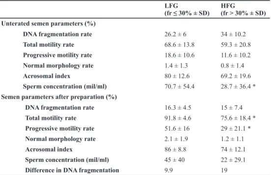
Firstly, the semen parameters (morphology, mo-tility and concentration) were compared between HFG and LFG in untreated and prepared semen samples. In untreated semen samples, lower sperm concentrations were observed in the HFG than LFG (p<0.05). However, parameters including morphology, acrosomal index, total motility and progressive motility rates were found similar be-tween the groups (Table 1).
Table 1: Mean values ± SD of semen parameters in LFG and HFG groups
HFG
(fr > 30% ± SD) LFG
(fr ≤ 30% ± SD) Unterated semen parameters (%)
34 ± 10.2 26.2 ± 6
DNA fragmentation rate
59.3 ± 20.8 68.6 ± 13.8
Total motility rate
11.6 ± 10.2 18.6 ± 10.6
Progressive motility rate
0.8 ± 1.4 1.4 ± 1.3
Normal morphology rate
69.2 ± 19.6 80 ± 12.6
Acrosomal index
28.7 ± 36.4 * 70.7 ± 54.4
Sperm concentration (mil/ml)
Semen parameters after preparation (%)
15 ± 7.4 16.3 ± 4.5
DNA fragmentation rate
75.6 ± 18.4 * 91.8 ± 4.6
Total motility rate
29 ± 21.1 * 51.6 ± 16
Progressive motility rate
1.2 ± 1.1 2.1 ± 1.9
Normal morphology rate
74 ± 12.1 86 ± 8.8
Acrosomal index
22 ± 29.1 45 ± 40
Sperm concentration (mil/ml)
19 9.9
Difference in DNA fragmentation
After sperm preparation, motility rates (total and progressive motility) declined in the HFG (p<0.05) as shown in Table 1. Additionally, the fragmenta-tion levels significantly reduced in both groups af-ter semen preparation (9.9 % in the LFG and 19% in the HFG).
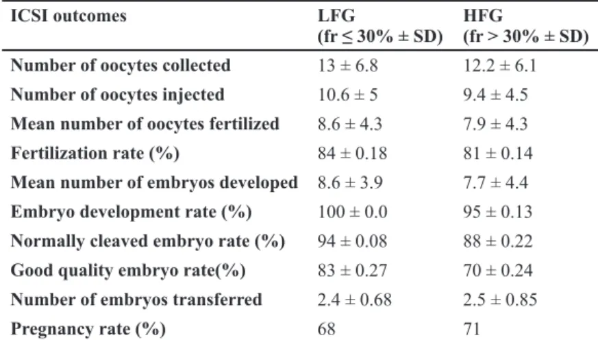
Secondly, ICSI outcome were compared between LFG and HFG groups. No significant differences were observed in parameters including fertilization (84% and 81%), embryo development (100% and 95%), normally cleaved (94% and 88%) and good quality embryo rates (83% and 70%) between LFG and HFG respectively (p>0.05) (Table 2). In addi-tion, pregnancy rates were not different between the groups (68% and 71% respectively) (p>0.05) (Table 2).
The values of parameters indicates mean ± stand-ard deviation of data.
Discussion
Conflicting results are present on the effects of sperm DNA fragmentation on semen parameters and the requirement for sperm chromatin assays in routine laboratory examinations. Some studies reported no correlation between sperm DNA frag-mentation and semen parameters (24, 25) whereas others observed a negative correlation in some and/or all parameters (26, 27). Recently, Cohen-Bacrie et al., 2009 reported a negative correlation between rapid progression rate and DNA frag-mentation determined by TUNEL, but no corre-lation between sperm DNA fragmentation, sperm morphology (head, acrosome and intermediate piece) and concentration (28). The chromatin condensation has been suggested a valuable pa-rameter to assess male fertility and used in routine laboratory investigations of semen in the cases of assisted reproduction (29).
Several methods are available to detect sperm DNA fragmentation including Halosperm, an im-proved SCD test developed by Férnandez (20). There is limited data in the literature comparing the differences in semen parameters of patients having different DNA fragmentation levels deter-mined by Halosperm. By using this technique, a negative correlation was found between all semen parameters (motility, morphology, concentration) and DNA fragmentation levels determined by Ha-losperm (30). In our study, we compared the differ-ences between the LFG and HFG in untreated and prepared semen. The sperm concentrations in the untreated semen samples and the motility rates (to-tal and progressive motility) reduced after semen preparation in the HFG however other parameters did not change. Thus, reduction of sperm concen-tration may indicate an apoptotic process present in those semen samples. In addition we found both total and progressive motility increased more in LFG than HFG after semen preparation suggesting that semen samples having lower DNA fragmenta-tion levels benefits more from semen preparafragmenta-tion by means of sperm motility.
Sperm DNA fragmentation levels reduced signifi-cantly in both groups after preparation proposing that semen preparation by density gradient tech-nique helps to eliminate the sperm cells having fragmented DNA therefore decrease the fragmen-tation levels in semen.
In the second part of the study, we compared ICSI outcome of the groups including fertilization, em-bryo development, emem-bryo quality, emem-bryo cleav-age and pregnancy rates. No correlation were reported between DNA fragmentation and fertili-zation rate using SCSA or comet assay (4, 8, 31), on the contrary, an inverse relationship were found in the studies utilizing Halosperm and SCD test Table 2: Mean values ± SD of ICSI outcome in LFG and HFG groups
HFG
(fr > 30% ± SD) LFG
(fr ≤ 30% ± SD) ICSI outcomes
12.2 ± 6.1 13 ± 6.8
Number of oocytes collected
9.4 ± 4.5 10.6 ± 5
Number of oocytes injected
7.9 ± 4.3 8.6 ± 4.3
Mean number of oocytes fertilized
81 ± 0.14 84 ± 0.18
Fertilization rate (%)
7.7 ± 4.4 8.6 ± 3.9
Mean number of embryos developed
95 ± 0.13 100 ± 0.0
Embryo development rate (%)
88 ± 0.22 94 ± 0.08
Normally cleaved embryo rate (%)
70 ± 0.24 83 ± 0.27
Good quality embryo rate(%)
2.5 ± 0.85 2.4 ± 0.68
Number of embryos transferred
71 68
Pregnancy rate (%)
(30, 32). In our results we found no differences in fertilization rates, embryo development, embryo quality (good and bad quality), and embryo cleav-age (normally and abnormally cleaved) rates be-tween the groups. According to these results it may be suggested that embryo’s developmental poten-tial is not affected by DNA fragmentation level of the semen until day 3 of development as the embryonic genome is activated after day 3 (33), that oocytes and embryos can repair sperm DNA damage (34) and because of the patient population selected. It may also be considered that, the sperm having fragmented DNA were eliminated and the best sperm was chosen with higher probability to have an intact DNA for the ICSI procedure. Our results confirm the results of other studies where different methods used for DNA fragmentation as-sessment. These investigators concluded that fer-tilization, embryo development and subsequent pregnancy were not affected by DNA fragmenta-tion of the sperm populafragmenta-tion (35, 36).
Additionally, we examined the relationship be-tween DNA fragmentation and pregnancy out-come. A recent meta-analysis and several other studies including studies using Halosperm and SCD test did not observe a relationship between sperm DNA damage and pregnancy rates (30, 32, 35, 37). On the contrary, in other studies a nega-tive correlation between sperm DNA fragmenta-tion and pregnancy rates were reported (9, 38). We observed no differences in pregnancy rates between the groups after ICSI. The lack of associ-ation between sperm DNA damage and pregnan-cy rates may be the result of selecting best sperms for ICSI and choosing the best scoring embryos for transfer. This may reduce the risk to use a sperm having fragmented DNA and therefore po-tential adverse effects of sperm DNA damage on pregnancy rates may not be observed.
Conclusion
Our results show that, semen parameters includ-ing initial sperm concentration and motility after preparation were decreased in patients having high DNA fragmentation whereas no differences were observed in other parameters. Semen prepa-ration by density gradient technique decreased the sperm percentage of fragmented DNA, caus-ing a higher probability to choose a sperm with intact DNA. None of the ICSI outcome parame-ters including fertilization, embryo development, quality and cleavage and pregnancy rates were af-fected by sperm DNA fragmentation levels. More detailed studies are required to observe the effects of sperm DNA fragmentation on semen
param-eters and ICSI outcome.
Acknowledgements
Study’s financial support is done by Ferticenter IVF Unit. There is no conflict of interest in this article.
References
1. Oosterhuis GJ, Mulder AB, Kalsbeek-Batenburg E, Lambalk CB, Schoemaker J, Vermes I. Measuring ap-optosis in human spermatozoa: a biological assay for semen quality? Fertil Steril. 2000; 74(2): 245-250. 2. Irvine DS, Twigg JP, Gordon EL, Fulton N, Milne PA, Aitken RJ. DNA integrity in human spermatozoa: rela-tionships with semen quality. J Androl. 2000; 21: 33-44. 3. Zini A, Bielecki R, Phang D, Zenzes MT. Correlations between two markers of sperm DNA integrity, DNA de-naturation and DNA fragmentation in fertile and infertile men. Fertil Steril. 2001; 75: 674-677.
4. Chohan KR, Griffin JT, Lafromboise M, De Jonge CJ, Carrell DT. Comparison of chromatin assays for DNA fragmentation evaulation in human sperm. J Androl. 2006; 27: 53-59.
5. Sakkas D, Mariethoz E, Manicardi G, Bizzaro D, Bi-anchi PG, BiBi-anchi U. Origin of DNA damage in ejaculat-ed human spermatozoa. Rev Reprod. 1999; 4: 31-37. 6. Sakkas D, Moffatt O, Manicardi GC, Mariethoz E, Tar-ozzi N, Bizzaro D. Nature of DNA damage in ejaculated human spermatozoa and the possible involvement of apoptosis. Biol Reprod. 2002; 66: 1061-1067.
7. Moustafa MH, Sharma RK, Thornton J, Mascha E, Abdel-Hafez MA, Thomas AJ, et al. Relationship be-tween ROS production, apoptosis and DNA denatura-tion in spermatozoa from patients examined for infertil-ity. Hum Reprod. 2004; 19: 129-138.
8. Larson KL, DeJonge CJ, Barnes AM, Jost LK, Even-son DP. Sperm chromatin structure assay parameters as predictors of failed pregnancy following assisted re-productive techniques. Hum Reprod. 2000; 15: 1717-1722.
9. Larson-Cook KL, Brannian JD, Hansen KA, Kasper-son KM, Aamold ET, EvenKasper-son DP. Relationship between the outcomes of assisted reproductive techniques and sperm DNA fragmentation as measured by the sperm chromatin structure assay. Fertil Steril. 2003; 80(4): 895-902.
10. Virant-Klun I, Tomazevic T, Meden-Vrtovec H. Sperm single-stranded DNA, detected by acridine orange stain-ing, reduces fertilization and quality of ICSI-derived em-bryos. J Assist Reprod Gen. 2002; 19: 319-328. 11. Carrell DT, Liu L, Peterson CM. Sperm DNA frag-mentation is increased in couples with unexplained re-current pregnancy loss. Arch Androl. 2003; 49: 49-55. 12. Virro MR, Larson -Cook KL, Evenson DP. Sperm chromatin structure assay (SCSA) parameters are relat-ed to fertilization, blastocyst development, and ongoing pregnancy in in vitro fertilization and intracytoplasmic sperm injection cycles. Fertil Steril. 2004; 81: 1289-1295.
pregnancy rates in in vitro fertilization and intracytoplas-mic sperm injection, but might be related to spontaneous abortion rates. Fertil Steril. 2008; 90(2): 352-359. 14. Li Z, Wang L, Cai J, Huang H. Correlation of sperm DNA damage with IVF and ICSI outcomes: A systematic review and meta-analysis. J Assist Reprod Gen. 2006; 23: 367-376.
15. Gorczyca W, Gong J, Darzynkiewicz Z. Detection of DNA strand breaks in individual apoptotic cells by the in situ terminal deoxynucleotidyl transferase and nick translation assays. Cancer Res. 1993; 53: 945-951. 16. Manicardi GC, Bianchi PG, Pantano, S Azzoni P, Biz-zaro D, Bianchi U, et al. Presence of endogenous nicks in DNA of ejaculated human spermatozoa and its rela-tionship to chromomycin A3 accessibility. Biol Reprod. 1995; 52: 864-867.
17. Hughes CM, Lewis SE, Mc Kelvey-Martin VJ, Thomp-son WA. CompariThomp-son of baseline and induced DNA dam-age in human spermatozoa from fertile and infertile men using a modified comet assay. Mol Hum Reprod. 1996; 2: 613-619.
18. Férnandez JL, Goyanes VJ, Ramiro-Dı´az J. Appli-cation of FISH for in situ detection and quantifiAppli-cation of DNA breakage. Cytogenet Cell Genet. 1998; 82: 251-256.
19. Fernández JL, Goyanes VJ, Ramiro-Díaz J, Gos-álvez J. Application of FISH for in situ detection and quantification of DNA breakage. Cytogenet Cell Genet. 1998; 82(3-4): 251-256.
20. Férnandez JL, Muriel L, Goyanes V, Segrelles E, Gosálvez J, Enciso M, et al. Simple determination of hu-man sperm DNA fragmentation with an improved sperm chromatin dispersion test. Fertil Steril. 2005; 84: 833-842.
21. Hoshi K, Katayose H, Yanagida K, Kimura Y, Sato A. The relationship between acridine orange fluorescence of sperm nuclei and the fertilizing ability of human sperm. Fertil Steril. 1996; 66: 634-639.
22. Van Steirteghem AC, Lin J, Joris H, Nagy Z, Jans-senenswillen C, Tournaye H, et al. Higher success rate by intracytoplasmic sperm injection than by subzonal insemination. Report of a second series of 300 consecu-tive treatment cycles. Hum Reprod. 1993; 8(7): 1055-1060.
23. Ziebe S, Petersen K, Linderberg S, Andersen AG, Gabrielsen A, Andersen AN. Embryo morphology or cleavage stage: how to select the best embryos for transfer after in-vitro fertilization. Hum Reprod. 1997; 12(7): 1545-1549.
24. Khalili MA, Aghaie-Maybodi F, Anuari M, Talebi AR. Sperm nuclear DNA in ejeculate of fertile and infertile men: correlation with semen parameters. Urol J. 2006; 3(3): 154-159.
25. Sills ES, Fryman JT, Perloe M, Michels KB, Tucker MJ. Chromatin fluorescence chracteristics and standard semen analysis parameters: correlations observed in andrology testing among 136 males referred for infertility evaulation. J Obstet Gynaecol. 2004; 24(1): 74-77. 26. Muratori M, Marchiani S, Tamburrino L, Tocci V, Failli
P, Forti G et al. Nuclear staining identifies two popula-tions of human sperm with different DNA fragmentation extent and relationship with semen parameters. Hum Reprod. 2008; 23(5): 1035-1043.
27. Erenpreiss J, Elzanaty S, Giwercman A. Sperm DNA damage in men from infertile couples. Asian J Androl. 2008; 10 (5): 786-790.
28. Cohen-Bacrie P, Belloc S, Ménézo YJ Clement P, Hamidi J, Benkhalifa M. Correlation between DNA dam-age and sperm parameters: a prospective study of 1,633 patients. Fertil Steril. 2009; 91(5):1801-1805.
29. Hammadeh ME, Zeginiadov T, Rosenbaum P, Georg T, Schmidt W, Strehler E. Predictive value of sperm chromatin condensation (aniline blue staining) in the assessment of male fertility. Arch Androl. 2001; 46(2): 99-104.
30. Velez de la Calle JF, Muller A, Walschaerts M, Cla-vere JL, Jimenez C, Wittemer C, et al. Sperm deoxyri-bonucleic acid fragmentation as assesed by the sperm chromatin dispersion test in assisted reproductive tech-nology programs:results of a large prospective multi-center study. Fertil Steril. 2008; 90: 1792-1799. 31. Morris ID, Ilott S, Dixon L, Brison DR. The spectrum of DNA damage in human sperm assessed by single cell gel electrophoresis (Comet assay) and its relation-ship to fertilization and embryo development. Hum Re-prod. 2002; 17(4): 990-998.
32. Muriel L, Garrido N, Férnandez JL, Remohí J, Pel-licer A, de los Santos MJ, et al. Value of the sperm deox-yribonucleic acid fragmantation leves as measured by the sperm chromatin dispersion test, in the outcome of in vitro fertilization and intracytoplasmic sperm injection. Fertil Steril. 2006; 85: 371-383.
33. Braude P, Bolton V, Moore S. Human gene expres-sion first occurs between the four-and eight-cell stages of preimplantation devolopment. Nature. 1988; 332: 459-461.
34. Ahmadi A, Ng SC. Devolopmental capacity of dam-aged spermatozoa. Hum Reprod. 1999; 14: 2279-2285.
35. Karydis S, Asimakopoulos B, Papadopoulos N, Vakalopoulos I, Al-Hasani S, Nikolettos N. ICSI outcome is not associated with the incidence of spermatozoa with abnormal chromatin condensation. In Vivo. 2005; 19(5): 921-925.
36. Abu-Hassan D, Koester F, Shoepper B, Schultze-Mosgau A, Asimakopoulos B, Diedrich K et. al. Comet assay of cumulus cells and spermatozoa DNA status, and the relationship to oocyte fertilization and embryo quality following ICSI. Reprod Biomed Online. 2006; 12(4): 447-452.
37. Gandini L, Lombardo F, Paoli D Caruso F, Eleuteri P, Leter G, et al. Full-term pregnancies achieved with ICSI despite high levels of sperm chromatin damage. Hum Reprod. 2004; 19: 1409 -1417.