○ ○ ○ ○ ○ ○ ○ ○ ○ ○ ○ ○ ABSTRACT○ ○ ○ ○ ○ ○ ○
Original Ar
ticle
○ ○ ○ ○ ○ ○ ○ ○ ○ ○ ○ ○ ○ ○ ○ ○ ○ ○ ○ ○ INTRODUCTION
Some tumor groups challenge routine his-topathological classification because of the absence of cell morphological differentiation that characterizes their lymphoid, epithelial or mesenchymal origin.1,2 These are poorly dif-ferentiated or undifdif-ferentiated tumors and can occur with relative frequency in the head and neck. They can arise in mucosa as well as in salivary glands, soft tissues or lymph nodes.3,4 The diagnosis and classification of such tumors are fundamental because suitable therapy and prognosis for each case depends upon precise histopathological diagnosis.5,6
The introduction of the immunohisto-chemical method, by Coons et al. in 1942,7 has become a powerful complementary tool in tumor analysis. It has increased the possi-bilities for histogenetic diagnosis of undiffer-entiated tumors. Through the identification of specific cellular components of cell patterns, using a specific panel of monoclonal or polyclonal antibodies, the immunohisto-chemical method has transformed the diag-nosis of these tumors. Diagnoses that used to be made on the basis of subjective informa-tion can now be accomplished using objec-tive criteria. However,there are only a few ref-erences in the literature to the immunohisto-chemical technique applied to the identifica-tion of undifferentiated head and neck tumors.8-11
In this retrospective analysis, we evaluated the occurrence of undifferentiated head and neck tumors in our service and the way in which they were distributed, according to cell pattern, patient’s age and tumor localization.
The role of immunohistochemical techniques in determining the conclusive diagnosis was also evaluated.
○ ○ ○ ○ ○ ○ ○ ○ ○ ○ ○ ○ ○ ○ ○ ○ ○ ○ ○ ○ METHODS
We reviewed 43 biopsies performed in the Departments of Otolaryngology and Head and Neck Surgery of the Universidade Estadual de Campinas, from January 1990 to December 1997, that were diagnosed as un-differentiated tumors. The criteria for inclu-sion were tumors located in the head and neck, histopathological diagnosis of undifferentiated tumors in sections stained using hematoxylin-eosin (HE), and sufficient quantity of mate-rial in the paraffin section for the immuno-histochemical technique to be performed. Tumors with evident differentiation seen in sections stained using HE and specimens with insufficient material for the immunohisto-chemical technique were excluded.
All the biopsies utilized were fixed in for-malin 10%, embedded in paraffin and stained with hematoxylin-eosin. They all had a diag-nosis of undifferentiated tumor as seen under optical microscopy, and these tumors were reviewed and distributed according to their cell pattern, into five groups:
1. Round cell tumors 2. Spindle cell tumors 3. Pleomorphic cell tumors 4. Myxoid tumors 5. Epithelioid cell tumors.
We applied an immunohistochemical panel with monoclonal antibodies (Table 1), in accordance with the
avidin-biotin-peroxi-Undifferentiated
head and neck tumors:
the contribution of
immunohistochemical
technique to differential
diagnosis
Department of Otorhinolaryngology, Hospital das Clínicas, Universidade
Estadual de Campinas, Campinas, São Paulo, Brazil
CONTEXT: Undifferentiated head and neck and skull base tumors are not unusual. They can arise in mucosa as well as in salivary glands, soft tissues or lymph nodes. Suitable therapy and prognosis for each case depends upon precise histopatho-logical diagnosis.
OBJECTIVE: To evaluate the role of immunohistochemi-cal techniques in determining the conclusive diag-nosis. The occurrence of these tumors in our serv-ice and the way in which they were distributed according to cell pattern, patient’s age and tumor location was also evaluated.
TYPE OF STUDY: Cross-sectional study.
SETTING: Hospital das Clínicas, Universidade Estadual de Campinas, Campinas, São Paulo, Brazil.
PARTICIPANTS: 43 biopsies performed between Janu-ary 1990 and December 1997, diagnosed as un-differentiated head and neck tumors.
PROCEDURES: We applied an immunohistochemical panel in accordance with the avidin-biotin-peroxidase complex method. The final diagnosis was achieved after new analysis in conjunction with biopsies stained using the hematoxylin-eosin technique.
MAIN MEASUREMENTS: This study evaluated undif-ferentiated tumors in head and neck, and the way in which they were distributed, according to cell pattern, patient’s age and tumor location.
RESULTS: The most frequent locations for undifferenti-ated tumors were the lymph nodes, 20.9%; phar-ynx and neck, 16.3%; paranasal sinus, 14%; and nose, 11.6%. They were most prevalent during the seventh decade of life (34.9%), and twice as preva-lent in men as in women. The immunohistochemi-cal technique allowed conclusive diagnosis for 60.5% of the tumors and was suggestive for 20.9% of the biopsies. The most prevalent cell pattern was round cells (51.2%), followed by epithelioid cells (20.9%), spindle cells (16.3%), myxoid pattern (9.3%) and pleomorphic cells (2.3%).
CONCLUSION: Our results demonstrate the fundamen-tal role of the immunohistochemical technique for conclusive diagnosis of undifferentiated tumors.
KEY WORDS: Immunohistochemical. Undifferentiated tumors. Diagnosis. Head neck. Avidin-biotin-per-oxidase.
• Walter Adriano Bianchini • Albina Maria Altemani • Jorge Rizzato Paschoal
São Paulo Medical Journal — Revista Paulista de Medicina
245
Original Ar
ticle
dase complex method (ABC),12 and with re-spect to the patient’s age, tumor location and cell pattern. The immunohistochemical pan-els employed for the analysis of undifferenti-ated tumors are given in Table 1.
In brief, the biopsy specimens were depa-raffinized and rehydrated, and endogenous peroxidase activity was blocked using 3% aqueous hydrogen peroxide in methanol. The slides were sequentially incubated with appro-priate non-immune serum and approappro-priate primary antiserum, overnight at 4º C. This was followed by incubation for 30 minutes with the biotinylated secondary antibody at 27º C and the avidin-biotin complex, before incubation with diaminobenzidine in phos-phate buffer.
The final diagnosis was achieved after new microscopic analysis in conjunction with sec-tions stained using the hematoxylin-eosin technique. The results were distributed accord-ing to cell pattern, patient’s age and tumor location.
○ ○ ○ ○ ○ ○ ○ ○ ○ ○ ○ ○ ○ ○ ○ ○ ○ ○ ○ ○ RESULTS
Undifferentiated tumors in the head and neck represented 1.1% of all tumors diagnosed in our service (43 cases out of 3,840 consecu-tive biopsies). There were 29 male (67.5%) and 14 female cases (32.5%), i.e. a propor-tion of 2:1, and patients’ ages ranged from 2 to 89 years. These tumors were most preva-lent during the seventh decade of life (34.9%) (Table 2). The lymph nodes (20.9%), phar-ynx and neck (16.3%), paranasal sinus (14%) and nose (11.6%) constituted the majority of sites, followed by oral cavity, ear, skin and lar-ynx (Table 2).
The most prevalent cell pattern was round cells (51.2%), followed by epithelioid cells (20.9%), spindle cells (20.9%), myxoid pat-tern (9.3%) and pleomorphic cells (2.3%) (Table 2).
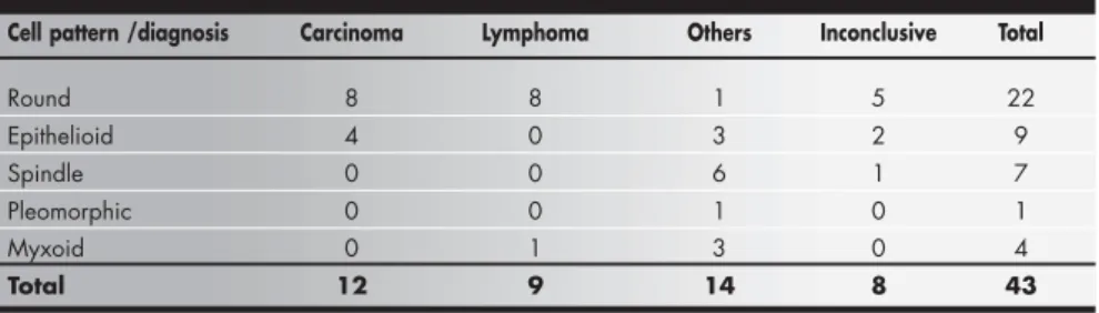
Diagnostic guidance was possible from 35 (81.4%) biopsies, and conclusive in 26 cases (60.5%). In 8 cases (18.6%), diagnosis was not possible. In the tumors in which diagno-sis was possible, we found 12 carcinomas (27.9%), 9 lymphomas (20.9%) and 14 other types of tumors (32.6%) (Table 3).
○ ○ ○ ○ ○ ○ ○ ○ ○ ○ ○ ○ ○ ○ ○ ○ ○ ○ ○ ○ DISCUSSION
The immunohistochemical technique has revolutionized surgical pathology knowledge, because the correct diagnosis of the neoplasia is essential for successful therapy and prognosis. Undifferentiated tumors represent 5-10% of all
diagnosed tumors.13 In our service, specifically in the head and neck region, the frequency was 1.1%. Undifferentiated tumors were found in all age groups, and were most prevalent in adult and elderly patients, with predominance among
the male population (2:1). These data are sup-ported by Azar et al. (1982),14 Coindre et al. (1986)15 and Vege et al. (1994).13
Round cell tumors, the most prevalent cell pattern found in our sample (51.16%), are a
Table 1. Immunohistochemical panels employed for the analysis of undifferentiated tumors
Epithelial AE1/AE3
MNF116 CAM5.2 CEA EMA
Lymphoid LCA CD15
CD 68 CD30
CD 45RO Lysozyme
CD 20
Mesenchymal Vimentin Chromogranin
Desmin 1A4
Myoglobin HHF-35
S 100 HMB-45
Factor VIII NSE
GFAP
Table 2. Immunohistochemical diagnosis and patient’s age, cell pattern and location in neoplasias of the head and neck
Variables CONCLUSIVE SUGGESTIVE INCONCLUSIVE Total
AGE
0-10 3 0 0 3
10-20 2 1 1 4
20-30 4 1 0 5
30-40 1 3 1 5
40-50 1 0 2 3
50-60 5 1 1 7
60-70 9 3 3 15
Over 70 1 0 0 1
Total 26 9 8 43
CELL PATTERN *
Epithelioid 5 2 2 9
Spindle 1 1 5 7
Myxoid 4 0 0 4
Pleomorphic 0 0 1 1
Round 16 5 1 22
Total 26 8 9 43
LOCATION
Oral cavity 4 0 0 4
Pharynx 3 4 0 7
Larynx 0 0 1 1
Lymph nodes 8 1 0 9
Nose 2 2 1 5
Ear 0 1 1 2
Skin 1 0 1 2
Neck 3 0 4 7
Paranasal sinus 5 0 1 6
Total 26 8 9 43
*p = 0.0357 (chi-squared test).
São Paulo Medical Journal — Revista Paulista de Medicina
246
group of highly aggressive malignant tumors composed of relatively small and monotonous undifferentiated cells. These tumors typically arise during childhood but can arise in adults and are often present in undifferentiated tumors. Conclusive diagnosis was most diffi-cult in cases of spindle cell tumors (14.3%), whereas good diagnostic rates were seen in cases of myxoid tumors (100%), round cell tumors (72.7%) and epithelioid cell tumors (62.5%). Conclusive diagnosis was not possible in the single case of pleomorphic cell tumor.
The most frequent location is the lymph
1. Caverivière P, AL Saati T, Voigt JJ, Delsol G. Diagnostic des
tumeurs indifférenciées à l’aide d’anticorps monoclonaux utilisables sur coupes en paraffine.[Diagnosis of undifferenti-ated tumors by means of monoclonal antibodies on paraffin
sections]. Presse Med 1985;14(32):1691-5.
2. Gatter KC, Alcock C, Heryet A, et al. The differential
diagno-sis of routinely processed anaplastic tumors using monoclonal antibodies. Am J Clin Pathol 1984;82(1):33-43.
3. Enzinger FM, Weiss SW. Immunohistochemistry of soft tissue
lesions. In: Enzinger FM, Weiss SW, editors. Soft Tissue Tumors. St. Louis: Mosby 1995. p.139-63.
4. Goodman MI, Pilch BZ. Tumors of the respiratory tract. In:
Fletcher CDM, editor. Diagnosis Histopathology of tumors. Edinburgh: Churchill Livingstone; 1995. p.79-126.
5. Michaels L. Neuroectodermal tumors. In: Michaels L, ed.
Ear, Nose and Throat Histopathology. Berlin: Springer-Verlag; 1987. p.89.
6. Triche TJ. Diagnosis of small round cell tumors of childhood.
Bull Cancer 1988;75(3):297-310.
○ ○ ○ ○ ○ ○ ○ ○ ○ ○ ○ ○ ○ ○ ○ ○ ○ ○ ○ ○ ○ ○ ○ ○ ○ ○ ○ ○ ○ ○ ○ ○ ○ ○ ○ ○ ○ ○ ○ ○ ○ ○ ○ ○ ○ ○ ○ ○ ○ ○ ○ ○ ○ ○ ○ ○ ○ ○ ○ ○ ○ ○ ○ ○ REFERENCES
7. Coons A, Creech JJ, Jones RN, Berlinger E. The
demonstra-tion of pneumococcal antigen in tissue by the use of fluorescent antibody. J Immunol 1942;45:159-70.
8. Abemayor E, Kessler DJ, Ward PH, Fu YS. Evaluation of poorly
differentiated head and neck neoplasms. Immunocytochemis-try techniques. Arch Otolaryngol Head Neck Surg 1987;113(5):506-9.
9. Darrouzet V, Stoll D, Benassayag C, Deminiere C. Apport de
l’immunohistochimie dans le diagnostic des cancers cervico-faciaux.[Contribution of immunohistochemistry to the diag-nosis of cervicofacial cancers]Rev Laryngol Otol Rhinol 1989;110(2):201-4.
10. Gallo O, Graziani P, Fini-Storchi O. Undifferentiated carci-noma of the nose and paranasal sinuses. An immunohisto-chemical and clinical study. Ear Nose Throat J 1993; 72(9):588-90, 593-5.
11. Milroy CM, Ferlito A. Immunohistochemical markers in the diagnosis of neuroendocrine neoplasms of the head and neck. Ann Otol Rhinol Laryngol 1995;104(5)413-8.
12. Hsu SM, Raine L, Fanger H. Use of avidin-biotin-peroxidase complex (ABC) in immunoperoxidase techniques: a compari-son between ABC and unlabeled antibody (PAP) procedures. J Histochem Cytochem 1981;29(4):577-80.
13. Vege DS, Soman CS, Joshi UA, Ganesh B, Yadav JN. Undiffer-entiated tumors: an immunohistochemical analysis on biop-sies. J Surg Oncol 1994;57(4):273-6.
14. Azar HA, Espinoza CG, Richman AV, Saba SR, Wang T. “Un-differentiated” large cell malignancies: an ultrastructural and immunocytochemical study. Hum Pathol 1982;13(4):323-33. 15. Coindre JM, Tanguy F, Merlío JP, De Mascarel I, De Mascarel A, Trojani M. The value of immunohistological techniques in undifferentiated cancers. Tumori 1986;72(6):539-44. 16. Gatter KC, Alcock C, Heryet A, Mason DY. Clinical
impor-tance of analysing malignant tumours of uncertain origin with immunohistological techniques. Lancet 1985;1(8441):1302-5. 17. Gatter KC, Mason DY. The use of monoclonal antibodies for histopathologic diagnosis of human malignancy. Semin Oncol 1982;9(4):517-25.
Table 3. Cell pattern and tumor types in neoplasias of the head and neck
Cell pattern /diagnosis Carcinoma Lymphoma Others Inconclusive Total
Round 8 8 1 5 22
Epithelioid 4 0 3 2 9
Spindle 0 0 6 1 7
Pleomorphic 0 0 1 0 1
Myxoid 0 1 3 0 4
Total 12 9 14 8 43
nodes,13 the majority with the round cell pat-tern,7 where there were five lymphomas, three carcinomas and one inconclusive case. There is greater incidence of lymphoid tumors in this location and more difficulty in achieving con-clusive diagnosis by conventional techniques because of the large morphological patterns. Metastases to lymph nodes in the head and neck are also frequent. The immunohistochemical technique was useful in conclusive diagnosis of the tumors in the oral cavity (100%), lymph nodes (88.9%) and paranasal sinuses (83.3%). Gatter et al.16,17 and Coindre et al.15
dem-onstrated greater incidence of lymphomas in undifferentiated tumors in general. In our study, however, carcinomas were the most fre-quent tumors (27.9%), a result that is similar to findings by Darrouzet et al.9 This is prob-ably because carcinomas are the most frequent tumors in the head and neck, which was the subject of our investigation.
Our results showed diagnostic guidance in 35 (81.4%) patients. It was not possible in eight patients (18.6%), probably due to limi-tations of the technique, antigen changes dur-ing tissue fixation or true absence of cellular differentiation.
○ ○ ○ ○ ○ ○ ○ ○ ○ ○ ○ ○ ○ ○ ○ ○ ○ ○ ○ ○ CONCLUSIONS
In conclusion, this paper supports the no-tion that the immunohistochemical technique has a fundamental role in the investigation and definition of undifferentiated tumor origin, thus determining correct guidance for treat-ment and possibly improving the prognosis for head and neck oncological patients.
São Paulo Medical Journal — Revista Paulista de Medicina
247
Tumores indiferenciados de cabeça e pescoço: contribuição da técnica imunoistoquímica para o diagnóstico diferencial
CONTEXTO: As neoplasias indiferenciadas em cabeça e pescoço e base do crânio não são raras. Ocorrem tanto em mucosas como em glându-las salivares, em partes moles e em linfonodos. O diagnóstico histopatológico preciso é funda-mental na conduta terapêutica ideal e na carac-terização do prognóstico de cada caso.
OBJETIVOS: Avaliar a ocorrência destas neoplasias em nosso serviço, sua distribuição conforme o padrão celular, a idade do paci-ente e a localização do tumor, avaliando-se a freqüência dos casos em que o exame imu-noistoquímico foi decisivo para o diagnósti-co diferencial diagnósti-conclusivo.
TIPO DE ESTUDO: Estudo de corte transversal.
LOCAL: Hospital das Clínicas, Universidade Es-tadual de Campinas, Campinas, São Paulo, Brasil.
PARTICIPANTES: Foram estudadas 43 biópsias de neoplasias indiferenciadas diagnosticadas no ambulatório da Disciplina de Otorrino-laringologia, no período de 1990 a 1997.
PROCEDIMENTOS: Aplicou-se um painel imunoistoquímico conforme o método com-plexo avidina-biotina-peroxidase (ABC),
de-○ ○ ○ ○ ○ ○ ○ ○ ○ ○ ○ ○ ○ ○ ○ ○ ○ ○ ○ ○ ○ ○ ○ ○ ○ ○ ○ ○ ○ ○ ○ ○ ○ ○ ○ ○ ○ ○ ○ ○ ○ ○ RESUMO
Walter Adriano Bianchini,MD, MSc. Department of Otorhinolaryngology, Universidade Estadual de Campinas, Campinas, São Paulo, Brazil.
Albina Messias Altemani, MD,PhD. Professor, Depart-ment of Pahtology, Universidade Estadual de Campinas, Campinas, São Paulo, Brazil.
Jorge Rizzato Paschoal, MD,PhD. Professor, Depart-ment of Otorhinolaryngology, Universidade Estadual de Campinas, Campinas, São Paulo, Brazil.
Conflict of interest: Not declared
Sources of funding: Not declared
Date of first submission: September 30, 2002
Last received: December 4, 2002
Accepted: February 14, 2003
Address for correspondence:
Walter Adriano Bianchini
Rua Major Sólon, 685 – Cambuí Campinas/SP – Brasil – CEP 13024-091 Tel./Fax (+55 19) 3255-1966 E-mail: [email protected]
COPYRIGHT © 2003, Associação Paulista de Medicina
○ ○ ○ ○ ○ ○ ○ ○ ○ ○ ○ ○ ○ ○ ○ ○ ○ ○ ○ ○ Publishing information
pendendo da idade dos pacientes, da localiza-ção do tumor e do padrão citoarquitetural das células neoplásicas. O laudo final foi emitido após nova análise conjunta com as lâminas coradas pela técnica da hematoxilina e eosina.
VARIÁVEIS ESTUDADAS: Distribuição das neoplasias indiferenciadas de cabeça e pesco-ço, conforme o padrão celular, a idade do paciente e a localização do tumor.
RESULTADOS: Os locais de ocorrência mais co-muns foram os linfonodos, 20.9%; faringe e pescoço, 16.3%; seios paranasais, 14.0% e ca-vidade nasal, 11.6%. Estas neoplasias foram mais prevalentes na sétima década de vida (34.9%), sendo duas vezes mais prevalentes em homens que em mulheres. O exame imunoistoquímico permitiu o diagnóstico con-clusivo em 60.5% dos tumores e o sugeriu em 20.9%. Os padrões citoarquiteturais mais co-muns foram: células redondas, 51.2%; células epitelióides, 20.9%; células fusiformes, 16.3%; mixóides, 9,30% e células pleomórficas, 2.3%.
CONCLUSÃO: Esses achados demonstram o papel fundamental do exame imunoisto-químico no diagnóstico conclusivo nestas neoplasias.
PALAVRAS-CHAVE: Imunoistoquímica. Neoplasias indiferenciadas. Diagnóstico. Ca-beça e pescoço. Avidina-biotina peroxidase.