* Extracted from the dissertation “Utilização do método Hálux-Calcâneo na identiicação de desvios de crescimento em recém-nascidos”, Universidade Federal de Goiás, 2013.
1 Universidade Federal de Goiás, Faculdade
de Enfermagem, Goiânia, GO, Brazil.
2 Universidade Federal de Goiás,
Programa de Pós-Graduação em Enfermagem, Goiânia, GO, Brazil.
3 Universidade Federal do Mato Grosso,
Programa de Pós-Graduação em Enfermagem, Cuiabá, MT, Brazil.
ABSTRACT
Objective: Comparing foot length measurements of newborns in high and low risk pregnancies at a public hospital in Goiânia, GO, Brazil. Method: A cross-sectional study carried out between April, 2013 and May, 2015, with a sample consisting of 180 newborns; 106 infants of women from high-risk pregnancies and 74 of women from low-risk pregnancies. Data were descriptively analyzed. Foot length measurement was performed using a stif transparent plastic ruler, graduated in millimeters. he length of both feet was measured from the tip of the hallux (big toe) to the end of the heel. Results: A statistically signiicant relationship was found between the foot length and newborn’s weight, between the cephalic and thoracic perimeters in the high-risk group and between the cephalic perimeter in the control group. Conclusion: here is a need for creating cut-of points to identify newborns with intrauterine growth disorders using foot length.
DESCRIPTORS
Infant Newborn; Gestacional Age; Pregnancy, High-Risk; Infant, Low Birth Weight; Maternal-Child Nursing.
Foot length measurements of newborns of
high and low risk pregnancies*
Medida do comprimento hálux-calcâneo de recém-nascidos em gestações de alto e baixo risco
Medida de longitud hallux-calcáneo de recién nacidos en gestaciones de alto y bajo riesgo
Ana Karina Marques Salge1, Érika Lopes Rocha2, Maria Aparecida Munhoz Gaíva3, Thaíla Correa Castral1, Janaína
Valadares Guimarães1,Raphaela Maioni Xavier2
How to cite this article:
Salge AKM, Rocha EL, Gaíva MAM, Castral TC, Guimarães JV, Xavier RM. Foot length measurements of newborns of high and low risk pregnancies. Rev Esc Enferm USP. 2017;51:e03200. DOI: http://dx.doi.org/10.1590/S1980-220X2016016703200
Received: 05/09/2016 Approved: 12/12/2016
Corresponding author:
Ana Karina Marques Salge Faculdade de Enfermagem – Universidade Federal de Goiás
Rua 227 Qd 68, S/N – Setor Leste Universitário CEP 74605-080 – Goiânia, GO, Brazil anasalge@gmail.com
INTRODUCTION
When the growth potential of the fetus sufers any interference in high-risk pregnancies, anthropometric mea-sures are one of the irst signs that can alert professionals to the presence of an adverse situation. In addition, some neonatal clinical conditions including premature birth and low birth weight are directly associated with the occur-rence of high-risk pregnancies where neonatal mortality is the main consequence(1-3).
Gestational age (GA) and newborn (NB) weight at birth are described in the literature as essential for eval-uating the pattern of intrauterine growth and develop-ment, to predict the diiculties of extrauterine adaptation, diseases in the neonatal period and to estimate the risk of (neonatal) death(3-4).
In this sense, in searching for simple and reliable meth-ods that may help in distinguishing between the narrow biological variability of the fetuses, studies have used foot length (hallux-calcaneus length – HCL) to estimate gesta-tional age with greater precision(5-10).
HCL is a quantiiable easy-to-apply morphometric parameter, and because it contains only one variable to be measured, it is less susceptible to misinterpretation(11-12).
HCL can be measured using equipment speciically developed for this purpose, or simply using a non-elastic tape or a graduated ruler(13).Usually the centimeters between the center of the heel to the tip of the hallux are measured(10). here is a clear need for a rapid and efective method to determine GA and birth weight which can be used in newborns with diseases, preterm/premature infants or those living in areas of poverty and communities with diicult access, since these groups present greater diiculties in evaluating these parameters(10). It is hypothesized that the HCL measurement could be a good parameter for new-born clinical evaluation, especially in cases of high-risk pregnancy. In this perspective, the present investigation is justiied by ofering knowledge about the importance of the HCL method to subsidize systematization and reorienta-tion of safe care to newborns.
Considering that despite the advantages of this method, it is still little used in our reality, so the objective of this study was to compare foot length (HCL) measurements in newborns of high and low risk pregnancies in a public hospital in Goiânia, GO, Brazil.
METHOD
his is a cross-sectional study developed in the maternity ward of a public federal hospital, a part of the (Brazilian) Uniied Healthcare System (SUS) of Goiânia, Goiás, Brazil, from April, 2013 to May, 2015.
he sample consisted of 180 newborns born between April, 2013 and May, 2015, and met the inclusion criteria; of which 106 were children of high-risk pregnancy women with the clinical diagnoses of Hypertensive Disorders of Pregnancy (HDP) and Diabetes Mellitus (DM), and 74 newborns born to women of low-risk pregnancy (con-trol group).
he study included live newborns of up to 24 hours of life whose mothers showed the clinical criteria for classiica-tion as a high-risk pregnancy, among them: Hypertensive Disorders of Pregnancy (HDP), Gestational Diabetes (GD) and Diabetes Mellitus type I and II (DM1 or DM2), in addition to having medical records available for data collec-tion and with complete informacollec-tion containing birth weight and gestational age. As HCL increases signiicantly during the irst ive days of extrauterine life(7), we chose to perform the measurements within the irst 24 hours after birth as recommended by the literature, and each newborn had the HCL measured only once. Newborns with congenital mal-formations in the lower limbs were excluded from the study. HCL was measured by one of the researchers on both feet of the newborn using a transparent and stif plastic 30-cm ruler graduated in millimeters, and adopting the length of the tip of the hallux to the end of the heel(8-12).
Mothers’ and newborns’ clinical data were obtained from the medical records of each patient through the institution’s own forms/ilescontaining the following items: maternal and fetal underlying diseases; gestational age (determined by the date of the last menstruation, the ultrasound performed in the irst trimester and by the Capurro method); obstetric history; possible maternal obstetric and fetal/neonatal com-plications; neonatal anthropometric measures (birth weight, cephalic and thoracic perimeters); and Apgar score in the 1st and 5th minutes.
Quantitative data such as mean gestational age and anthropometric measurements were descriptively analyzed by frequency distribution, means, and standard devia-tion. Proportions such as the relationship between weight, cephalic perimeter, thoracic perimeter and Apgar score with the newborn’s HCL from the mothers in the control group and those from high-risk pregnancies were compared by the c2 test, followed by the Fisher’s exact test or Yates correction test. Statistically signiicant diferences were observed in which the p value was lower than 5% (p<0.05).
he study development complied with national and international standards of research ethics involving human subjects. he results presented here are part of the matrix project “Avaliação da resposta imunológica materna e fetal em gestantes com doença hipertensiva”(Evaluation of the maternal and fetal immune response in pregnant women with hyper-tensive disease), and approved by the Ethics Committee on Human and Animal Medical Research of the Hospital das Clínicas of the Federal University of Goiás, under opinion number 101/2008. he women who met the inclusion crite-ria established for this study were clariied as to the purpose of the investigation and the nature of the data collection, and those who agreed to participate signed the Free and Informed Consent Form (TCLE).
RESULTS
Salge AKM, Rocha EL, Gaíva MAM, Castral TC, Guimarães JV, Xavier RM control group (0.002), and in the high-risk pregnancy group
(0.001). SGA newborns predominated in both groups, with
60.9% in the control group and 49% in the high-risk preg-nancy group (Table 1).
Table 1 – Distribution of newborns (NB) of women in high-risk pregnancies and control group, according to birth weight classification in a public federal maternity hospital – Goiânia, GO, Brazil, 2013-2015.
nB control group nB high-risk pregnancy group
nB n (%) gA in days (mean±SD) p-value nB n (%) gA (mean±SD) p-value
AgA 22(29.7) 264.4±2.2 0.008 AgA 35(33) 264.2±14.4 0.086
SgA 45(60.9) 278.8±5.1 0.002* SgA 52(49) 259±15.9 0.001*
lgA 7(9.4) 288.6±3.7 0.762 lgA 19 (18) 270.9±14.4 0.026
Total 74(100) 277.3±1.8 Total 106(100) 264.4±5.5
AGA: Adequate for gestational age; LGA: Large for gestational age; SGA: Small for gestational age; GA: gestational age; SD: standard deviation; n: number of cases;
p- value: Chi-square or Fisher’s exact test.
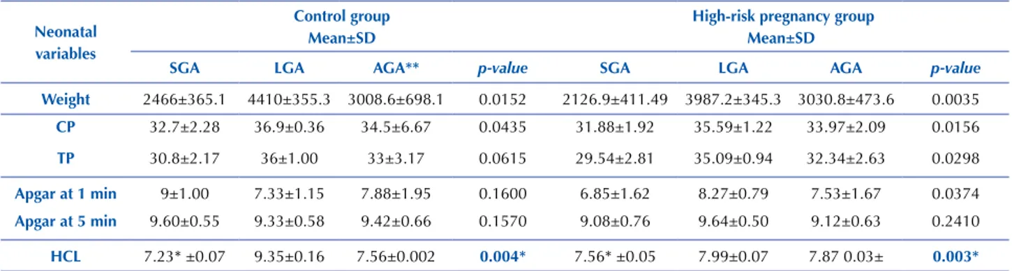
Table 2 describes the relationship between weight, cephalic perimeter (CP), thoracic perimeter (TP) and Apgar score with HCL in infants born to mothers in the control group and for mothers of high-risk pregnancies. Newborns
with SGA had the lowest HCL measurements, with a sta-tistically signiicant relationship between the HCL of new-borns with SGA in the control group (p = 0.004) and in the high-risk pregnancy group (p = 0.003).
Table 2 – Relationship between foot length (HCL) and the neonatal variables in newborns of high-risk pregnancies and of mothers in the control group in a public federal maternity hospital – Goiânia, GO, Brazil, 2013-2015.
neonatal variables
Control group Mean±SD
High-risk pregnancy group Mean±SD
SgA lgA AgA** p-value SgA lgA AgA p-value
Weight 2466±365.1 4410±355.3 3008.6±698.1 0.0152 2126.9±411.49 3987.2±345.3 3030.8±473.6 0.0035
CP 32.7±2.28 36.9±0.36 34.5±6.67 0.0435 31.88±1.92 35.59±1.22 33.97±2.09 0.0156
TP 30.8±2.17 36±1.00 33±3.17 0.0615 29.54±2.81 35.09±0.94 32.34±2.63 0.0298
Apgar at 1 min 9±1.00 7.33±1.15 7.88±1.95 0.1600 6.85±1.62 8.27±0.79 7.53±1.67 0.0374
Apgar at 5 min 9.60±0.55 9.33±0.58 9.42±0.66 0.1570 9.08±0.76 9.64±0.50 9.12±0.63 0.2410
HCl 7.23* ±0.07 9.35±0.16 7.56±0.002 0.004* 7.56* ±0.05 7.99±0.07 7.87 0.03± 0.003*
SD: standard deviation; SGA: Small for gestational age; LGA: Large for gestational age; AGA: Adequate for gestational age; SD: standard deviation; n: number of cases;
p-value: Chi-square or Fisher’s exact test; CP: Cephalic perimeter; TP: horacic perimeter
DISCUSSION
Foot length assessment is an important element for the fetal structural screening at all gestational ages. As it is a linear body measure, it can be closely related to gestational age, weight, length and perimeters(6,9-10).
Weight is the most frequently associated measure of growth. Birth weight establishes whether a child is small, large, or adequate for gestational age(10). An accurate assess-ment of growth in the neonatal period is very important and allows insight into whether the fetus was subjected to abnormal intrauterine conditions that resulted in delayed or accelerated growth(12).
A statistically signiicant relationship was found between gestational age and infants with SGA in the control group and in the high-risk pregnancy group, and between the HCL of SGA newborns in the control group and in the high-risk pregnancy group. Several studies have also shown a strong correlation between foot length growth and birth weight(7-9,13-14). According to the literature, the HCL value
suggested for adequate fetal growth would be 7.1 to 7.3 cm for SGA infants, 7.5 to 7.7 cm for AGA infants, and from 7.8 to 7.9 cm for LGA infants(13).
SGA infants from control group had a mean HCL of 7.23 cm, similar to that described in three other studies which observed control groups values of 7.13 cm(15), 7.24 cm(13) and 7.20 cm(9).
Regarding AGA infants, the mean HCL of the control group was 7.56 cm and 7.87 cm for the high-risk group, similar values to those found in other studies of 7.6 cm(15), 7.8 cm(8) and 7.92 cm(13). It may be noted that all cut-of points for AGA were equal to or greater than the cut-of points for SGA.
2000 to 8.5% in 2011(16). In a hospital-based recruitment conducted in Tanzania, a 15% prevalence of LBW infants was identiied(17). On the other hand, a study developed in Uganda showed a slightly lower percentage of LBW, being 12%(7). Two studies conducted in Nepal, where 95% of births are homebirths and 75% of the population live below the poverty line, discrepant percentages of LBW were identiied: 28.6%(18) prevalence in the irst study;and more recently of 6.7% prevalence in the second study(10).
he ideal maximum period to perform HCL measure-ment is still controversial. Some authors state that HCL maintains a strong correlation with weight from birth up to the ifth day after birth, being a good predictor for the evalu-ation of low birth weight(16-18). However, a study revealed that newborn’s feet signiicantly increase in length/size during the irst ive days of life, suggesting that further research needs to be developed to determine the maximum period for this measure(7). HCL of infants adequate for gestational age was 7.56 cm in the control group and 7.87 cm in the high-risk group. According to the literature, HCL of newborns with adequate weight may range from 7.40 cm to 7.99 cm(7).
Regarding the Apgar data in the irst minute, the high-risk group obtained a higher number of newborns with averages lower than eight in comparison to infants from the control group. Regarding the relationship between HCL and Apgar, this was only signiicant in the irst minute for the high-risk pregnancy group, and did not present a strong correlation with the other newborns. here are reports in the literature of an association between low val-ues of the Apgar score in the irst and ifth minutes and congenital orthopedic problems, and not directly related to the HCL measurement(19). No studies were found that directly related HCL to cephalic and thoracic perimeters values, and Apgar indexes.
It is observed that HCL is routinely used in pediatric and forensic necropsies in order to establish GA in fetuses and stillbirths. Although not used in neonatal nursing care, HCL along with other criteria for clinically evaluating new-borns (such as weight, Apgar score, cephalic, thoracic and abdominal circumferences) can positively contribute to a
more overall analysis of a newborn’s conditions at birth, their ability to adapt to extrauterine life and to measure GA(20).
Considering that HCL is easy to apply since it has only one variable to be measured and is therefore less susceptible to errors, and that the material used for the measurement is easily found in any maternity or health service, this mor-phometric parameter should be more used by professionals such as nurses, pediatricians/neonatologists, members of the nursing team and pathologists who work in maternity and child areas.
his study presents some limitations for being a prospec-tive study and for using data from medical records in which there was no standardization of the records, the informa-tion was often written down in an incomplete or inadequate manner and in illegible handwriting, and often did not con-tain records of the nursing team. hus, a large amount of information is missing in this type of study.
CONCLUSION
A statistically signiicant relationship was found between HCL and weight at birth in SGA, LGA and AGA infants of all gestational ages in both groups, as well as between HCL and CP and TP in the high-risk group, and between the HCL and CP in the control group.
he heterogeneity of the results points to the need to establish cutof points to identify newborns with possible alterations/disorders in low and high-risk pregnancies, as well as to deine the mean HCL that is to be accepted as normal for the neonatal population, so that this param-eter can be used as a reliable instrument for measuring GA in newborns.
Weight, cephalocaudal length and perimeters are com-monly evaluated during the anthropometric evaluation of newborns. However, the combined use of anthropometric measures that compare two or more aspects of growth may provide more consistent information regarding the risk of morbidity. For this reason, the HCL measurement may aid in the anthropometric evaluation of newborns and provide a more comprehensive view on the quality of fetal and neo-natal growth and development.
RESUMO
Objetivo: Comparar as medidas do comprimento hálux-calcâneo de recém-nascidos em gestações de alto e baixo risco em um hospital público de Goiânia, GO. Método: Estudo transversal, realizado no período de abril de 2013 a maio de 2015, cuja amostra constituiu-se de 180 recém-nascidos, 106 ilhos de mulheres com gestação de alto risco e 74 de mulheres com gestação de baixo risco. Os dados foram analisados descritivamente. A medida do comprimento hálux-calcâneo foi realizada utilizando-se de régua plástica transparente rígida, graduada em milímetros. Foram medidos ambos os pés, aferindo-se o comprimento da ponta do hálux até a extremidade do calcâneo. Resultados: Foi encontrada relação estatisticamente signiicante entre o comprimento hálux-calcâneo e o peso do recém-nascido, entre os perímetros cefálico e torácico no grupo de alto risco e entre o perímetro cefálico no grupo controle. Conclusão: Existe necessidade da criação de pontos de corte para identiicar recém-nascidos com desvios de crescimento intrauterino utilizando-se do comprimento hálux-calcâneo.
DESCRITORES
Recém-Nascido;Idade Gestacional;Gravidez de Alto Risco; Recém-Nascido de Baixo Peso; Enfermagem Materno-Infantil.
RESUMEn
Salge AKM, Rocha EL, Gaíva MAM, Castral TC, Guimarães JV, Xavier RM de manera descriptiva. La medida de la longitud hallux-calcáneo se realizó mediante regla de plástico rígido transparente, graduada en milímetros. Se midieron en ambos pies, las longitudes de la punta del hallux hasta el inal del calcáneo. Resultados: Se encontró una relación estadísticamente signiicativa entre la longitud hallux-calcáneo y el peso del recién nacido, entre las circunferencias cefálica y torácica en el grupo de alto riesgo y entre la circunferencia cefálica en el grupo control. Conclusión: Existe la necesidad de crear puntos de corte para identiicar los recién nacidos con desviaciones de crecimiento intrauterino utilizando la longitud desde el hallux hasta el calcáneo.
DESCRIPTORES
Recién Nacido; Edad Gestacional; Embarazo de Alto Riesgo; Recién Nacido de Bajo Peso; Enfermería Maternoinfantil.
REFERENCES
1. Hirst JE, Ha LT, Jeffery HE. The use of fetal foot length to determine stillborn gestational age in Vietnam. Int J Gynaecol Obstet. 2012;116(1):22-5.
2. Dekker GA. Management of preeclampsia. Pregnancy Hypertens. 2014;4(3):246-47.
3. Koullali B, Oudijk MA, Nijman TA, Mol BW, Pajkrt E. Risk assessment and management to prevent preterm birth. Semin Fetal Neonatal Med. 2016;21(2):80-8.
4. Juárez SP. Notes on vital statistics for the study of perinatal health. Gac Sanit. 2014;28(6):505-07.
5. Feresu SA, Wang Y, Dickinson S. Relationship between maternal obesity and prenatal, metabolic syndrome, obstetrical and perinatal complications of pregnancy in Indiana, 2008-2010. BMC Pregnancy Childbirth. 2015;15:266.
6. Mukherjee S, Roy P, Mitra S, Samanta M, Chatterjee S. Measuring new born foot length to identify small babies in need of extra care: a cross-sectional hospital based study. Iran J Pediatr. 2013;23(5):508-12.
7. Nabiwemba E, Marchant T, Namazzi G, Kadobera D, Waiswa P. Identifying high-risk babies born in the community using foot length measurement at birth in Uganda. Child Care Health Dev. 2013;39(1):20-6.
8. Marchant T, Penfold S, Mkumbo E, Shamba D, Jaribu J, Manzi F, et al. The reliability of a newborn foot length measurement tool used by community volunteers to identify low birth weight or premature babies born at home in southern Tanzania. BMC Public Health. 2014;14:859-63.
9. Ashish KC, Nelin V, Vitrakoti R, Aryal S, Målqvist M. Validation of the foot length measure as an alternative tool to identify low birth weight and preterm babies in a low-resource setting like Nepal: a cross-sectional study. BMC Pediatr. 2015;15:43.
10. Wyk LV, Smith J. Postnatal foot length to determine gestational age: a pilot study. J. Trop Pediatr. 2016;62(2):144-51.
11. Elizabeth NL, Christopher OG, Patrick K. Determining an anthropometric surrogate measure for identifying low birth weight babies in Uganda: a hospital-based cross sectional study. BMC Pediatr. 2013;13:54.
12. Thawani R, Dewan P, Faridi MM, Arora SK, Kumar R. Estimation of gestational age, using neonatal anthropometry: a cross-sectional study in India. J Health Popul Nutr. 2013;31(4):523-30.
13. James DK, Dryburgh EH, Chiswick ML. Foot length-a new and potentially useful measurement in the neonate. Arch Dis Child. 1979;54(3):226-30.
14. Das S, Bapat U, More NS, Alcock G, Fernandez A, Osrin D. Nutritional status of young children in Mumbai slums: a follow-up anthropometric study. Nutr J. 2012;11:100.
15. Gohil JR, Sosi M, Vani SN, Desai AB. Footlength measurement in the neonate. Indian J Pediatr. 1991;58(5):675-77.
16. United Nations Children’s Fund (UNICEF). Committing to Child Survival: a Promise Renewed. Progress Report [Internet]. New York: UNICEF; 2014 [cited 2016 Jan 25]. Available from: http://www.apromiserenewed.org/APR_2014_web_15Sept14.pdf
17. Marchant T, Jaribu J, Penfold S, Tanner M, Schellenberg JA. Measuring newborn foot length to identify small babies in need of extra care: a cross sectional hospital based study with community follow-up in Tanzania. BMC Public Health. 2010;10:624.
18. Mullany LC, Darmstadt GL, Khatry SK, Leclerq SC, Tielsch JM. Relationship between the surrogate anthropometric measures, foot length and chest circumference and birth weight among newborns of Sarlahi, Nepal. Eur J Clin Nutr. 2007;61(1):40-6.
19. Chotigavanichaya C, Leurmsumran P, Eamsobhana P, Sanpakit S, Kaewpornsawan K. The incidence of common orthopaedic problems in newborn at Siriraj Hospital. J Med Assoc Thai. 2012;95(9):S54-61.
20. Zago AFR, Paravidine LM, Siqueira LMS, Balbão LM, Reis MA, Castro ECC. Estudo comparativo entre o comprimento hálux-calcâneo e outros métodos de avaliação de idade gestacional em recém-nascidos. Pediatr Mod. 2000;36(6):388-91.