C
URRENTO
PINIONPrenatal imaging findings in fetal Zika
virus infection
Edward Araujo Ju´nior
a, Francisco H.C. Carvalho
b,
Gabriele Tonni
c, and Heron Werner
dPurpose of review
The aim of this review is to report the most recent observations concerning intrauterine Zika virus (ZIKV) infection and associated neuroimaging.
Recent findings
ZIKV outbreak in Brazil in 2015 was associated with an impressive registration of cases of congenital microcephaly in women with symptoms suggestive of ZIKV infection. Clinical and laboratory testing for ZIKV and hypothetic etiopathogenetic mechanisms are described. Diagnostic tests on blood, urine and amniotic fluid should be performed in all mothers with symptoms suggestive of intrauterine ZIKV infection. ZIKV causes multiple teratogenic malformations, mainly affecting the developing brain.
Summary
Neuroimaging investigation contributes to the prenatal detection of microcephaly and other brain abnormalities in cases of intrauterine ZIKV infection. Neuroimaging is based antenatally on two-dimensional and three-two-dimensional ultrasound and fetal MRI, whereas computed tomography scan is performed postnatally. Although neuropathology associated with intrauterine ZIKV infection is characterized by nonspecific findings of brain disorder, reduced cortical gyration and white-matter hypomyelination or dysmyelination and cerebellar hypoplasia have been consistently observed in the majority of fetuses and newborns. Prenatal or postnatal genetic workup should be carried out to exclude cases of primary microcephaly. Follow-up should rely upon MRI and computed tomography scan as well as neuropediatrician to better define developmental outcome in survivors.
Keywords
computed tomography, intrauterine infection, microcephaly, MRI, Zika virus
INTRODUCTION
One of the most outstanding viremia of the year 2000 may be the Zika virus (ZIKV) infection. There are data consistent with over 4 million people being infected in the Americas by ZIKV, although 80% of them are asymptomatic [1]. In adults, ZIKV infec-tion is characterized by an erythematous rash and fever that may end, in the worse cases, in signs and symptoms overlapping with those of Guillan–Barre` syndrome. ZIKV was first isolated in a Uganda forest
from a sentinel monkey (Macacamulata) in 1947
and has been active in several African and Asian countries as the first case of infection in humans through which ZIKV spread to the Pacific, especially to Yap Island [1] (Federated State of Micronesia) in 2007, French Polynesia (2008–2014) and more recently to the Americas. The infection is caused by a flavivirus and is related to dengue virus (DENV), chikungunya virus (CHIKV) and yellow fever.
ZIKV has been isolated from Aedes africanus,Aedes
luteocephalus, Aedes aegypti, Aedes albopictus, Aedes furciferandAedes vittatusmosquitoes, although A. aegyptiis the main vector in the Brazilian outbreak [1–5].
When French Polynesia identified its first case in 2013, it was further estimated that 11.5% (32 000) of
a
Department of Obstetrics, Paulista School of Medicine, Federal Uni-versity of Sa˜o Paulo (EPM-UNIFESP), Sa˜o Paulo, Sa˜o Paulo,
bDepartment of Maternal and Child, Federal University of Ceara´
(UFC), Fortaleza, Ceara´, Brazil,cDepartment of Obstetrics and
Gyne-cology, Guastalla Civil Hospital, AUSL, Reggio Emilia, Italy and
dDepartment of Radiology, Clı´nica de Diagno´stico por Imagem (CDPI),
Rio de Janeiro, Rio de Janeiro, Brazil
Correspondence to Prof Edward Araujo Ju´nior, PhD, Department of Obstetrics, Paulista School of Medicine, Federal University of Sa˜o Paulo (EPM-UNIFESP), Rua Belchior de Azevedo, 156 apto. 111 Torre Vitoria, Sa˜o Paulo CEP 05089-030, Brazil. Tel: +55 11 37965944;
fax: +55 11 37965944; e-mail: [email protected]
Curr Opin Obstet Gynecol2017, 29:95–105
the population had ZIKV-like symptoms. When the diagnostic test based on rRT-PCR (real-time reverse transcription-PCR) became available (2013–2014), surveillance found that 383 cases (2.8% of the coun-try’s population) were serologically confirmed. At the time of ZIKV infection spreading in Yap in 2007, Duffyet al.[1] identified 185 cases of suspected ZIKV infections; a blood sample taken 10 days after the onset of symptoms resulted in 49 (26%) confirmed and 59 (32%) probable cases of ZIKV infection. The serologic test was based on an ELISA for IgM anti-bodies against ZIKV and DENV. The median age of patients with ZIKV infections was 36 years (range, 1–76) with 61% being women with sex-specific attack rates of 17.9 per 1000 women and 11.4 per 1000 men [1]. It may be estimated that between 500 000 and 1.5 million of Brazilians were infected with ZIKV during the 2015 outbreak [6,7].
The autochthonous circulation of ZIKV in the Americas was first confirmed on Easter Island (Chile) in 2014 [8], whereas in Brazil the first labora-tory confirmation was in mid-July 2015 [9,10]. The Brazilian Ministry of Health has estimated that between 440 000 and 1 300 000 cases of ZIKV infec-tions occurred in Brazil in the same year especially in the northeast state of Bahia (93% of cases overall [11&
] in which the attack rate has been calculated to range between 4.4 and 25 per 1000 inhabitants [12]). Notification of ZIKV cases is now conducted on behalf of the Brazilian health authority via the Notifiable Diseases Information System [13], with 22 out of 24 Brazilian administrative states register-ing autochthonous ZIKV infection [14].
ZIKV is a single stranded, icosahedral, positive-sense RNA virus with a 10.7 kb genome encoding a single polyprotein that is cleaved into three structural proteins: capsid; precursor membrane/
membrane and E, envelope, and seven nonstruc-tural proteins [15]. There are at present two main hypotheses according to which ZIKV would act. One is a direct viral effect in which glycoprotein E attaches to the cellular receptors of the host, fol-lowed by a process of endocytosis, viral fusion and virus release into the cytoplasm [16&&
]. The second may be secondary to a placental perturbation caused by ZIKV at the chorionic villi. ZIKV might cause the perturbed synthesis of neuropeptides and cytokines that are essential for normal fetal brain development [17]. It may be postulated that ZIKV accesses the embryo via placental macrophages that are more permissive than trophoblast cells to ZIKV [18&
]. Laboratory confirmation of ZIKV infection relies on RT-PCR in blood, urine or both in symptomatic patients and in amniotic fluid in the case of con-genital ZIKV infection [19&
,20&
]. When suspected ZIKV infection is associated with microcephaly, cytomegalovirus, rubella virus, herpes simplex virus, DENV, CHIKV and toxoplasmosis must be investi-gated for cross-reactivity. In addition, anti-ZIKV antibodies are highly cross-reactive with other fla-viviruses such as West Nile virus, Japanese encepha-litis and yellow fever or induced from yellow fever and Japanese encephalitis vaccines [7].
Ultrasound and ultrasound-targeted f-MRI (fetal MRI) are diagnostic in the prenatal detection of brain abnormalities caused by ZIKV. The first pub-lication of a possible association between a third trimester prenatal diagnosis (29.2 and 30.1 weeks’ gestation, respectively) of microcephaly and ZIKV infection was reported by Oliveira Meloet al.[21&
] in January 2016. Brain malformations were character-ized by ventriculomegaly, brain calcifications, cor-pus callosum and cerebellar dysgenesis (mainly affecting the vermis), thalamic damage and severe brain atrophy leading to lissencephaly.
In this review, we present the main imaging prenatal diagnosis techniques for the assessment of the central nervous system (CNS) of fetuses affected by ZIKV intrauterine infection.
TWO-DIMENSIONAL ULTRASOUND
FINDINGS IN ZIKA VIRUS INTRAUTERINE
INFECTION
Following the initial suspicion of a link between ZIKV and fetal microcephaly, observations have emerged favoring evidence of this relationship, both in animal and human studies [19&
–21&
]. The neuro-tropism of the virus has been confirmed suggesting a role in all phases of neurodevelopment: proli-feration, migration, organization and myelination [22&&
,23&
,24&&
,25&
,26&
]. As a result, many other CNS disorders have been reported. Structural changes
KEY POINTS
Main brain malformations in Zika virus intrauterine infection are the following: ventriculomegaly, brain calcifications, corpus callosum and cerebellar dysgenesis (mainly affecting the vermis), thalamic damage and severe brain atrophy leading to lissencephaly.
Prenatal and postnatal neuroimaging in the case of Zika intrauterine infection is possible using two-dimensional and three-two-dimensional ultrasound, computed tomography and MRI.
beyond the CNS have also been reported. The fre-quency of microcephaly [27,28] has been reported in only a few articles that described fetal findings in pregnancies that have been or were presumed
to be affected by ZIKV [20&
,29,30]. Tables 1 and 2 summarize the main ultrasonographic findings described in fetuses from pregnancies with confirmed or suspected ZIKV infection.
Table 1. Case reports describing two-dimensional ultrasound neuroimgaing findings in pregnancies with suspected/confirmed intrauterine Zika virus infection
Author Clinical symptoms Confirmed ZIKV intrauterine infection Gestational age at ultrasound Ultrasound neuroimaging findings Extra-CNS US findings Oliveira Melo
et al.[21& ]
Not reported Yes – positive amniotic fluid
30 weeks Microcephaly, calcifications involving white matter of the frontal lobe and cerebellum, corpus callosum dysgenesis and cerebellar vermis and
enlargement of the cisterna magna
–
Case 1 rRT-PCR
Oliveira Melo
et al.[21&
] Not reported Yes – positive amnioticfluid 29 weeks Microcephaly, severe unilateralventriculomegaly with midline deviation and thinning of the parenchyma, calcifications around the lateral ventricles and IVth ventricle, nonvisualization of the corpus callosum
Ocular asymmetry (microphthalmia), cataract and intraocular calcification
Case 2 rRT-PCR
Mlakaret al.
[24&&
] Yes – 13 weeks Yes – rRT-PCR atautopsy specimens 32 weeks Microcephaly, ventriculomegaly,calcifications, and cerebellar hypoplasia
Fetal growth restriction, diffuse calcifications in the placenta Werneret al.
[27] Yes – 10 weeks Yes – positive serologyat 31 weeks 33 weeks Bilateral ventricular enlargement,periventricular calcifications and lissencephaly
Abnormal profile and nuchal edema Werneret al.
[28] Yes – 12 weeks No 32 weeks Microcephaly and diffuse braincalcifications – Calvetet al.
[19&
] Yes – 18 weeks Yes – positiveamniotic fluid 21 weeks Microcephaly, ventricularenlargement, cerebellar hypoplasia and absence of cerebellar vermis
–
rRT-PCR Driggerset al.
[29]
Yes – 11 weeks Yes – positive rRT-PCR in maternal serum
19 weeks Thin cerebral cortex with increased extra-axial space, dilation of the third ventricle, enlargement of both frontal horns and apparent dysgenesis of the corpus callosum
–
Moronet al.
[30] Yes – ‘Early inthe second trimester’
No –"IgG positive but IgM negative using indirect ELISA assay. rRT-PCR assay for ZIKV in umbilical cord blood was negative
32 weeks Microcephaly, brain cortical atrophy, ventricular enlargement, parenchymal calcifications, lissencephaly and pachygyria, increased subarachnoid space
Redundant cranial skin fold, calcifications in the placenta
Sarnoet al.
[31&
] No Yes – positive rRT-PCRat autopsy specimens (brain, medulla oblongata); positive AF and in CSF
21 weeks Severe microcephaly,
hydranencephaly, intracranial calcifications and destructive lesions of posterior fossa
Hydrops (hydrothorax, ascites and subcutaneous edema) Culjatet al.
[32& ]
Not reported Yes – positive serum IgM and newborn CSF
Diffuse parenchymal calcifications, hydrocephalus ex vacuo and cerebellar hypoplasia
–
Perezet al.
[33&
] Yes – 17 weeks Yes – positive rRT-PCRin maternal serum and AF
19 weeks Bilateral hydrocephalus Arthrogryposis multiplex congenita
The first prospective study was published by
Brasil et al. [27] with 42 pregnant women from
Rio de Janeiro, Brazil, who presented symptoms suggestive of ZIKV and were confirmed with rRT-PCR between 6 and 35 weeks of gestation. There was fetal/placental impairment in 12 pregnancies (28.6%). The main two-dimensional ultrasono-graphic findings described were microcephaly, cerebral and cerebellar calcifications, ventriculo-megaly, brachycephaly cerebellar atrophy, agenesis vermis, mega cisterna magna, club foot, olygohy-dramnios/anhydramnios, fetal growth restriction, intrauterine fetal death and placental insufficiency as assessed by Doppler study. Four of the 12 (33.3%) pregnancies presented Doppler changes. Doppler waveform abnormalities were reported in only one case out of 52 (1.9%) in the series by Sarno
et al.[30] and had not been previously seen by other authors, even in cases with associated fetal growth restriction. Microcephaly was still found in ZIKV infections occurring at 26 weeks. Only considering the ZIKV infections that occurred in the first tri-mester, the frequency of microcephaly was 22%,
much higher than the 1% found in French Poly-nesia [28].
THREE-DIMENSIONAL ULTRASOUND, MRI,
COMPUTED TOMOGRAPHY AND
THREE-DIMENSIONAL PHYSICAL MODEL
FINDINGS IN ZIKA VIRUS INTRAUTERINE
INFECTION
ZIKV infection has been associated with several malformations of the CNS, especially microcephaly [19& –21& ,23& ,24&& ,25& ,31& –33&
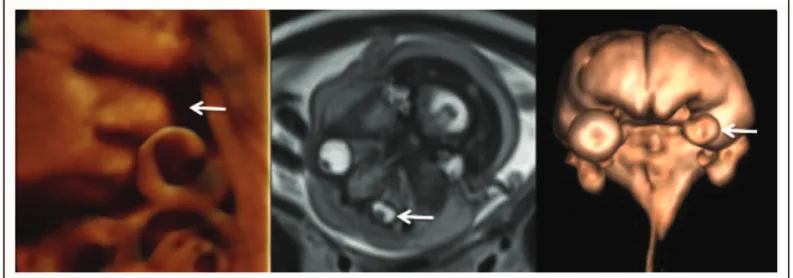
,34]. Imaging tech-nology advancements have led to tremendous improvements in fetal evaluation. Two main tech-nologies are mostly used to obtain images inside the uterus during pregnancy – ultrasound and MRI, whereas computed tomography (CT) scan is better performed postnatally. Ultrasound is currently the first method for fetal scanning as it is safer, patient-friendly, useful and cost-effective. When ultrasound provides equivocal results, f-MRI can be used to aid in diagnosis (Fig. 1). CT may also be performed but only after 30 weeks of gestation and in cases
Table 2. Case series describing two-dimensional ultrasound neuroimaging findings in pregnancies with suspected/confirmed Zika virus intrauterine infection
Reference
Size and sampling characteristics Confirmed ZIKV intrauterine infection Gestational age at ultrasound Ultrasound
neuroimaging findings Extra CNS findings Meloet al.
[34]
11 cases of fetuses with CNS changes at ultrasound and confirmed ZIKV. Clinical symptoms suggestive ofFlavivirusin 90.1%
Yes – positive rRT-PCR in amniotic fluid in 6 (54.5%). Other cases confirmed with rRT-PCR in newborn’s CSF or in autopsy specimens
17–35 weeks Ventricular enlargement (90.1%), usually asymmetric, severe cerebral atrophy (81.8%), hypoplasia of the cerebellar vermis and cerebellum (81.8%), calcifications in the brain, cerebellum and brainstem
Fetal akinesia deformation sequence or
arthrogryposis (27.3%), microphthalmia and bilateral cataracts (9.1%) and fetal growth restriction
Carvalho
et al.[20& ]
19 cases of microcephaly (highly probable cases – had specific neuroimaging findings and negative laboratory results for other congenital infections). Clinical symptoms suggestive ofFlavivirusin 68.4%
Confirmed in 2 cases by positive amniotic fluid rRT-PCR
22–39 weeks Cerebral calcifications (73.7%), unilateral or bilateral ventricular enlargement (52.6%), cerebellar calcifications (31.6%), increased cisterna magna (26.3%), cerebellar vermis dysgenesis/agenesis (15.8%) and cerebral atrophy/increased subarachnoid space (15.8%)
Fetal growth restriction (26.3%), arthrogryposis/ contractures (15.8%), olygohydramnios (10.5%), cardiac calcifications (5.3%) and ocular calcifications (5.3%)
Sarnoet al.
[35]
52 cases of microcephaly, excluding other causes of congenital infections. Maternal exanthematic disease was present in the majority (86.5%) of cases, 67.3% in the first trimester of pregnancy
78.3% were classified as confirmed cases according to WHO criteria for diagnosis of ZIKV-related fetal microcephaly
19–40 weeks Ventricular enlargement (65.4%), cerebral periventricular/basal ganglia calcifications (44.2%), posterior fossa abnormalities (32.7%) and corpus callosum
dysgenesis (3.8%)
Fetal growth restriction (48.1%), congenital talipes (17.3%), arthrogryposis (9.6%), cardiac calcifications/ pericardial effusion, (3.8%), olygohydramnios (11.5%) and intrauterine fetal death (7.7%)
associated with skeletal dysplasia [35]. As antenatal detection of microcephaly represents a complex and challenging case, combined investigations have been proposed to assist ultrasound diagnosis (Fig. 2) [32&
,36]. Three-dimensional ultrasound may not add important information in this particu-lar setting. However, dedicated three-dimensional software has been developed to assist biometry
obtained by real-time two-dimensional scanning, introducing fetal volume data sets. Although not all three-dimensional volume data sets are appro-priate for further offline extraction of measure-ments, especially when the fetal head is in the anteroposterior position, good agreement between the two techniques was observed, even if three-dimensional ultrasound was more time consuming
FIGURE 1.Three-dimensional ultrasound, MRI and three-dimensional reconstruction from MRI show unilateral microphthalmia
(arrows) in a 37-week fetus with Zika virus infection.Note:the difficulty to see the microphthalmia in three-dimensional ultrasound due to fetal position.
[37]. Very recently, Rizzo et al. [38] have demon-strated that with the development of novel three-dimensional software, namely five-three-dimensional CNS, 98.3% of axial cephalic planes could be suc-cessfully reconstructed and all the basic biometric head and brain measurements as well as fetal head volume could be calculated. Moreover, a good inter-class correlation with two-dimensional ultrasound was seen (>0.92). Contrary to the observation by
Sarriset al.[37], five-dimensional CNS was also able to reduce scanning time [38]. These results are in agreement with those of Tsaiet al.[39] that previously demonstrated that a three-dimensional volume analysis algorithm based on the universal facial surface template model containing the geometric shape information of a fetal craniofacial structure, constructed from a fetal phantom, could precisely
measure craniofacial structures. Nevertheless,
Tonniet al.[40] have shown that cerebral mid-line
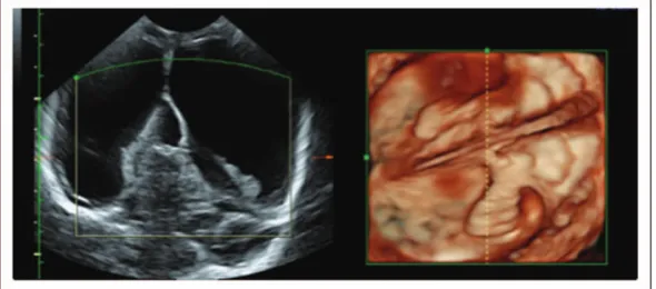
anomalies, especially those involving the corpus cal-losum and the cerebellar vermis, can be accurately reformatted using a reslicing technique. One of the main advantages of three-dimensional ultrasound is that all three orthogonal planes can be ‘interrogated’ and postprocessed offline and sent to experts for consultation using digital communication in medi-cine (Fig. 3 and Fig. S1, http://links.lww.com/COOG/ A34). f-MRI and postnatal CT scan may be added to two-dimensional–three-dimensional ultrasound to further improve diagnostic accuracy as it allows ana-tomical images to be obtained with higher resolution of soft tissue contrast (Figs 4 and 5). Moreover, MRI is not influenced as ultrasound is by increased maternal habitus, decreased amniotic fluid or unfavorable fetal positioning. Moreover, f-MRI could improve the diagnostic accuracy of the severity of the brain abnor-malities; this technical characteristic correlates well with advancing gestational age [27,32&
,41].
Neuropathology associated with intrauterine ZIKV infection is characterized by nonspecific find-ings of brain disorder (diffuse cortical calcification,
mid-line echo abnormalities encompassing callosal dysgenesis to cerebellar vermis anomalies, reduced gyration, pachygyria and lissencephaly) that over-laps with those seen in other viral congenital infec-tions. Reduced cortical gyration and white-matter hypomyelination or dysmyelination and cerebellar hypoplasia in the majority of newborns suggest that ZIKV is associated with a disruption in brain devel-opment rather than destruction of the brain. Neuronal and glial proliferation as well as neuronal migration appear to be affected [42&
].
The contribution of f-MRI to the diagnosis of polymicrogyria, laminar necrosis and brainstem anomalies, compared with ultrasound, is well estab-lished [43,44]. Moreover, it may also confirm ultrasound in cases of microcephaly, defined sono-graphically as head circumference below the third centile and at MRI as cerebral biparietal diameter and fronto-occipital diameters below the third centile [45]. Specifically, f-MRI should describe, better than ultrasound, the following brain characteristics: pachygyria, cerebral ventricular enlargement due to white matter hypoplasia (mainly affecting the posterior aspect of the lateral ventricles), malfor-mations of cortical development and sulcation, cor-pus callosum dysgenesis (agenesis, hypogenesis and hypoplasia), evaluation of myelination (normal or delayed) according to the stage of development at which changes in myelination appear on T1-weighted and T2-T1-weighted images [46,47]. One of the main hallmarks of brain imaging is widespread calcifications in the junction between the cortical and subcortical white matter, abnormal cortical
development (pachygyria and polymicrogyria),
especially involving the frontal lobe, ventriculome-galy and enlarged cisterna magna, cerebellar and brainstem hypoplasia, moderate and severe lissen-cephly and delayed myelination. It is noteworthy that the predominance of the frontal lobes for pachy-gyria and polymicropachy-gyria has not been observed in cases of other congenital infections of the CNS [46].
FIGURE 3.(a) Transvaginal ultrasound shows brain calcifications (arrows) and microcephaly in a 37-week fetus. (b) Prenatal
In addition, enlargement of the supratentorial arachnoid space may cause a severely enlarged head circumference, thereby making a small brain volume [46]. MRI and CT scans offer high sensitivity and specificity for brain disorder (Figs 6 and 7 and Fig. S2, http://links.lww.com/COOG/A34). Nonetheless, the use of three-dimensional volumes generated by MRI and CT alongside additive manufacturing technologies has increased expo-nentially, producing virtual physical models of con-genital anomalies (Fig. S3, http://links.lww.com/ COOG/A34). This novel technology is adopted to assist medical diagnosis, allowing physicians to accurately visualize and define relevant internal and external anatomical details as well as convert-ing the reconstructed anatomy into a set of digital three-dimensional data [36,41].
These files are then converted into three-dimensional files with a Standard Triangulation Language (STL) extension, which basically consists
of X, Y and Z coordinates. Once the STL file is
generated, the next step is the horizontal slicing
of the entire three-dimensional volumetric
file, using the appropriate software. Thus, the
supporting structures will be calculated, if needed (Fig. S4, http://links.lww.com/COOG/A34). The building process starts with the sequential depo-sition of material layers, which range from microns to fractions of millimeters, depending on the type of technology selected [48,49].
There are currently several systems of additive manufacturing/three-dimensional printing tech-nologies that are commercially available. Although they use different material processes, they are all based on the same principle of physical materializa-tion by layer deposimaterializa-tion. Three-dimensional print-ing may also provide a new didactic approach to learning when applied to modern medicine.
Three-dimensional virtual physical models of
congenital anomalies obtained from three-dimen-sional ultrasound, MRI and CT scan data have been used to enhance parents’ understanding of several congenital malformations [32&
,36,41, 48,49], especially if parents are blind [50]. It is our
belief that three-dimensional virtual physical
models could be an imaging repository of the pre-natal and postpre-natal diagnosis of microcephaly-associated ZIKV infection.
FIGURE 4. T2-weighted MRI of the fetus in sagittal, axial and coronal view at 37 weeks. Note: microcephaly, ventricular
CONCLUSION
The spectrum of microcephaly-associated intrauter-ine ZIKV infection and multiple brain abnormalities raises important issues when managing a mother
with potential congenital ZIKV transmission
(Fig. 7). Prenatal neuroimaging is mainly based on two-dimensional–three-dimensional ultrasound, although f-MRI may be added as complementary diagnostic armamentarium [20&
,21&
,27,32&
]. The
contribution of f-MRI to the diagnosis of
FIGURE 5.(a) Postnatal computed tomography (a1 and a2) and MRI (a3, T1-weighted image and a4, susceptibility-weighted
polymicrogyria, laminar necrosis and brainstem anomalies, compared with ultrasound, is well estab-lished [47,48]. Moreover, f-MRI may be useful to confirm ultrasound findings in case of microcephaly [45]. Brain calcifications, one of the most common neuroimaging features, are seen better with a CT scan rather than MRI [20&
,21&
,44,46]; this finding should be taken into consideration in newborns undergoing neuroimaging follow-up. Considering that f-MRI is a costly diagnostic investigation that may not be avail-able at all medical facilities and also considering the
fact that fetuses referred for an ultrasound-targeted f-MRI represent a subset of fetuses requiring a thorough MRI neuroimaging investigation, it is mandatory that f-MRI be interpreted by a pediatric neuroradiol-ogist. MRI represents the most sensitive postnatal investigation to assess brain abnormalities, the latter occurring in nearly 76% of cases of microcephaly-associated intrauterine ZIKV infection. Brain dis-orders include white matter anomalies (40%), corpus callosum anomalies (31%), infratentorial lesions (15%) and gyration defects (14%) [51]. Genetic
FIGURE 6. Postnatal two-dimensional and three-dimensional transfontanelle ultrasound showing cortical atrophy and
ventricular dilatation.
workup aids in distinguishing between primary microcephaly, especially the type associated with simplified gyral pattern and brainstem hypoplasia, from secondary microcephaly to viral infections or
Toxoplasmosis. A molecular search should be carried out for mutations in genes involved in the regulation of the developing forebrain and hindbrain likeLIS1or
MCPH1 genes [52] or in genes related to neuronal migration and synaptic structural support [calcium/ calmodulin-dependent serine protein kinase (CASK, X-linked)] as well as gene encoding for vesicle
mem-brane transport likeRAB3GAP 1orRAB3GAP2
(War-burg-microcephaly, autosomal dominant) [53]. The genetic panel should be consideredancillaryto diag-nostic investigation and performed in all cases of antenatally detected microcephaly with associated brain disorder. The disease might not be microce-phaly-associated intrauterine ZIKV infection but per-haps a novel syndrome characterized by a wide phenotype in which microcephaly with associated brain disorder may reflect a severe form or end-stage of the clinical spectrum of congenital ZIKV infection. We do agree with Sa`fadi’s statement that ‘the true burden of the congenital disease associated with ZIKV infection is underestimated’ [54]. In summary, the following considerations could be drawn: prenatal neuroimaging is based on two-dimensional–three-dimensional ultrasound; f-MRI may enhance the sensitivity of the antenatal detection of brain abnor-malities associated with ZIKV intrauterine infection; genetic workup to exclude primary microcephaly should be carried out and postnatal follow-up in newborns with pathologic neuroimaging should be based on MRI and CT. Future therapeutic options and development of vaccines [55,56] might be the possible scenario for this severe viral congenital infection.
Acknowledgements
We would like to thank Dr Andrea Rossi and Dr Ana Sofia Cruz for their assistance with the study.
Financial support and sponsorship
None.
Conflicts of interest
There are no conflicts of interest.
REFERENCES AND RECOMMENDED
READING
Papers of particular interest, published within the annual period of review, have been highlighted as:
& of special interest && of outstanding interest
1. Duffy MR, Chen TH, Hancock WT, et al. Zika virus outbreak on Yap Island, Federated States of Micronesia. N Engl J Med 2009; 360:2536– 2543.
2. Diagne CT, Diallo D, Faye O,et al.Potential of selected SenegaleseAedes
spp. mosquitoes (Diptera: Culicidae) to transmit Zika virus. BMC Infect Dis 2015; 15:492.
3. Grard G, Caron M, Mombo IM,et al.Zika virus in Gabon (Central Africa) – 2007: a new threat fromAedes albopictus? PLoS Negl Trop Dis 2014; 8:e2681.
4. Wong PS, Li M, Chong C,et al. Aedes (Stegomyia) albopictus(Skuse): a potential vector of Zika virus in Singapore. PLoS Negl Trop Dis 2013; 7:e2348.
5. Diallo D, Sall A, Diagne C,et al.Zika virus emergence in mosquitoes in Southeastern Senegal, 2011. PLoS One 2014; 9:e109442.
6. Paixa˜o ES, Barreto F, Teixeira MG,et al.History, epidemiology, and clinical
manifestations of Zika: a systematic review. Am J Public Health 2016; 106:606–612.
7. Boeuf P, Drummer HE, Richards JS,et al.The global threat of Zika virus to
pregnancy: epidemiology, clinical perspectives, mechanisms, and impact. BMC Med 2016; 14:112.
8. Tognarelli J, Ulloa S, Villagra E,et al.A report on the outbreak of Zika virus on Easter Island, South Pacific, 2014. Arch Virol 2016; 161:665–668.
9. Zanluca C, Melo V, Mosimann A,et al.First report of autochthonous transmis-sion of Zika virus in Brazil. Mem Inst Oswaldo Cruz 2015; 110:569–572.
10. Campos GS, Bandeira A, Sardi S. Zika virus outbreak, Bahia, Brazil. Emerg Infect Dis 2015; 21:1885–1886.
11.
&
Faria NR, da Silva Azevedo RS, Kraemer MUG,et al.Zika virus in the Americas: early epidemiological and genetic findings. Science 2016; 352:345–349.
This manuscript used a phylogenetic molecular approach to investigate the molecular epidemiology of Zika virus (ZIKV) in the Americas.
12. Ministry of Health of Brazil, Secretary of Health of the State of Bahia. Epidemiological situation of arboviruses. Bull Epidemiol 2015; 11.
13. Silva MM, Rodrigues MS, Paploski IA,et al.Accuracy of dengue reporting by national surveillance system, Brazil. Emerg Infect Dis 2016; 22:336–339.
14. Ministe´rio da Sau´de do Brasil. Boletim Epidemiolo´gico 47:7: Semana
epi-demiolo´gica (SE) 04. Secretaria de Vigilaˆncia em Sau´de; 2016 ; Available from: http://portalsaude.saude.gov.br/index.php/situacao-epidemiologica-dados-dengue. [Accessed 30 January 2016; in Portuguese]
15. Lindenbach BD, Rice CM. Molecular biology of flaviviruses. Adv Virus Res 2003; 59:23–61.
16.
&&
Adibi JJ, Marques ET Jr, Cartus A, Beigi RH. Teratogenic effects of the Zika virus and the role of the placenta. Lancet 2016; 387:1587–1590. This manuscript theorizes the pathogenetic mechanisms of the placenta causing brain defects.
17. Montiel JF, Kaune H, Maliqueo M. Maternal-fetal unit interactions and eu-therian neocortical development and evolution. Front Neuroanat 2013; 7:22.
18.
&
Adibi JJ, Zhao Y, Cartus AR,et al.Placental mechanics in the Zika-micro-cephaly relationship. Cell Host Microbe 2016; 20:9–11.
Pathogenetic mechanisms of intrauterine ZIKV infection.
19.
&
Calvet G, Aguiar RS, Melo AS,et al.Detection and sequencing of Zika virus from amniotic fluid of fetuses with microcephaly in Brazil: a case study. Lancet Infect Dis 2016; 16:653–660.
Genetic detection of ZIKV in amniotic fluid.
20.
&
Carvalho FH, Cordeiro KM, Peixoto AB,et al.Associated ultrasonographic findings in fetuses with microcephaly because of suspected Zika virus (ZIKV) infection during pregnancy. Prenat Diagn 2016; 36:882–887.
Detection of associated fetal malformations.
21.
&
Oliveira Melo AS, Malinger G, Ximenes R,et al.Zika virus intrauterine infection causes fetal brain abnormality and microcephaly: tip of the iceberg? Ultra-sound Obstet Gynecol 2016; 47:6–7.
First prenatal detection of microcephaly in third trimester of pregnancy associated with ZIKV infection.
22.
&&
Martines RB, Bhatnagar J, Keating MK,et al.Notes from the field: evidence of Zika virus infection in brain and placental tissues from two congenitally infected newborns and two fetal losses – Brazil, 2015. MMWR Morb Mortal Wkly Rep 2016; 65:159–160.
Evidence of a correlation between intrauterine ZIKV infection and disorder.
23.
&
Rubin EJ, Greene MF, Baden LR. Zika virus and microcephaly. N Engl J Med 2016; 374:984–985.
Association between ZIKV infection and microcephaly.
24.
&&
Mlakar J, Korva M, Tul N,et al.Zika virus associated with microcephaly. N Engl J Med 2016; 374:951–958.
First isolation of ZIKV RNA in brain tissue.
25.
&
Schuler-Faccini L, Ribeiro EM, Feitosa IM,et al.Possible association between Zika virus infection and microcephaly – Brazil, 2015. MMWR Morb Mortal Wkly Rep 2016; 65:59–62.
Insight of etiologic factors of brain disorder in ZIKV infection.
26.
&
Hughes BW, Addanki KC, Sriskanda AN,et al.Infectivity of immature neurons to Zika virus: a link to congenital Zika syndrome. EBioMedicine 2016; 10:65–70. Demonstrated that undifferentiated neurons of the fetus are highly vulnerable to ZIKV infection, especially before 17 weeks’ gestation.
27. Brasil P, Pereira JP Jr, Raja Gabaglia C,et al.Zika virus infection in pregnant women in Rio de Janeiro – preliminary report. N Engl J Med 2016; 375:2321–2334.
29. Melo AS, Aguiar RS, Amorim MM, et al.Congenital Zika virus infection: beyond neonatal microcephaly. JAMA Neurol 2016; 73:1407–1416.
30. Sarno M, Aquino M, Pimentel K,et al.Progressive lesions of central nervous system in microcephalic fetuses with suspected congenital Zika virus syn-drome. Ultrasound Obstet Gynecol 2016. [Epub ahead of print]
31.
&
Werner H, Sodre´ D, Hygino C,et al.First-trimester intrauterine Zika virus infection and brain pathology: prenatal and postnatal neuroimaging findings. Prenat Diagn 2016; 36:1–5.
Neuropathology with antenatal and postnatal imaging.
32.
&
Werner H, Fazecas T, Guedes B,et al.Intrauterine Zika virus infection and microcephaly: correlation of perinatal imaging and three-dimensional virtual physical models. Ultrasound Obstet Gynecol 2016; 47:657–660. First description of three-dimensional virtual models of ZIKV infection-associated microcephaly.
33.
&
Driggers RW, Ho CY, Korhonen EM,et al.Zika virus infection with prolonged maternal viremia and fetal abnormalities. N Engl J Med 2016; 374:2142– 2151.
Demonstration that although ZIKV RNA may be found in different fetal organs and the placenta, the virus can be isolated only from brain tissue.
34. Moron AF, Cavalheiro S, Milani H, et al.Microcephaly associated with maternal Zika virus infection. BJOG 2016; 123:1265–1269.
35. Cassart M, Massez A, Cos T,et al.Contribution of three-dimensional com-puted tomography in the assessment of fetal skeletal dysplasia. Ultrasound Obstet Gynecol 2007; 29:537–543.
36. Werner H, Lopes Dos Santos JR, Ribeiro G,et al.Combination of ultrasound, magnetic resonance imaging and virtual reality technologies to generate immersive fetal 3D visualizations during pregnancy for fetal medicine studies. Ultrasound Obstet Gynecol 2016. [Epub ahead of print]
37. Sarris I, Ohuma E, Ioannou C,et al.Fetal biometry: how well can offline measurements from three-dimensional volumes substitute real-time two-di-mensional measurements? Ultrasound Obstet Gynecol 2013; 42:560–570.
38. Rizzo G, Aiello E, Pietrolucci ME,et al.The feasibility of using 5D CNS
software in obtaining standard fetal head measurements from volumes acquired by three-dimensional ultrasonography: comparison with two-dimen-sional ultrasound. J Matern Fetal Neonatal Med 2016; 29:2217–2222.
39. Tsai PY, Chen HC, Huang HH,et al.A new automatic algorithm to extract craniofacial measurements from fetal three-dimensional volumes. Ultrasound Obstet Gynecol 2012; 39:642–647.
40. Tonni G, Grisolia G, Sepulveda W. Second trimester fetal neurosonography: reconstructing cerebral midline anatomy and anomalies using a novel three-dimensional ultrasound technique. Prenat Diagn 2014; 34:75–83.
41. Werner H, dos Santos JR, Fontes R,et al.Additive manufacturing models of fetuses built from three-dimensional ultrasound, magnetic resonance imaging and computed tomography scan data. Ultrasound Obstet Gynecol 2010; 36:355–361.
42.
&
Hazin AN, Poretti A, Turchi Martelli CM,et al.Computed tomographic findings in microcephaly associated with Zika virus. N Engl J Med 2016; 374:193– 195.
Description of neuroradiographic findings in ZIKV infection-associated microce-phaly.
43. Blondiaux E, Garel C. Fetal cerebral imaging – ultrasound vs. MRI: an update. Acta Radiol 2013; 54:1046–1054.
44. Guillemette-Artur P, Besnard M, Eyrolle-Guignot D. Prenatal brain MRI of fetuses with Zika virus infection. Pediatr Radiol 2016; 46:1032–1039.
45. Tilea B, Alberti C, Adamsbaum C,et al.Cerebral biometry in fetal magnetic
resonance imaging: new reference data. Ultrasound Obstet Gynecol 2009; 33:173–181.
46. de Fatima Vasco Aragao M, van der Linden V, Brainer-Lima AM,et al.Clinical
features and neuroimaging (CT and MRI) findings in presumed Zika virus related congenital infection and microcephaly: retrospective case series study. BMJ 2016; 353:i1901.
47. Dreha-Kulaczewski SF, Brockmann K, Henneke M,et al.Assessment of myelination in hypomyelinating disorders by quantitative MRI. J Magn Reson Imaging 2012; 36:1329–1338.
48. Werner H, Lopes J, Tonni G, Araujo Ju´nior E. Physical model from 3D ultrasound and magnetic resonance imaging scan data reconstruction of lumbosacral myelomeningocele in a fetus with Chiari II malformation. Childs Nerv Syst 2015; 31:511–513.
49. Werner H, Rolo LC, Araujo Ju´nior E, Dos Santos JR. Manufacturing models of fetal malformations built from 3-dimensional ultrasound, magnetic resonance imaging, and computed tomography scan data. Ultrasound Q 2014; 30: 69–75.
50. Werner H, Lopes J, Tonni G, Araujo Ju´nior E. Maternal-fetal attachment in blind women using physical model from three-dimensional ultrasound and magnetic resonance scan data: six serious cases. J Matern Fetal Neonatal Med 2016; 29:2229–2232.
51. Soares de Oliveira-Szejnfeld P, Levine D, Melo AS,et al.Congenital brain
abnormalities and Zika virus: what the radiologist can expect to see prenatally and postnatally. Radiology 2016; 282:203–218.
52. Rajab A, Manzini MC, Mochida GH,et al.A novel form of lethal microcephaly
with simplified gyral pattern and brain stem hypoplasia. Am J Med Genet 2007; 143A:2761–2767.
53. Seltzer EL, Paciorkowski AR. Genetic disorders associated with postnatal microcephaly. Am J Med Genet C Semin Med Genet 2014; 166C:140–155.
54. Sa`fadi MA. Zika virus: what have we learned? Am J Perinatol 2016; 33:1029– 1031.
55. Marston HD, Lurie N, Borio LL, Fauci AS. Considerations for developing a Zika virus vaccine. N Engl J Med 2016; 375:1209–1212.