Submitted on: 01/19/2010 Accepted on: 06/28/2010
Corresponding author:
Fábio Humberto Ribeiro Paes Ferraz
Hospital Regional da Asa Norte.
SQN 108, bloco C, aparta-mento 107
Asa Norte – Brasília – DF CEP: 70744-030
Email: fabionefro@gmail. com
We declare no conflict of interest.
Authors
Fabio Humberto Ribeiro Paes Ferraz1 Carla Guapindaia Braga Martins2 Janaína Costa Cavalcanti2
Frederico Lopes de Oliveira2
Renata Miguel Quirino1 Rosana Chicon1 Sergio Raimundini Cavechia1
1Service of Nephrology
of the Hospital Regional da Asa Norte, HRAN, Brasília - DF
2Residence of Internal
Medicine of the Hospital Regional da Asa Norte, HRAN, Brasília -DF
A
BSTRACTIntroduction: Glomerular diseases are a fre-quent etiology of chronic kidney disease, espe-cially in the developing countries. Objective: To determine the profile of such glomeru-lopathies in a public hospital located in the city of Brasilia, Federal District. Methods: 121 renal biopsies in different patients were performed by the Renal Division of Hospi-tal Regional da Asa Norte (HRAN) between August 2005 and May 2009. Eight renal biopsies in renal-transplant patients were excluded and the medical records of 113 remaining patients were analyzed. Analyzed data: sex, age, laboratory exams, glomeru-lar syndrome, clinical diagnosis, degree of interstitial fibrosis, immunosuppressants use, need for dialysis and clinical outcome. Results: The age average was 34.9 ± 16.2 years-old, a predominance of male patients (51.3%). Major glomerular syndromes were: nephrotic syndrome (41.6%) and the rapid-ly-progressive glomerulonephritis (35.4%). Among primary glomerulopathies focal glo-merulosclerosis (26.8%) followed by IgA nephropathy (25%) were predominant; and among the most prevalent secondary glomer-ulopathies we had lupus nephritis (50%) and diffuse exudative proliferative glomerulone-phritis (34.2%).The majority of the patients used immunosuppressants (68.1%) and al-most one third of them (29.2%) needed di-alysis during their hospitalization. Progressed to chronic dialysis therapy 13.3% of the patients and 10.6% died. Conclusion: This study may contribute to better epidemiologi-cal understanding of glomerular diseases in the Federal District, guiding the adoption of public policies aiming the quick clinical treat-ment of such diseases.
Keywords: nephropathy, nephritis, glomeru-lonephritis, nephrotic syndrome, lupus syn-drome, focal segmental glomerulosclerosis, IgA nephropathy, chronic kidney disease [J Bras Nefrol 2010;32(3):248-255]©Elsevier Editora Ltda.
Profile of glomerular diseases in a public hospital of
Federal District, Brazil
I
NTRODUCTIONChronic kidney disease is currently un-derstood as a public health problem, be-cause of its increasing prevalence, high morbidity and mortality, and high costs for maintaining patients with chronic kidney disease (CKD) stage 5 in different modalities of renal replacement therapy (RRT) (hemodialysis, peritoneal dialysis, and kidney transplantation).1-3 Currently, over one million people worldwide are estimated to be on any form of chronic dialysis therapy, and Latin America ac-counts for almost one quarter of such pa-tients.4-6 The cost of that treatment is high: Brazilian data have shown that more than 10% of the budget of the Health Ministry is destined to maintain RRT programs, while North-American data point to a cost of 29 billion dollars per year to treat patients that need RRT.7,8
In Brazil, as in several other countries, glomerular diseases are a frequent etiolo-gy of chronic kidney failure, 9,10 and kid-ney biopsy plays a fundamental role in the correct histopathological and etiological diagnosis and even in the prognosis of su-ch cases.11,12
Glomerular diseases often have an in-sidious and asymptomatic course, which determines diagnostic delay, contributing to a poorer kidney and patient’s clinical survival.13
Epidemiological studies of glomerular diseases, even when conducted regionally, are important because they contribute to better understanding the incidence of those pathologies and allow the adoption of differentiated strategies aiming at new forms of prevention and treatment.
undergoing kidney biopsy at a secondary referral hospital for internal medicine of the Brazilian Federal District, the Hospital Regional da Asa Norte (HRAN).
M
ATERIALS ANDM
ETHODSThe study consisted of a retrospective analysis of the medical records of all patients undergoing kidney biopsy in primitive kidneys from August 2005 to May 2009, who remained hospitalized and cared for at the Service of Nephrology of the HRAN.
During that four-year period, 121 kidney biopsies were performed, eight of which were excluded from the study because they were performed in transplan-ted kidneys. Thus, 113 biopsies constitutransplan-ted the object of study.
Data analyzed were divided into four major groups:
a) Clinical data on admission: including data re-ferring to age, sex, and kidney biopsy indication.
b) Laboratory data: including serum levels of urea, creatinine, and albumin, as well as hemoglobin and hematocrit values.
c) Histopathological data: referring to the results of the kidney biopsy and the degree of interstitial fi-brosis reported by the pathologist.
d) Data of treatment and clinical outcome: refer-ring to the need for using immunosuppressive agents, for performing dialysis during hospitalization, and the clinical outcome of patients until the end of stu-dy (conservative follow-up, death, chronic dialysis, or loss to follow-up).
The indications for kidney biopsy were in accordan-ce with the Brazilian Consensus of Glomerulopathies13 and were as follows: urinary abnormalities; suspicion of rapidly progressive glomerulonephritis (RPGN); nephritic syndrome; nephrotic syndrome; and unde-termined kidney failure.
Urinary abnormalities were defined as the pre-sence of micro- or macroscopic hematuria associa-ted or not with some degree of proteinuria. Rapidly progressive glomerulonephritis was defined as the abrupt loss of kidney function over a few days or weeks in association with active urinary sediment, presence of proteinuria, and normal or enlarged si-ze kidneys on ultrasonography. Nephritic syndrome was defined as the sudden start of hematuria asso-ciated with oliguria, edema, and arterial hyperten-sion associated or not with a mild deficit in kidney function (the latter defined as creatinine elevation up to 1.5 mg/dL). Nephrotic syndrome was defi-ned as 24-hour proteinuria > 3.5 g in association
with hypoalbuminemia and variable edema. Arterial hypertension was defined as systolic blood pressure > 140 mm Hg and/or diastolic blood pressure > 90 mm Hg. Undetermined kidney failure was defined as kidney function loss with no apparent cause in the presence of unaltered urinary sediment and normal or enlarged kidney size on ultrasonography.
The histological diagnoses were divided into three groups: primary glomerulopathies; secondary glome-rulopathies; and others. The latter group comprised the following: tubulointerstitial nephrites; acute tubu-lar necrosis; normal kidney; and insufficient samples.
Histological analysis comprised light microsco-py (LM) and direct immunofluorescence (IF). The biopsies were performed by the Nephrology staff of HRAN, stored in a solution for transportation and mailed to the Laboratory of Pathology of the Federal University of São Paulo (UNIFESP) for analysis. For LM, the fragments were fixed in Bouin’s solution, embedded in paraffin, underwent semi-serial and se-quential histological sections, and stained according to the following methods: hematoxylin-eosin (H&E) staining; Jones’ silver stain; periodic acid Schiff stain (PAS); and Masson trichrome staining. For IF, the sam-ples underwent cryostat sectioning and were incuba-ted with fluorescein conjugaincuba-ted antisera, anti human immunoglobulins A, G, and M, Kappa and Lambda light chains, C1q and C3d complement fractions, and fibrinogen.
The WHO recommendations modified in 1995 were used for the histological classification of the glomerulopathies.14
Assessment of chronicity was considered based on the percentage of sclerosed glomeruli, tubular atrophy, and semiquantitative analysis of the degree of intersti-tial fibrosis described by the pathologist (mild fibrosis defined as present in up to 20% of the sample; mode-rate fibrosis, from 20% to 50%; and severe fibrosis, more than 50%).
STATISTICALANALYSIS
Table 1
Parameters Values
Age (years) 34.9 ± 16.3
Sex (M/F) % 51.3 / 48.7
Kidney biopsy indication %
a) Nephrotic syndrome 41.6
b) RPGN 35.4
c) Urinary abnormalities 14.2
d) Nephritic syndrome 5.3
e) undetermined KF 3.5
Hemoglobin – g/dL 11.5 ± 2.6
Hematocrit 33.6 ± 7.9
Serum urea – mg/dL 83 (17 - 300)a Serum creatinine – mg/dL 3.3 (0.5 - 13.7)a Serum albumin – g/dL 2.9 ± 0.7 Histopathological diagnosis %
a) primary glomerulopathy 46 b) secondary glomerulopathy 33.6
c) others 20.4
Degree of interstitial fibrosis
a) absent 39,8
b) mild 32.7
c) moderate 18.6
d) extensive 8.8
Immunosuppression (yes/no) % 68.1 / 31.9 Dialysis need (yes/no) % 29.2 / 70.8 Clinical outcome %
a) conservative 73.4
b) chronic dialysis 13.3
c) death 10.6
d) lost to follow-up 2.7
a Median (minimum – maximum).
ADMISSION, LABORATORY, HISTOPATHOLOGICAL DATA, USEOFIMMUNOSUPPRESSION, DIALYSIS NEED, ANDCLINICALOUTCOMEOFTHEPATIENTS UNDERGOINGKIDNEYBIOPSY (N=113)..
All significance probabilities (p values) presented are of the bilateral type and values below 0.05 we-re considewe-red statistically significant. The statistical analysis of data was performed by use of the SAS sof-tware, version 9.2 (Statistical Analysis System, Cary, NC, USA).
R
ESULTSOn admission, the mean age of the population studied was 34.9 ± 16.2 years, and male patients prevailed (58/113; 51.3%) (Table 1).
The major indications for kidney biopsy were as follows: nephrotic syndrome (47/113; 41.6%); RPGN (40/113; 35.4%); urinary abnormalities (16/113; 14.2%); nephritic syndrome (6/113; 5.3%); and un-determined kidney failure (4/113; 3.5%) (Table 1).
Laboratory data were as follows: mean hemoglo-bin, 11.5 ± 2.6 g/dL; hematocrit, 33.6 ± 7.9; urea, 83.0 ± 53.1 g/dL; creatinine, 3.3 ± 3.3 g/dL; serum albumin, 2.9 ± 0.7 g/dL (Table 1).
Regarding the histopathological diagnoses, prima-ry glomerulopathies (52/113; 46%) predominated, followed by secondary glomerulopathies (38/113; 33.6%) and others (20.4%) (Table 1).
Regarding the degree of interstitial fibrosis descri-bed by the pathologist, the findings were as follows: absent (45/113; 39.8%); mild (37/113; 32.7%); mo-derate (21/113; 18.6%); and severe (10/113; 8.8%) (Table 1).
Most patients used immunosuppressive agents throughout hospitalization (77/113; 68.1%), and almost one third of the patients required dialysis (33/113; 29.2%). The major clinical outcomes until the end of the study were as follows: conservative treatment (83/113; 73.8%); chronic dialysis (15/113; 13.3%); death (12/113; 10.6%); and loss to follow- up after hospital discharge (3/113; 2.7%) (Table 1).
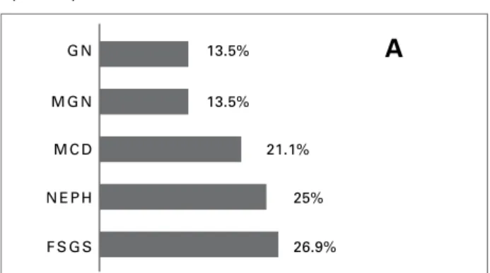
Regarding primary glomerulopathies (n = 52), focal segmental glomerulosclerosis (FSGS) predomi-nated (14/52; 26.9%), followed by IgA nephropathy (13/52; 25%), minimal change disease (MCD) (11/52; 21.1%), membranous glomerulopathy (MGN) (7/ 52; 13.5%), and undetermined chronic glomerulonephri-tis (CGN) (7/52; 13.5%) (Figure 1).
Regarding secondary glomerulopathies (n = 38), lupus nephritis predominated (19/38; 50%), followed by diffuse proliferative glomerulonephritis (DPGN) (13/38; 34.2%), pauci-immune glomerulonephritis (PAUCI GN) (3/38; 8%), and hypertensive nephros-clerosis (3/38; 8%) (Figure 1). In the cases of lupus nephritis, class IV predominated (11/19; 57.9%),
followed by class V (4/19; 21%), class III (3/19; 15.8%), and classes I/II (1/19; 5.3%).
In the group denominated “others” (n = 23), the major diagnoses were as follows: normal kidney (8/23; 34.8%); insufficient material (7/23; 30.4%); tubulointerstitial nephritis (6/23; 26.1%); and acute tubular necrosis (2/23; 8.7%).
When comparing indication for biopsy and histo-logical finding, the following was observed:
• Patients undergoing biopsy due to nephrotic syn-drome (n = 47) had the following results: FSGS (27,6% -13/47); MCD (23,4% - 11/47); lupus ne-phritis (17% - 8/47); MGN (14,9% -7/47); others (10,6% - 5 /47); hypertensive nephrosclerosis (4,2% - 2/47); and IgA nephropathy (2.1% - 1/47) (Figure 2).
• Patients undergoing biopsy due to RPGN (n = 40) had the following results: lupus nephritis (27.5% - 11/40); PDGN (25% - 10/40); others (12.5% - 5/40); IgA nephropathy (10% - 4/40); PAUCI GN (7.5% -3/40); and FSGS (2.5% - 1/40) (Figure 2). • Patients undergoing biopsy due to urinary abnor-malities (n = 16) had the following results: IgA ne-phropathy (43.7% - 7/16); others (43.7% - 7/16); DPGN (6.3% - 1/16); and hypertensive nephros-clerosis (6.3% - 1/16) (Figure 2).
• Patients with nephritic syndrome (n = 6) had the following histological results: others (50% - 3/6); DPGN (33.3% - 2/6); and IgA nephropathy (16.7% - 1/6) (Figure 2).
• Patients with undetermined kidney failure (n = 4) had the following results: others (75% - 3/4) and chronic GN (25% - 1/4) (Figure 2).
Regarding the patients whose indication for biop-sy was RPGN, except for the subgroup “others” and the isolated case of FSGS, all others (85% -34/40) showed cell crescent formation, and in half such ca-ses (42.5% - 17/40) the crescents occupied more than 50% of the biopsy sample, characterizing crescen-tic glomerulopathies. It is worth emphasizing that two patients with PAUCI GN (66.7% - 2/3) were diagnosed as having systemic vasculitis (Wegener’s granulomatosis).
Comparing patients having primary and secondary glomerulopathies, no statistically significant differen-ce was observed regarding age, sex, use of immuno-suppressive medication, frequency of dialysis during hospitalization, and hematocrit and serum albumin levels. However, patients with primary glomerulopa-thies had higher levels of hemoglobin (11.7 ± 2.5 g/ dL vs. 10.7 ± 2.8 g/dL; p = 0.0393) and a higher inci-dence of chronic dialysis as clinical outcome (21.1% vs. 5.2 %; p = 0.0342), while patients with secondary glomerulopathies had a higher frequency of death as clinical outcome (21% vs. 5%; p = 0.0477) (Table 3).
D
ISCUSSIONOur study aimed at delineating the profile of patients suspected of having glomerular disease undergoing kidney biopsy at a public hospital of the Brazilian Federal District.
Suspicion of glomerular disease was confirmed in almost 80% of the cases undergoing biopsy. This emphasizes that a high degree of clinical suspicion is required so that a quick diagnosis can be established and treatment initiated.13
Our study identified that glomerular diseases predominated and that the major indication for kid-ney biopsy was the presence of nephrotic syndrome. Primary glomerulopathies, as compared to secondary ones, were also found to predominate, FSGS being the most frequently found among the primary glomerulo-pathies, while lupus nephritis was the most common among the secondary ones. Those data are in accor-dance with those of several national15-18 and Latin American19,20 studies.
Figure 1.A. Distribution of primary glomerulopathies (n = 52). B. Distribution of secondary glomerulopathies (n = 38).
G N 13.5%
13.5%
21.1%
25%
26.9% M G N
M C D
N E P H
F S G S
A
PAUCI GN = Pauci-immune glomerulonephritis; HYP NSC = Hypertensive nephrosclerosis; DPGN = Diffuse proliferative glomerulonephritis; SLE NEPH = Lupus nephritis.
8%
8%
50% 34.2%
PA U C I G N
H Y P N S C
D P G N
S L E N E P H
B
Table 2 shows several specific data of each glome-rulopathy found in our study, which are in accordan-ce with the literature,12 as follows:
• a large number of cases of FSGS evolving to chro-nic dialysis need;
• among patients with nephrotic syndrome, FSGS, MCD, and MGN predominated;
• patients with MGN belonged to an older age group; • lupus nephritis affected mainly younger
individu-als and women;
• a high percentage of patients with PAUCI GN pre-senting as RPGN and evolving to death.
Many of the correlations between clinical symp-tomatology and biopsy result observed in our study are in accordance with classical data in the literature, such as the strong association between nephrotic syn-drome and FSGS and between urinary abnormalities and IgA nephropathy. The fact that a large number
of patients presenting with RPGN had a histological report of lupus nephritis can be explained by the pre-dominance of class IV, with a traditionally more ag-gressive presentation.21,12
The second most common histological finding among patients with urinary abnormalities was non--glomerular diseases (subgroup “others”). This can be explained both by the fact that tubulointerstitial dise-ases can mimic glomerular disedise-ases and by the biopsy findings described as normal kidney in this subgroup that could correspond to thin basement membrane di-sease (TMD), since such pathology requires electron microscopy to be diagnosed (not performed in our study). In some studies, TMD exceeds IgA nephro-pathy as the major cause of urinary abnormalities.22
In our study, the most prevalent primary glomeru-lopathies in decreasing order were: FSGS; IgA nephro-pathy; MCD; and MGN. Such glomerulopathies have
FSGS IgA MCD MGN Chronic GN SLE DPGN PAUCI GN HYP NSC
Parameters (n = 14) (n = 13) (n = 11) (n = 7) (n = 7) (n = 19) (n = 13) (n = 3) (n = 3)
Sex
a) Male 7 ( 50%) 8 (61.5%) 7 (63.6%) 4 (57.1%) 4 (57.1%) 1 (5.3%) 12 1 (33.3%) 3 (100%)
b) Female 7 ( 50%) 5 (38.5%) 4 (36.4%) 3 (42.8%) 3 (42.8%) 18 (94.7%) 1 (7.7%) 2 (66.7%) 0 Age (years) 34.5 ± 14.7 33.1± 11.4 34± 21 51.8 ± 15.5 35.3 ± 17.5 25.1± 7.1 34.3± 16 34.3 ± 28.3 52.6± 16.2
Biopsy indication
a) Urinary abnormalities 0 7 (53.8%) 0 0 0 0 1 (7.7%) 0 1 (33.3%)
b) RPGN 1 (7%) 4 (30.8%) 0 0 6 (85.7%) 11 (57.9%) 10 (76.9%) 3 (100%) 0 c) Undetermined KF 0 0 0 0 1 (14.3%) 0 0 0 0 d) Nephrotic syndrome 13 (93%) 1 (7.7%) 11 (100%) 7 (100%) 0 8 (42.1%) 0 0 2 (66.7%) e) Nephritic syndrome 0 1 (7.7%) 0 0 0 0 2 (15.4%) 0 0
Interstitial fibrosis
a) No 4 (28.5%) 6 (46.2%) 5 (45.5%) 2 (28.6%) 0 5 (26.3%) 9 (69.2%) 0 0
b) Mild 4 (28.5%) 4 (30.8%) 6 (54.5%) 4 (57.1%) 0 8 (42.1%) 1 (7.7%) 1 (33.3%) 2 (66.7%) c) Moderate 5 (36%) 2 (15.4%) 0 1 (14.3%) 3 (42.8%) 4 (21%) 2 (15.4%) 1 (33.3%) 1 (33.3%)
d) Severe 1 (7%) 1 (7.7%) 0 0 4 (57.1%) 2 (10.5%) 1 (7.7%) 1 (33.3%) 0 Immunosuppressive
drug use 10 (71.4%) 9 (69.2%) 8 (72.7%) 3 (42.8%) 5 (71.4%) 13 (68.4%) 8 (61.5%) 3 (100%) 2 (66.7%) Dialysis 3 (21.4%) 3 (23%) 2 (18.2%) 0 7 (100%) 5 (26.3%) 5 (38.5%) 2 (66.7%) 0
Clinical outcome
a) Conservative 11 (78.5%) 11 (84.6%) 8 (72.7%) 7 (100%) 1 (14.3%) 11 (57.9%) 11 (84.6%) 1 (33.3%) 3 (100%)
b) Chronic dialysis 3 (21.4%) 2 (15.4%) 0 0 6 1 (5.3%) 1 (7.7%) 0 0 c) Death 0 0 3 (27.3%) 0 0 5 (26.3%) 1 (7.7%) 2 (66.7%) 0
d) Lost to follow-up 0 0 0 0 0 2 (10.5%) 0 0 0
Table 3 COMPARISONOFTHEDEMOGRAPHICANDLABORATORYDATA, IMMUNOSUPPRESSIVETREATMENT, ANDCLINICALOUTCOMEOF PATIENTSWITHPRIMARYANDSECONDARYGLOMERULOPATHIESUNDERGOINGBIOPSYAT HRAN (N = 90).
Parameters Primary( n = 52) Secundary (n = 38) p value
Age (years) 36.5 ± 16.6 31.1 ± 15 0.1193a
Male sex % 57.6 44.7 0.2243b
Presence of ibrosis % 67.3 63 0.6824b
Immunosuppression % 76.9 73.6 0.7244b
Dialysis need % 28.8 31.5 0.7799b
Outcome: chronic dialysis % 21.1 5.2 0.0342b
Outcome: death % 5.7 21 0.0477a
Hemoglobin – g/dL 11.7 ± 2.5 10.7 ± 2.8 0.0393a
Hematocrit 34.5 ± 8.1 31.6 ± 7.7 0.1012a
Albumin – g/dL 2.9 ± 0.6 2.8 ± 0.6 0.6754a
a = Student t test; b = chi-square test; c = Fisher exact test.
greatly variable prevalence worldwide, depending on the country of origin.23-27 Several studies have repor-ted that FSGS has become the major primary glome-rulopathy causing nephrotic syndrome worldwide.28,29
The presence of DPGN (in our study, highly rela-ted to acute post streptococcal glomerulonephritis) as the second major cause of secondary glomerulopathy has also been reported in some national studies,17 but not at a such high proportion as that found in our stu-dy. Although we do not have a consistent explanation for that, it is worth emphasizing the highest inciden-ce of post streptococcal glomerulonephritis in deve-loping countries and the significant percentage (over 5%) of cases evolving to rapidly progressive forms in adult patients.30-31 Some studies question the benigni-ty of “acute glomerulonephritis” over time, especially when related to epidemic outbreaks caused by some specific bacterial strains.32,33
In our study, patients with primary glomerulopa-thies most frequently had chronic dialysis as clinical outcome as compared with the subgroup of patients with secondary glomerulopathies. That emphasizes the insidious course of those diseases, mainly in their initial phases. Many patients, especially those with FSGS, were referred to our service after a significant loss of kidney function, therefore evolving more rapi-dly to end-stage kidney failure.
Regarding the immunosuppressive treatment, apparently little difference was observed between bo-th groups wibo-th glomerular diseases. However, in bo-the subgroup of secondary glomerulopathies, many deaths occurred, which can be attributed to the fact that that subgroup comprised many patients with lupus nephri-tis (mainly class IV), in addition to patients with syste-mic vasculites and DPGN evolving to crescent forma-tion. That profile of patients contributed to the finding Figure 2. Correlation between biopsy indication and
histopathological findings.
nefrotic syndrome
RPGN
UA
Nefritic sd
Undet KF
Others Chronic GN
75%
25%
Others DPGN IgA NEPH
50% 33.3%
16.7% IgA NEPH Others DPGN HYP NSC
43.7% 43.7%
6.3% 6.3%
SLE NEPH
DPGN 25%
Others 12.5%
IgA 10%
PAUCI GN 7.5%
FSGS 2.5% 27.5%
HYP NSC
IgA NEPH Others
MGN SLE NEPH MCD FSGS 27.6% 23.4%
17% 14.9% 10.6%
that the second glomerular syndrome requiring kidney biopsy in our study was the suspicion of RPGN, a fact not observed in other national studies.15-17 Several stu-dies have shown the high impact of lupus nephritis on morbidity and mortality, especially in the presence of other risk factors, such as non-Caucasian race and low socioeconomic level, frequent in developing countries and among the patients of our study.34,35 A study com-paring patients with lupus nephritis and patients with other primary glomerulopathies undergoing the same immunosuppression has reported death as an outcome more frequent among lupus patients, showing the pact of the underlying autoimmune disease on the im-munosuppression degree of those patients.36 Therefore, the most immunosuppressed profile of patients with se-condary glomerulopathies may have contributed to the greater frequency of death as a clinical outcome, regar-dless of the form or type of immunosuppression used.
C
ONCLUSIONOur study provides important epidemiological infor-mation about the profile of patients with glomerular diseases at a public hospital of the Federal District in Brazil. However, further studies are required at other services in the Federal District and in other states of the West-Central region to the better understanding of the behavior of glomerulopathies in that Brazilian region.
A
CKNOWLEDGEMENTSTo Natasha Ferraroni, MD, and professors Luis Moura, MD, and Marcello Franco, MD (both from UNIFESP), for the histopathological analysis. We also thank Frederico Moreira and the ESTAT team of the University of Brasília for the statistical analysis.
R
EFERENCES1. Salgado Filho N, Brito DJA. Doença Renal Crônica: a grande epidemia deste milênio. J Bras Nefrol 2006; 28:1-5.
2. Sesso R, Lopes AA, Thome FS, Bevilacqua JL, Romão Junior JE, Lugon J. Relatório do censo brasileiro de diálise 2008. J Bras Nefrol 2008; 30:233-8.
3. Bastos MG, Carmo WB, Abrita RR et al. Doença Renal Crônica: problemas e soluções. J Bras Nefrol 2004, 26:202-15.
4. Hafez MH, Abdellatif DA, Elkhatib MM. Prevention of renal disease progression and renal replacement therapy in emerging countries. Artif Organs 2006; 30:501-9.
5. Cusumano AM, Romao JE, Poblete Badal H et al.
Latin-American Dialysis and Kidney Transplantation Registry: data on the treatment of end-stage renal dis-ease in Latin America. G Ital Nefrol 2008; 25:547-53.
6. Cusumano A, Garcia-Garcia G, Di Gioia C et al. End-stage renal disease and its treatment in Latin America in the twenty-first century. Ren Fail 2006; 28:631-7. 7. Sesso R. Epidemiologia da doença renal crônica no
Brasil. In: Barros E, Manfro RC, Thomé FS, Gonçalves LF. Nefrologia: Rotinas, Diagnóstico e Tratamento. 3ed. Porto Alegre: Artmed, 2006. p.39-46.
8. Hamer RA, El Nahas AM. The burden of chronic kid-ney disease: is rising rapidly worldwide. BMJ 2006; 332:563-4.
9. Oliveira MB, Romao Jr JE, Zatz R. End-stage renal disease in Brazil: epidemiology, prevention, and treat-ment. Kidney Int Suppl 2005; 68:S82-86.
10. Maisonneuve P, Agodoa L, Gellert R et al. Distribution of primary renal diseases leading to end-stage renal failure in the United States, Europe, and Australia/ New Zealand: results from an international compara-tive study. Am J Kidney Dis 2000; 35:157-65.
11. Cohen AH, Nast CC. Clinical utility of kidney biopsies in the diagnosis and management of renal disease. Am J Nephrol 1989; 9:309-15.
12. Barros RT, Alves MAR, Kirsztajn GM, Sens YAS, Dantas M. Glomerulopatias: patogenia, clínica e trata-mento. 2. ed. Editora Sarvier, São Paulo, 2006.
13. Sociedade Brasileira de Nefrologia (SBN). Consenso Brasileiro de Glomerulopatias. J Bras Nefrol 2005; 27(2-Supl.1).
14. Churg J, Sobin LH, editors. Renal disease: classifica-tion and atlas of glomerular diseases. Tokyo: Igaku-Shoin , 1995.
15. Cardoso AC, Mastroianni-Kirsztajn G. Padrões his-topatológicos das doenças glomerulares no Amazonas. J Bras Nefrol 2006; 28:39-4.
16. Carmo PAV, Carmo WB, Bastos MG, Andrade LCF. Estudo das Doenças Glomerulares na Zona da Mata Mineira. J Bras Nefrol 2008; 30:15-2.
17. Malafronte P, Mastroianni-Kirsztajn G, Betôncio GN et al. Paulista Registry of glomerulonephritis: 5-year data report. Nephrol Dial Transplan 2006; 21:3098-105. 18. Polito MG, de Moura LA, Kirsztajn GM. An overview
on frequency of renal biopsy diagnosis in Brazil: clinical and pathological patterns based on 9,617 native kidney biopsies. Nephrol Dial Transplant 2010;25(2):490-6. 19. Arias LF, Henao J, Giraldo RD, Carvajal N, Rodelo J,
Arbeláez M. Glomerular diseases in a Hispanic popu-lation: review of a regional renal biopsy database. São Paulo Med J 2009; 127:140-4.
20. Mazzuchi N, Acosta N, Caorsi H et al. Frequency of diagnosis and clinic presentation of glomerulopathies in Uruguay. Nefrologia 2005; 25:113-20.
21. Korbet SM, Lewis EJ, Schwartz MM, Reichlin M, Evans J, Rohde RD. Factors predictive of outcome in severe lupus nephritis. Lupus Nephritis Collaborative Study Group. Am J Kidney Dis 2000; 35:904-14. 22. Van Paassen P, Van Breda Vriesman PJ, Van Rie H,
Tervaert JW. Signs and symptoms of thin basement membrane nephropathy: a prospective regional study on primary glomerular disease-The Limburg Renal Registry. Kidney Int 2004; 66:909-13.
23. Chang JH, Kim DK, Kim HW et al. Changing
24. Covic A, Schiller A, Volovat C et al. Epidemiology of renal disease in Romania: a 10 year review of two re-gional renal biopsy databases. Nephrol Dial Transplant 2006; 21:419-24.
25. Rychlík I, Jancová E, Tesar V et al. The Czech regis-try of renal biopsies. Occurrence of renal diseases in the years 1994-2000. Nephrol Dial Transplant 2004; 19:3040-9.
26. Li LS, Liu ZH. Epidemiologic data of renal diseases from a single unit in China: analysis based on 13,519 renal biopsies. Kidney Int 2004; 66:920-3.
27. Panichi V, Pasquariello A, Innocenti M et al. The Pisa experience of renal biopsies, 1977-2005. J Nephrol 2007; 20:329-35.
28. Oliveira MB, Penna DO, Saldanha LB, Mota ELA, Barros RT, Romão Jr JE. Primary glomerular diseases in Brazil (1979-1999): is the frequency of focal and segmental glomerulosclerosis increasing? Clin Nephrol 2004; 61:90-7.
29. Hass M, Spargo B, Coventry S. Increasing incidence of focal segmental glomerulosclerosis among adult ne-phropathies: a 20-year renal biopsy study. Am J Kidney Dis 1995; 26:740-50.
30. Rodriguez-Iturbe, B. The current state of poststrepto-coccal glomerulonephritis. J Am Soc Nephrol 2008; 19:1855-64.
31. Barros RT, Vieira Jr JM. Glomerulonefrites secundárias às infecções bacterianas. In: Barros RT, Alves MAR, Kirsztajn GM, Sens YAS, Dantas M. Glomerulopatias: patogenia, clínica e tratamento. 2. ed. Editora Sarvier, São Paulo, 2006.
32. Sesso R, Pinto SWL. Epidemic glomerulonephritis due to Streptococcus zooepidemicus in Nova Serrana, Brazil. Kidney Int 2005; 97:132-6.
33. Sesso R, Pinto SWL. Five-year follow-up of patients with epidemic glomerulonephritis due to strepto-coccus zooepidemicus. Nephrol Dial Transpl 2005; 20:1808-12.
34. Barr RG, Seliger S, Appel GB et al. Prognosis in proli-ferative lupus nephritis: the role of socio-economic sta-tus and race / ethnicity. Nephrol Dial Transplant 2003; 18:2039-46.
35. Korbet SM, Lewis EJ, Schwartz MM, Reichlin M, Evans J, Rohde RD. Factors predictive of outcome in severe lupus nephritis. Lupus Nephritis Collaborative Study Group. Am J Kidney Dis 2000; 35:904-14. 36. Balbi AL, Barbosa RA, Lima MCP, de Almeida DB.