J. bras. pneumol. vol.39 número4
Texto
Imagem
Documentos relacionados
Ousasse apontar algumas hipóteses para a solução desse problema público a partir do exposto dos autores usados como base para fundamentação teórica, da análise dos dados
The gLite middleware facilitates the users with high level services for scheduling and running computational jobs, for accessing, moving and sharing big data with collaborators
A CT scan of the chest revealed a massive collection with an air-fluid level occupying nearly all of the right hemithorax, resulting in compression of the adjacent
The probability of attending school four our group of interest in this region increased by 6.5 percentage points after the expansion of the Bolsa Família program in 2007 and
didático e resolva as listas de exercícios (disponíveis no Classroom) referentes às obras de Carlos Drummond de Andrade, João Guimarães Rosa, Machado de Assis,
Nodule in the right lung apex and percutaneous biopsy of the same: in A, positron emission tomography-CT (PET-CT) scan showing the nodule (arrow); in B, CT scan showing the point
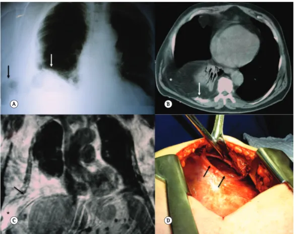
Curved coronal CT reconstruction showing a bronchopleural istula in the anterior segment of the right lower
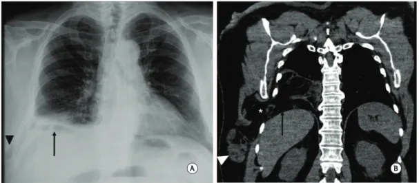
In B, axial contrast-enhanced CT of the chest scan showing a heterogeneous hypervascular mass (asterisk) iniltrating the right serratus anterior and pectoralis muscles, as well as
