ISSN 1806-3713 © 2016 Sociedade Brasileira de Pneumologia e Tisiologia
http://dx.doi.org/10.1590/S1806-37562016000000228
Clusters of small nodules with no
conluence
Edson Marchiori1,2, Bruno Hochhegger3,4, Gláucia Zanetti2,5
1. Universidade Federal Fluminense, Niterói (RJ) Brasil. 2. Universidade Federal do Rio de Janeiro, Rio de Janeiro (RJ) Brasil. 3. Santa Casa de Misericórdia de Porto Alegre, Porto Alegre (RS) Brasil.
4. Universidade Federal de Ciências da Saúde de Porto Alegre, Porto Alegre (RS) Brasil. 5. Faculdade de Medicina de Petrópolis, Petrópolis (RJ) Brasil.
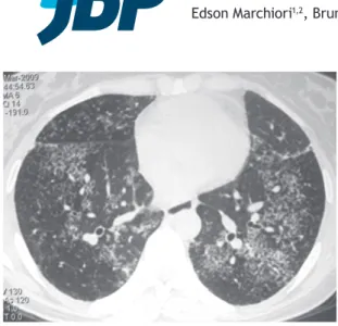
Figure 1. HRCT scan showing clusters of small nodules with
no conluence in the lower lobes of both lungs.
CLINICAL HISTORY
A 36-year-old man presented with a two-month history of dry cough, fever, and weight loss (8 kg). Laboratory test results were normal. HRCT of the chest showed clusters of interstitial nodules with no conluence (Figure 1). BAL specimens were negative for tuberculosis and fungi. Open lung biopsy showed necrotic granulomas. Culture of lung tissue was positive for Mycobacterium tuberculosis.
DISCUSSION
This patient had multiple small interstitial nodules on HRCT. Small pulmonary nodules (or micronodules) are round opacities of soft tissue density and less than 1 cm in diameter. On the basis of their distribution in the lung parenchyma, they can be classiied as perilymphatic, centrilobular, or random micronodules.
A perilymphatic distribution occurs when the small nodules are located predominantly along the lung
lymphatic system (peribronchovascular interstitium, interlobular septa, and subpleural regions). A centrilobular distribution is characterized by nodules that are located within a few millimeters of the pleural surface and issures but do not touch them. A random pattern corresponds to small nodules randomly distributed in the secondary lobule and scattered uniformly throughout the lungs.
Some other patterns of distribution of small interstitial nodules have been described more recently. They include the sarcoid galaxy sign, the sarcoid cluster sign, and the nodular reversed halo sign. The reversed halo sign is deined as a round area of ground-glass attenuation surrounded by a ring of consolidation. The designation of nodular reversed halo sign is used when the walls of the dense peripheral ring are formed by nodules, instead of exhibiting the usual smooth appearance. Nodules are also frequently seen within the halo. Findings of this pattern are highly suggestive of active granulomatous disease, particularly tuberculosis or sarcoidosis.
The sarcoid galaxy sign corresponds to a large parenchymal nodule that is formed by the coalescence of small nodules and is surrounded by satellite nodules. The sarcoid cluster sign corresponds to a generally round or elongated group of small nodules with no conluence, as in the case presented here. The nodules are close to each other but do not group. These two signs were initially reported in patients with sarcoidosis, hence their names. However, they were subsequently described in patients with tuberculosis, thus requiring to be renamed. The names currently proposed for these new signs are “cluster of nodules with conluence” and “cluster of nodules without conluence”, respectively. From an anatomical and pathological standpoint, in the cases of both signs, the nodules correspond to granulomas, and the two major diagnoses that should be considered in the presence of these indings are active tuberculosis and sarcoidosis.
RECOMMENDED READING
1. Marchiori E, Zanetti G, Barreto MM, de Andrade FT, Rodrigues RS. Atypical distribution of small nodules on high resolution CT studies:
patterns and differentials. Respir Med. 2011;105(9):1263-7. http:// dx.doi.org/0.1016/j.rmed.2011.02.010
J Bras Pneumol. 2016;42(6):402-402
402