UNIVERSIDADE FEDERAL DA PARAÍBA CENTRO DE CIÊNCIAS EXATAS E DA NATUREZA
PROGRAMA DE PÓS-GRADUAÇÃO EM BIOLOGIA CELULAR E MOLECULAR LABORATÓRIO DE GENÉTICA DE MICRORGANISMOS
ANÁLISE EVOLUTIVA DAS SUBUNIDADES LIGADORAS DE SUBSTRATO PRESENTES NO SISTEMA DE OSMOPROTEÇÃO EM PROCARIOTOS
TARCISIO JOSÉ DOMINGOS COUTINHO
TARCISIO JOSÉ DOMINGOS COUTINHO
ANÁLISE EVOLUTIVA DAS SUBUNIDADES LIGADORAS DE SUBSTRATO PRESENTES NO SISTEMA DE OSMOPROTEÇÃO EM PROCARIOTOS
Prof. Dr. Sávio Torres de Farias
ORIENTADOR
JOÃO PESSOA – PARAÍBA 2012
C871a Coutinho, Tarcisio José Domingos.
Análise evolutiva das subunidades ligadoras de substrato presentes no sistema de osmoproteção em procariotos / Tarcisio José Domingos Coutinho.-- João Pessoa, 2012.
97f. : il.
Orientador: Sávio Torres de Farias
Dissertação (Mestrado) – UFPB/CCEN
TARCISIO JOSÉ DOMINGOS COUTINHO
Dissertação de mestrado avaliada em: 13/12/2012
BANCA EXAMINADORA
____________________________________________________
Prof. Dr. Sávio Torres de Farias – orientador (Departamento de Biologia Molecular – CCEN – UFPB)
____________________________________________________
Prof.ª Dr.ª Thaís Gaudencio do Rêgo – membro externo
(Unidade Acadêmica de Tecnologia do Desenvolvimento - CDSA – UFCG)
____________________________________________________
Prof. Dr. Luís Fernando Marques dos Santos – membro interno (Departamento de Biologia Molecular – CCEN – UFPB)
____________________________________________________
A Tarcisio Pessoa Coutinho (In memorian).
AGRADECIMENTOS
A Deus, “não tenho palavras para agradecer tua bondade, dia após dia me cercas com
fidelidade, nunca me deixes esquecer que tudo o que tenho, tudo o que sou e o que vier a ser vem de Ti, Senhor”.
À minha mãe, Eunice, meu exemplo de vida. Obrigado por tudo. Se hoje estou aqui é porque a senhora esteve sempre ao meu lado.
À minha irmã, Tatiany, que sempre foi uma força a me mostrar que podia ir mais longe. Valeu, mana!
À minha tia Tarcísia (segunda mãe). Não sei nem como agradecer por tanto que fez em meu favor. Obrigado, tia, por acreditar tanto em mim.
Ao meu tio Arnaldo Barreto (segundo pai). Seu carinho e atenção não têm limites. Obrigado por me permitir ser um membro de sua família.
Aos meus primos Aline, Liane e Barreto Filho (irmãos). Obrigado pelo carinho, vocês são um presente.
Aos meus primos Beto, Carla, Danilo, Caio e Tiago pelo incentivo, carinho e cuidado.
À minha mais nova família, a família Maul, que me recebeu de braços abertos e em especial à matriarca Dona Márcia, cujo carinho a mim demonstrado jamais poderia ser descrito em palavras.
A Ludmilla Maul, que entrou na minha vida sem querer e provocou uma verdadeira revolução. Estar aqui hoje tem uma participação fundamental desta incrível pessoa que conheci e com a qual pretendo dividir o resto dos meus dias. Te amo.
Ao meu orientador Sávio Torres de Farias. Não tenho palavras pra te agradecer. Obrigado por ter ido comigo até o fim.
Ao ilustre professor Luís Fernando, meu amigo, se posso assim dizer. Nunca esquecerei sua atitude no momento mais difícil desta caminhada. Estar aqui hoje é também sua responsabilidade.
Aos demais membros da banca: professora Thaís Gaudêncio e Teresa Cristina pela gentileza e disponibilidade de participarem deste momento tão especial.
Ao professor Plínio Delatorre pela incrível capacidade de questionamento que me ensinou a enxergar a pesquisa com outros olhos.
A todos os colegas do Programa de Pós-Graduação em Biologia Celular e Molecular, que trabalham diuturnamente em seus laboratórios realizando pesquisas de qualidade e que contribuirão muito para a continuidade do sucesso do Programa.
Ao atual coordenador do Programa de Pós-Graduação em Biologia Celular e Molecular, professor Eleonidas Moura, pelo trabalho em prol de melhorias para que o Programa seja melhor.
À minha turma de mestrado: Guilherme, Yara, Danielle. Foi um prazer dividir este momento com vocês.
Ao meu grupo de estudo: Nureyev, Jonathas, Suellen, Vanessa e Danielle. Minha visão sobre biologia molecular nunca mais será a mesma depois de tantas noites estudando com vocês. Foi um honra conhecê-los.
Ao meu amigo Jonathas. Minha visão e interpretação de moléculas concatenadas e de transposons nunca mais será a mesma depois de nossas discussões. Muito sucesso e um brinde com Santa Felicidade, você merece!
Ao meu amigo e irmão Nureyev, porque é isso que você é pra mim. Espero ter você sempre por perto. Jamais esquecerei tudo que vivemos, conversamos e compartilhamos ao longo desse tempo.
Ao meus colegas de laboratório: Priscilla Anne, Juliana, Danilo, Soraya, Francisco, Ariadne, Mayra, Janaína, Patrícia e Lobão pelo carinho. Em especial, Priscilla e Juliana, por terem tido muita paciência comigo enquanto estive na bancada.
Aos técnicos do laboratório de Genética de Micro-organismos Bosco e Severina. Ter vocês por perto deixou o final deste trabalho muito mais agradável. Obrigado pelo carinho.
À professora Daniela Priscila e ao meu amigo Luiz Cláudio. Nossas conversas sempre foram muito animadas e revigorantes. Embora estejam há pouco tempo em minha vida, mas já são especiais.
À família de tio Wilson (tio de Nureyev). Os finais de noite, especialmente depois de dias que não pareciam ter fim, sempre foram revigorantes, recheados de muita risada e carinho.
Ao meu amigo de infância e irmão, Luíz Antonio Junior. Obrigado por estar sempre comigo.
A Cláudia Gracielle e família, pois durante o tempo que estive com vocês, sempre fui incentivado a seguir em frente.
A Aquilino Ramalho, diretor do colégio Classe A, por ser um incentivador do crescimento profissional e por ter me compreendido tão bem, adequando os horários para que eu pudesse cursar as disciplinas do mestrado.
Ao professor e diretor do Colégio Motiva, Karamuh Martins, por ter compreendido tão bem minha decisão de deixar sua escola. Obrigado pelo incentivo.
Aos meus amigos professores Guilherme Stanford, Job Fonseca e Moabe Pina. Não poderia haver melhor equipe de biologia nesta cidade e será sempre uma honra dividir uma sala de aula com vocês.
Aos meus alunos, que durante o tempo deste mestrado, sofreram e se alegraram comigo. Sou muito feliz por ser professor e jamais deixarei de ser.
Toda caminhada começa com um primeiro passo, a natureza não tem pressa segue seu compasso e
inexoravelmente chega lá!
RESUMO
As subunidades ligadoras de substrato são componentes muito importantes do sistema de importação de soluto conhecido, como sistema de osmoproteção, que consiste em uma proteína de membrana pertencente à superfamlía ABC. Estas moléculas reconhecem substratos específicos que apresentam papéis fisiológicos diversos em procariotos, incluindo a colaboração para a sobrevivência destes organismos em ambientes com elevada concentração de sal. Utilizando o software MEGA 5.05, foi realizada uma análise filogenética de 431 sequências nucleotídicas destas subunidades, ortólogas entre si, coletadas a partir do banco de dados contido no site http://www.genome.jp/kegg/. Como resultado desta análise, foram geradas árvores filogenéticas que demonstraram claramente que houve a transferência horizontal de alguns dos genes devido ao compartilhamento por organismos diferentes. Foram geradas também duas prováveis sequências ancestrais que apresentam homologia com permeases que transportam colina, glicina betaína e carnitina, que são aminas trimetiladas, presentes atualmente em diversos procariotos. Portanto, este sistema provavelmente surgiu em organismos procarióticos com a função básica de captura de nutrientes e por desempenhar esta função basal, ao ser compartilhado com outros organismos, foi fixado aos genomas. No entanto, a partir da diversificação de habitats, por parte dos procariotos, este sistema colaborou de forma decisiva para a adaptação destes organismos aos mais diversos ambientes, incluindo, especialmente os ambientes que apresentavam uma elevada concentração de sal, atuando e sendo caracterizado atualmente como um sistema de osmoproteção.
ABSTRACT
Substrate-binding subunits are very important components of the solute importation system, known as the osmoprotectant system, which consists of a membrane protein belonging to the ABC superfamily. These molecules recognize specific substrates that have different physiological roles in prokaryotes, i.e. roles that contribute to the survival of these organisms in environments with high concentrations of salt. Using MEGA 5.05 software, this study performed a phylogenetic analysis of 431 nucleotide sequences of these subunits, orthologous to each other, collected from the database contained on the website http://www.genome.jp/kegg/. As a result of this analysis, phylogenetic trees were generated that clearly demonstrated that there was a horizontal transfer of some genes due to the sharing by different organisms. Also, two probable ancestral sequences were generated that showed homology with permeases that transport choline, glycine betaine and carnitine, which are trimethylamines currently present in various prokaryotes. Therefore, this system probably arose in prokaryotic organisms with the basic function of capturing nutrients, and by performing this basal function of being shared with other organisms, was fixed to the genome. However, because of the diversification of habitats by the prokaryotes, this system contributed decisively to the adaptation of these organisms to different environments, especially environments that had a high salt concentration; thus, acting and being currently characterized as a system of osmoprotection.
LISTA DE FIGURAS
Figura 1. Alguns solutos compatíveis...19
Figura 2. Síntese de metionina em Sinorhizobium meliloti...20
Figura 3. Vias metabólicas de síntese de glicina betaína...21
Figura 4. Esquema da estrutura básica de proteínas ABC importadoras e exportadoras....22
Figura 5. Estrutura básica de proteínas ABC e suas regiões conservadas...23
Figura 6. Alguns membros do sistema de importação osmoprotetora...26
Figura 7. Estrutura da subunidade ligadora de substrato de OpuC...28
LISTA DE TABELAS
Tabela 1: Principais famílias de importadores ABC presentes em procariotos...24
LISTA DE ABREVIAÇÕES E SIGLAS
Asp– ácido aspártico (aminoácido)
ATP–Adenosine triphosphate ( trifosfato de adonosina)
DNA–Deoxyribonucleic acid ( ácido desoxirribonucleico)
MEGA – Molecular Evolutionary Genetics Analysis (Análises Genéticas Moleculares
Evolutivas).
K+ - Íon potássio
mRNA - Messenger ribonucleic acid (ácido ribonucleico mensageiro)
Na+ - Íon sódio
NBD–Nucleotide binding domain (domínio de ligação de nucleotídeo)
SBP–Substrate binding domain (domínio de ligação do substrato)
Superfamília ABC–ATP Binding Cassete
TMD–Transmembrane domain (domínio transmembrana)
SUMÁRIO
1. INTRODUÇÃO………...15
1.1. Estresse e ambiente...16
1.2. Organismos halófilos e osmoadaptação...17
1.3. Glicina betaína...19
1.4. Superfamília de proteínas ABC...21
1.5. Sistema de osmoproteção...26
1.6. Subunidades ligadoras de substrato...27
1.7. Análise comparativa de genes...29
2. OBJETIVOS………...γβ 3. RESULTADOS E DISCUSSSÃO……….....34
4. CONCLUSÃO………...59
1. INTRODUÇÃO
1.1. ESTRESSE E AMBIENTE
O estresse ambiental é a principal força evolutiva que age sobre os seres vivos, uma vez que, promove alterações ambientais com impacto sobre os sistemas biológicos e processos evolutivos (NEVO, 2011).
A conservação de uma espécie depende primariamente de sua adaptação às mudanças abióticas que ocorrem em seu habitat, ou seja, deve haver um equilíbrio entre a fisiologia dos organismos e suas adaptações ao ambiente, por isso, a chave principal para compreensão das mudanças evolutivas deve ser de caráter ecológico, com destaque para a quantidade de energia necessária para manutenção de um organismo em ambiente estressante, o que pode ser utilizado como base para justificar vários padrões de mudanças na composição de seres vivos de um ambiente (PARSONS, 2005).
Organismos presentes em ambientes estressantes aumentam seu custo energético. Na sequência de uma perturbação ambiental grave, o balanço de energia intracelular muda de manutenção e produção para reparação e recuperação, por isso a organização metabólica destes organismos consiste em um fator fundamental para sobrevivência (PARSONS, 2005).
As células dos organismos vivos são expostas a vários tipos de estresses ao longo da vida (RÖSGEN; PETTITT; BOLEN, 2007) tais como: disponibilidade de nutrientes, variações de temperatura, pH e osmolaridade (SPANHEIMER; MULLER, 2008). Diante de tantas possibilidades de variações abióticas o maior desafio para uma célula é manter sua homeostase, ou seja, ajustar as propriedades físicas e químicas do meio intracelular em resposta a processos fisiológicos e alterações ambientais (WOOD, 2011).
1.2. ORGANISMOS HALÓFILOS E OSMOADAPTAÇÃO
Halófilos são organismos adaptados a viver em ambientes que apresentam variações nas condições de salinidade (PAUL et al., 2008) e são encontrados nos três domínios da vida: Archaea, Bacteria e Eukarya (MA et al., 2010).
De acordo com a concentração ótima de sal para crescimento, os halófilos são classificados nas seguintes categorias: ligeiramente halófilos – crescem otimamente em meio contendo de 0,2 à 0,5 M de sal; halófilos moderados - crescem otimamente em meio contendo de 0,5 à 2,5 M de sal e halófilos extremos - crescem otimamente em meio contendo de 2,5 à 5,2 M de sal (KUSHNER, 1985, apud VENTOSA; NIETO, 2005).
Para viver em ambientes com elevada concentração de sal são necessárias adaptações moleculares, dentre as quais, as relacionadas com proteínas, uma vez que, estas moléculas exercem papel fundamental na fisiologia celular (PAUL et al., 2008).Análises comparativas entre proteomas de procariontes halófilos e não halófilos têm comprovado a existência de uma assinatura molecular na composição dos aminoácidos em proteínas com adaptação à ambientes com elevada concentração de sal. Tais proteínas apresentam um aumento na quantidade de resíduos carregados negativamente, como ácido glutâmico e ácido aspártico, que estão mais distribuídos pela superfície da proteína, além disso, estas moléculas apresentam uma diminuição do conteúdo hidrofóbico total (TADEO et al., 2009).
Em ambientes com elevada concentração de sal, torna-se importante também, evitar a perda de água para o ambiente, uma vez que a água é a principal molécula relacionada com o metabolismo celular por propiciar um meio adequado para ocorrência das mais diversas reações químicas intracelulares e lidar com variações internas de água é essencial para a sua sobrevivência. A perda de água para o ambiente é uma ameaça comum, ocorrendo devido à evaporação, excreção de resíduos e osmose nos mais diversos ambientes. Quanto ao estresse hídrico, os organismos foram tradicionalmente divididos em dois grupos: osmorreguladores –
que utilizam transporte de íons para, homeostaticamente, regular a pressão osmótica intracelular e osmoconformadores – costumam utilizar solutos compatíveis para manter a pressão osmótica intracelular igual a do ambiente exterior (YANCEY, 2005).
A estratégia de importação de íons parece estar presente apenas em arqueas halófilas extremas da família Halobacteriaceae que inclui os gêneros Halobacterium, Haloarcula,
Haloquadratum, Halorhabdus, Natronobacterium e Natronococcus, em bactérias da ordem
acumulado, ocorrendo também importação de Na+, no entanto, o citoplasma destes organismos requer que a maioria de suas enzimas sejam ricas em aminoácidos ácidos e suas atividades dependentes da presença dos íons. A outra estratégia envolve o acúmulo e/ou síntese de moléculas com baixo peso molecular, denominadas, solutos orgânicos compatíveis que proporciona várias possibilidades de adaptação a ambientes que variam osmoticamente e isto pode ser comprovado pela grande variedade de microrganismos que dependem exclusivamente destes solutos para a osmoadaptação (EMPADINHAS; DA COSTA, 2008).
Os solutos compatíveis apresentam uma propriedade físico-química, em especial, que provavelmente foi alvo da seleção natural e que permitiu a animais, vegetais e microrganismos adaptarem-se a ambientes extremos. Esta propriedade, conhecida como efeito osmofóbico, permite que proteínas mantenham-se enoveladas mesmo o organismo estando em um ambiente estressante. Este efeito tem como referencia a interação desfavorável que ocorre entre o soluto compatível e o esqueleto de carbonos da cadeia polipeptídica, o que força a proteína manter sua conformação mesmo em condições ambientais adversas (BOLEN; BASKAKOV, 2001).
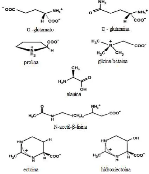
Os tipos de moléculas orgânicas que são caracterizados como solutos compatíveis incluem: polióis e derivados, açúcares e derivados, aminoácidos e derivados, betaínas, ectoínas e ocasionalmente alguns peptídeos (Figura 1). Normalmente, bactérias e eucariotos acumulam solutos neutros, enquanto, arqueas tendem a modificar muitos destes solutos, em moléculas carregadas negativamente. O fato destas moléculas não inibirem as funções celulares, embora possam atuar modulando a atividade de enzimas individuais, foi motivo pelo qual as mesmas foram designadas como solutos compatíveis e seu acúmulo intracelular ajuda a manter a turgescência, pressão e volume celular (ROBERTS, 2005).
Figura 1: Alguns solutos compatíveis (EMPADINHAS; DA COSTA, 2008)
Dentre os solutos compatíveis importados do meio por procariotos, as aminas trimetiladas, como a colina, carnitina e glicina betaína estão associadas à capacidade de osmoadaptação de muitos desses organismos e seus transportadores têm sido cada vez mais estudados (ZIEGLER; BREMER; KRÄMER, 2010).
1.3. GLICINA BETAÍNA
Esta molécula é particularmente eficaz como um citoprotetor, atuando contra vários tipos de estresse abiótico, sendo capaz de estabilizar enzimas e diversas outras proteínas mesmo quando os organismos estão expostos a condições desfavoráveis de temperatura e salinidade (CHEN; MURATA, 2008).
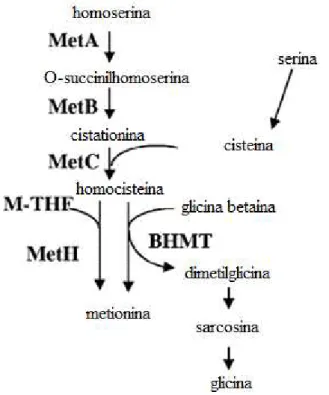
Outro aspecto importante, relacionado aos papéis fisiológicos que desempenha a glicina betaína, consiste em atuar como doadora de grupos metil (Figura 2), participando assim do ciclo da metionina (BARRA et al., 2006).
Figura 2: Sintese de metionina em Sinorhizobium meliloti. Enzimas envolvidas: MetA, homoserina O -succiniltransferase; MetB, cistationina gamasintase; MetC, cistationina beta-liase; MetH, 5-metiltetrahidrofolato- homocisteina metiltransferase; BHMT, betaina-homocisteina metil transferase. M-THF representa metiltetrahidrofolato. Modificado de Barra et al. 2006.
Os microrganismos apresentam duas vias diferentes para sintetizar glicina betaína, uma oxidativa e outra a partir da metilação da glicina (Figura 3). A via oxidativa ocorre normalmente utilizando um sistema proteico associado à membrana codificado a partir de genes organizados em um operon (bet) e seus componentes são: colina desidrogenase (betA), glicina betaína aldeído desidrogenase (betB), e um regulador (betI). A desidrogenase catalisa a oxidação de colina para o aldeído de betaína, que é então oxidado para betaína pelo produto do gene betB (CÁNOVAS et al., 2000; ROBERTS, 2005). As três reações de metilação da
metilação de glicina ou sarcosina formando sarcosina ou dimetilglicina e dimetilglicina sarcosina metiltransferase (sdm) que catalisa a metilação de sarcosina ou dimetilglicina formando betaína dimetilglicina ou glicina betaína (KIMURA et al., 2010).
Figura 3: Vias metabólicas de síntese de glicina betaína. Modificado de Roberts, 2005
1.4. SUPERFAMÍLIA DE PROTEÍNAS ABC
As proteínas pertencentes à superfamília ABC (ATP binding cassete) constituem um
dos maiores sistemas de superfamílias proteicas de sequências parálogas e são encontradas em todos os gêneros dos três domínios da vida. Estas proteínas utilizam energia da hidrólise do ATP para a realização de uma variedade impressionante de fenômenos biológicos essenciais, que incluem não apenas o transporte transmembrana, pelo qual são mais conhecidos, como também processos não relacionados ao transporte, tais como, alongamento da tradução e reparo de DNA (DAVDISON et al., 2008).
peptidios, lípidios, drogas hidrofóbicas, polissacarídeos e proteínas, incluindo toxinas, como a hemolisina A; a terceira categoria aparentemente não está envolvida no transporte, mas em outros processos fisiológicos como a tradução de mRNA e no reparo do DNA (DAVDISON et al., 2008).
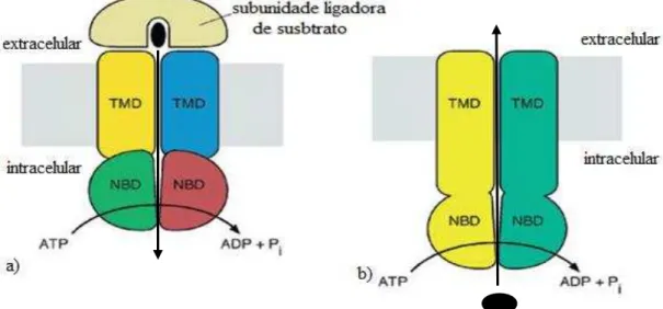
Independentemente do sentido do transporte (importação ou exportação), a natureza do substrato transportado (baixo ou alto peso molecular) e a função fisiológica, as proteínas da superfamília ABC são compostas de dois domínios transmembranares hidrofóbicos (TMD
– transmembrane domain) e dois domínios de ligação a nucleotídeos (NDB – nucleotide domais binding). Em importadores, além dos quatro domínios básicos há também a
participação de uma subunidade ligadora de substrato (SBP – substrate domais protein)
(Figura 4) que é a responsável pela captura do soluto específico que será transportado (VAN DER DOES; TAMPÉ, 2004).
Figura 4: Estrutura básica de proteínas ABC importadoras (a) e exportadoras (b). Modificado de Dawson; Hollestein; Lacher, 2007.
Os domínios transmembranares são duas subunidades altamente hidrofóbicas ou dois domínios proteicos que criam um canal pelo qual o substrato passa durante a translocação. Nos importadores, eles formam o sítio de encaixe para a subunidade ligadora de susbstrato e de transdução de sinal aos domínios de ligação de nucleotídeos que hidrolisam o ATP. Estes domínios atravessam a membrana várias vezes sendo muitos dos transportadores formados
por uma estrutura de doze segmentos α-hélice transmembranares por unidade funcional (BIEMANS-OLDEKINKEL; DOEVEN; POOLMAN, 2006).
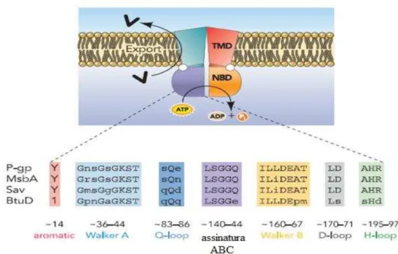
-folhas e seis α-hélices, onde há um motivo Walker A (GXXGXGKS/T, onde X é qualquer aminoácido) ou P-loop e um motivo Walker B (φφφφD, dos quais φ é um resíduo
hidrofóbico). O domínio helicoidal é constituído por três ou quatro hélices e a assinatura característica de proteínas ABC, também conhecidos como sequência LSGGQ. Neste domínio, também há um resíduo de glutamina em uma alça denominada Q loop, que está
envolvida na interação entre NBD e TMD, particularmente na região da hidrólise de nucleótideos para as alterações conformacionais do TMD durante a translocação do substrato. O motivo H, ou região de troca, contém um resíduo de histidina altamente conservado, que é também importante na interação do domínio com ATP (Figura 5) (REES; JOHNSON; LEWINSON, 2009).
Figura 5: Estrutura básica de proteínas ABC e seus domínios conservados. As letras maiúsculas representam resíduos invariáveis enquanto que as letras minúsculas representam resíduos variáveis.
Modificado de Linton, 2007.
Em procariotos as proteínas pertencentes a família ABC estão divididas em importadoras (Tabela 1) e exportadoras (Tabela 2), onde cada uma dessas categorias apresenta famílias de proteínas que são agrupadas normalmente com base na filogenia e no tipo de soluto que transporta (DAVIDSON; CHEN, 2004).
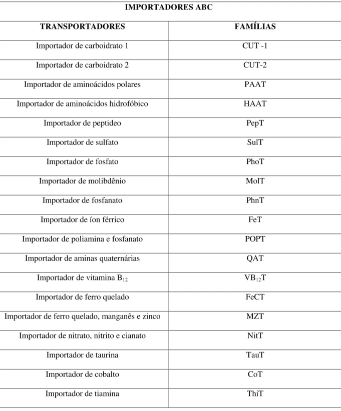
Tabela 1: Principais famílias de importadores ABC presentes em procariotos. Modificado de Milton; Saier, 2000.
IMPORTADORES ABC
TRANSPORTADORES FAMÍLIAS
Importador de carboidrato 1 CUT -1
Importador de carboidrato 2 CUT-2
Importador de aminoácidos polares PAAT
Importador de aminoácidos hidrofóbico HAAT
Importador de peptideo PepT
Importador de sulfato SulT
Importador de fosfato PhoT
Importador de molibdênio MolT
Importador de fosfanato PhnT
Importador de íon férrico FeT
Importador de poliamina e fosfanato POPT
Importador de aminas quaternárias QAT
Importador de vitamina B12 VB12T
Importador de ferro quelado FeCT
Importador de ferro quelado, manganês e zinco MZT
Importador de nitrato, nitrito e cianato NitT
Importador de taurina TauT
Importador de cobalto CoT
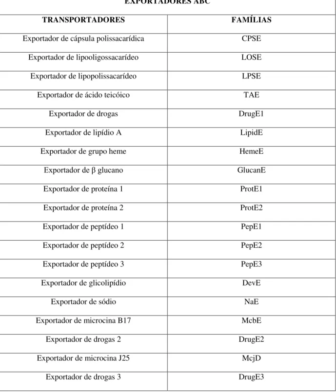
Tabela 2: Principais famílias de exportadores ABC presentes em procariotos. Modificado de Modificado de Milton; Saier, 2000.
EXPORTADORES ABC
TRANSPORTADORES FAMÍLIAS
Exportador de cápsula polissacarídica CPSE
Exportador de lipooligossacarídeo LOSE
Exportador de lipopolissacarídeo LPSE
Exportador de ácido teicóico TAE
Exportador de drogas DrugE1
Exportador de lipídio A LipidE
Exportador de grupo heme HemeE
Exportador de glucano GlucanE
Exportador de proteína 1 ProtE1
Exportador de proteína 2 ProtE2
Exportador de peptídeo 1 PepE1
Exportador de peptídeo 2 PepE2
Exportador de peptídeo 3 PepE3
Exportador de glicolipídio DevE
Exportador de sódio NaE
Exportador de microcina B17 McbE
Exportador de drogas 2 DrugE2
Exportador de microcina J25 McjD
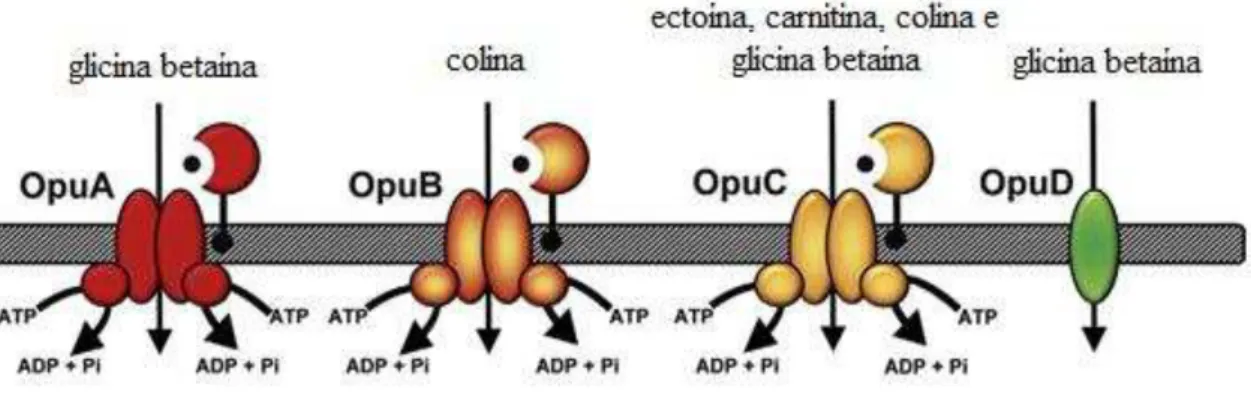
1.5. SISTEMA DE OSMOPROTEÇÃO
Os ambientes nos quais ocorrem variações frequentes do conteúdo de água, como por exemplo, camadas mais superficiais de solo, consistem em um desafio fisiológico para microrganismos e para que estes possam sobreviver em tais ambientes, eles necessitam de mecanismos adaptativos. Um desses mecanismos consiste em um sistema de importação de soluto compatível denominado sistema de importação osmoprotetora (Opu – osmoprotectant uptake) (Figura 6), que é formado por cinco membros, dos quais, três transportadores
pertencem à superfamília de proteínas ABC – OpuA (transporta glicina betaína e prolina betaína), OpuB (transporta apenas colina) e OpuC (transporta vários solutos compatíveis tais como glicina betaína, prolina betaína, carnitina, colina, -butirobetaina, colina-O-sulfato e ectoina), e os dois outros são os transportadores secundários OpuD e OpuE (DU et al., 2011).
Figura 6: Alguns membros do importador osmoprotetor. Modificado de Pittelkow et al., 2011.
As proteínas OpuA, OpuB e OpuC são, denominadas individualmente, como um sistema de osmoproteção (osmoprotectant system), muito comum em procariotos (KAPPES et
al. 1999). Dentre os seus membros, apenas OpuC apresenta uma grande diversidade de solutos transportados, incluindo ectoína e carnitina que são exclusivas deste transportador (DU et al., 2011). Entretanto o principal soluto a ser transportado é a glicina betaína pelo fato de apresentar uma elevada afinidade por essa molécula (CHEN; BEATTIE, 2007).
Outro fator observado quanto ao sistema de osmoproteção é que este transporta essencialmente aminas trimetiladas, o que pode sugerir uma ancestralidade comum para este sistema e que este tipo de soluto deve ter sido transportado desde o seu surgimento.
de salinidade do ambiente, especialmente em condições de elevada salinidade (ROBERTS, 2005). Esta especificidade no transporte deste tipo de amina está diretamente relacionada à presença da subunidade ligadora de substrato presente nos membros deste sistema (DAVIDSON et al., 2008).
1.6. SUBUNIDAES LIGADORAS DE SUBSTRATO
Proteínas (subunidades) ligadoras de substrato (SBP – substrate binding protein) ou
domínios de ligação ao substrato formam uma classe ou domínio de proteínas que são frequentemente associados a complexos proteicos de transporte de substâncias ou transdução de sinal ligados à membrana. Foram inicialmente associadas apenas às proteínas ABC, no entanto, atualmente já há comprovações de sua associação com outros complexos proteicos de membrana. Uma característica marcante destas moléculas é que elas apresentam pouca semelhança, no que diz respeito à sequência de resíduos de aminoácidos, mas suas estruturas tridimensionais são altamente conservadas. O núcleo destas moléculas é formado por dois domínios estruturalmente conservados sendo, entre estes domínios, a região de ligação do substrato, que ao se ligar, estabiliza a estrutura fechada da molécula (BERNTSSON et al., 2010).
Em bactérias gram-negativas, as subunidades ligadoras de substrato estão localizadas no periplasma celular, enquanto que em gram-positivas e arqueas estão ancoradas em lipídios de membrana ou fundidas aos domínios transmembrana (SMITS et al., 2008).
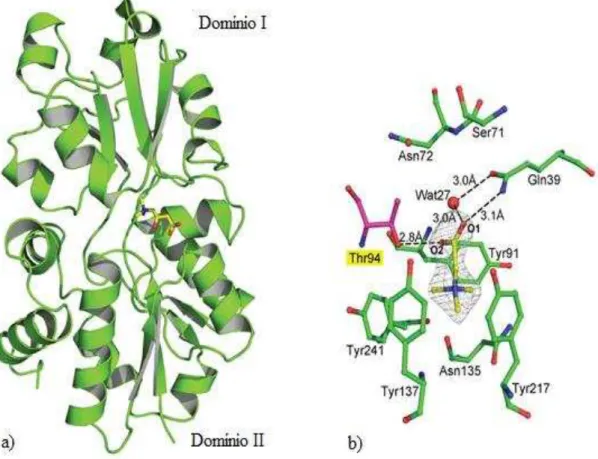
Quanto à estrutura das subunidades ligadoras de substrato características do sistema de osmoproteção, a região responsável pelo reconhecimento do substrato apresenta um “cinturão aromático” que é onde se encaixa a porção carregada postivamente da amina trimetilada
Figura 7: a) A subunidade ligadora de susbtrato de OpuC ligado à carnitina; b) participação dos resíduos aromáticos e não aromáticos na acomodação da glicina betaína em OpuC. Modificado de Du et al., 2011.
1.7. ANÁLISE COMPARATIVA DE GENES
A seleção natural promove a sobrevivência dos indivíduos mais aptos dentro de uma espécie. Ao longo de muitas gerações, isso pode resultar na manutenção de traços ancestrais (conservação através de seleção purificadora), ou o aparecimento de novas características benéficas (adaptação por seleção positiva). Ao nível genético, o efeito purificador ou positivo pode promover a evolução lenta ou rápida dos genes, o que permite identificar quais dessas forças evolutivas estão agindo sobre os seres vivos em um determinado ambiente (SHAPIRO; ALM, 2008).
O grau de seleção varia de acordo com o papel de um determinado gene na sobrevivência celular. Genes com pouca contribuição para a manutenção da homeostase celular são mais suscetíveis à inativação e mutações deletérias, enquanto que aqueles que são essenciais para o perfeito funcionamento do organismo são menos susceptíveis. Além disso, o grau da seleção que está atuando sobre um gene em particular, pode mudar ao longo do tempo e de acordo com um determinado contexto ecológico, por exemplo, a inativação ou perda de um ou mais genes pode aumentar o valor adaptativo de outros e as alterações na ecologia ou estilo de vida de um organismo podem tornar alguns genes redundantes. Tão importante quanto a intensidade de seleção é a sua eficácia, a qual depende, dentre outros fatores, do tamanho e estrutura da população. Em grandes populações, mutações deletérias em genes benéficos não são susceptíveis de fixar-se por acaso, mas em pequenas populações, mesmo genes úteis pode ser inativados e excluídos. Assim, os organismos que apresentam tamanhos populacionais pequenos são mais propensos à deterioração e perda de genes, mesmo para os que apresentam uma função benéfica para a célula (OCHMAN; DAVALOS, 2006).
incorporado. Genes que interferem em processos metabólicos periféricos ou que interagem, até mesmo complementando a atividade de genes naturais do genoma, são mais susceptíveis a serem fixados (ABBY; DAUBIN, 2007).
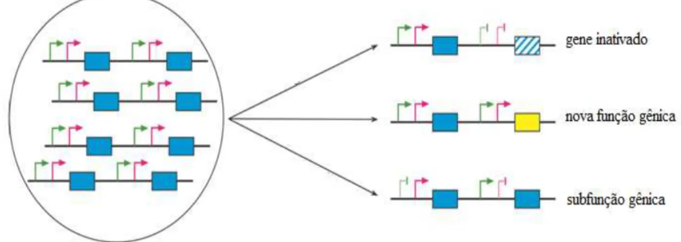
Existem três possibilidades para um gene que foi duplicado e fixado no genoma (Figura 8): a inativação, o desenvolvimento de uma nova função ou a manutenção do padrão de expressão do gene original, que será dividido entre as duplicatas que atuarão complementando uma à atividade da outra, o que caracteriza uma subfunção gênica (HURLES, 2004)
Figura 8: Possibilidades para um gene duplicado e fixado em um genoma. Modificado de Hurles, 2004.
O aumento do material genético representa também um aumento no custo energético para a maioria dos organismos. Desta forma, qual o motivo pelo qual os procariotos, normalmente retém os genes duplicados? A resposta a esta pergunta está nas novas funções que podem ser desempenhadas pelas duplicatas e que podem por exemplo permitir a adaptação destes organismos em ambientes estressantes (ZHANG, 2003)
A capacidade de adaptação é fundamental para a sobrevivência do organsimo em um ambiente estressante e para que o gene duplicado não seja suprimido, deve apresentar uma mutação que o torne vantajoso diante das novas condições ambientais, seja por responder diretamente a estas novas condições ou por apenas permitir um aumento na dosagem do gene como resposta a um aumento da procura de seu produto de transcrição (BRATLIE et al., 2010).
ancestral comum das espécies (REMM; STORM; SONNHAMMER, 2001), enquanto que
parálogos são genes que embora tenham um ancestal comum a duplicação que deu origem a estes genes ocorreu depois da especiação (BRATLIEet al., 2010).
A correta identificação de ortólogos é de fundamental importância tanto para os aspectos funcionais, quanto para os evolutivos da genômica comparativa, uma vez que, ortólogos ocupam o mesmo nicho funcional em diferentes organismos, por isso, esta categoria de genes é utilizada quando se quer fazer uma análise filogenética para averiguar a relação de parentesco entre espécies diferentes (MARAKOVA et al., 2007).
2. OBJETIVOS
2.1. OBJETIVO GERAL
Realizar uma análise evolutiva das subunidades ligadoras de soluto presentes no sistema de osmoproteção de procariotos.
2.2. OBJETIVOS ESPECÍFICOS:
Propor a história evolutiva deste sistema, analisando as possíveis estruturas moleculares ancestrais, as causas pelas quais este foi fixado e o tipo de seleção a que está sendo submetido.
O artigo Phylogenetic analysis of substrate-binding subunits in an osmoprotectant system apresenta uma análise evolutiva/filogenética das sequências nucleotídicas ortólogas de diversas subunidades ligadoras de substrato presentes em organismos procarióticos.
3. RESULTADO E DISCUSSÃO
Phylogenetic analysis of substrate-binding
subunits in an osmoprotectant system
Authors: Coutinho TJD¹, Rodrigues NF¹, and Farias ST¹.
1 – Departamento de Biologia Molecular, Universidade Federal da Paraíba, Brasil.
Abstract: Substrate-binding proteins are very important components of the solute uptake system, known as the osmoprotectant system, which consists of a membrane protein belonging to the ABC superfamily. These molecules recognize specific substrates that have different physiological roles in prokaryotes, i.e. roles that contribute to the survival of these organisms in environments with high concentrations of salt. Using MEGA software, this study performed a phylogenetic analysis of 431 nucleotide sequences of these subunits, orthologous to each other, collected from the database contained on the website http://www.genome.jp/kegg/. As a result of this analysis, phylogenetic trees were generated that clearly demonstrated that there was a horizontal transfer of some genes due to the sharing by different organisms. Also, two probable ancestral sequences were generated that showed homology with permeases that transport choline, glycine betaine and carnitine, which are trimethylamines currently present in various prokaryotes. Therefore, this system probably arose in prokaryotic organisms with the basic function of capturing nutrients, and by performing this basal function of being shared with other organisms, was fixed to the genome. However, because of the diversification of habitats by the prokaryotes, this system contributed decisively to the adaptation of these organisms to different environments, especially environments that had a high salt concentration; thus, acting and being currently characterized as a system of osmoprotection.
KEY WORDS: Transportation, Phylogenetic analysis, Glycine betaine, Osmoadaptation.
INTRODUCTION
Water is the most important molecule related to life and the capacity of an organism to cope with the internal variations of water for its survival. Water loss to the environment is a common threat, due to evaporation, excretion of waste and osmosis in various environments. As for water stress, organisms were traditionally divided into two groups: osmoconformers - usually using compatible solutes to maintain intracellular osmotic pressure equal to the outside environment; and osmoregulators – using ion transport to homeostatically regulate intracellular osmotic pressure (Yancey, 2005).
The types of organic molecules that are characterized as compatible solutes include: polyols and derivatives, sugars and derivatives, amino acids and derivatives, betaines, ecotins, and occasionally some peptides. Normally, bacteria and eukaryotes accumulate neutral solutes while archaea tend to modify many of the solutes accumulated by bacteria and eukaryotes, in negatively charged molecules. The fact that these molecules do not inhibit cellular functions, although they may act by modulating the activity of individual enzymes, was why they were designated as compatible solutes and their intracellular accumulation helps maintain cell turgidity, pressure and volume (Roberts, 2005).
In the natural environment, compatible solutes may be introduced into the medium after organism death, or by efflux processes which allow these molecules to become accessible to other organisms and be used for osmoadaptation or as a carbon source. In certain cases, capture systems are essential to the microorganisms that lack the biochemical machinery for synthesis of these compounds, and because of this, it is common to find transport systems (influx) with high affinity for these molecules that allow that they be rapidly imported and their intracellular levels be readily restored if there is a necessity or respond quickly to changes in salinity of the medium (Empadinhas and Da Costa, 2008).
Environments in which frequent variations occur in water content, for example, in the most superficial layers of soil, there is a physiological challenge for microorganisms, and for them to survive in such environments requires adaptive mechanisms. One such mechanism consists of a system for importing compatible solute called Osmoprotectant uptake (Opu), which is formed by five members, of which three transporters belong to the ABC superfamily of proteins (ATP-binding cassette) - OpuA (transports glycine betaine and proline betaine ), OpuB (transports only choline) and OpuC (transports several compatible solutes such as
glycine betaine, proline betaine, carnitine, choline, -butyrobetaine, choline-O-sulfate and ectoine), which use the energy released by ATP hydrolysis to import solutes (Du et al., 2011); a transporter of the BCCT family (betaine-choline-carnitine transporter) - OpuD that uses the energy of the driving force of sodium symport transport for importing solutes (Ziegler et al., 2010 ); and a transporter from the family of sodium/solute symport transporters - OpuE, which is specific for importing proline (Moses et al., 2012).
creating two categories related to the transport of substrate: exporters and importers. In the exporters, the TMDs and the NDBs are fused, while in the importers, besides the TMDs and NDBs being unfused subunits, there is also one more subunit which binds with high affinity to the substrates in order to deliver them to the TMDs (Oldham et al., 2008).
Of the subunits forming solute importers, the substrate-binding subunit SBP (substrate-binding protein) is extremely important for prokaryotes since it is fairly widespread in the genomes of these organisms (Thomas, 2010). Besides this, it provides an interesting structural diversity, since in Gram-negative bacteria it is located in the periplasmic space between the outer and inner membrane, and in Gram-positive bacteria it can be bound to lipoproteins of the extracellular face of the plasma membrane, to the transmembrane domain (TMD) of the ABC transporter (Davidson et al., 2008), or anchored in lipids of the external face of the plasma membrane (Biemans-Oldehinkel et al., 2006). On the other hand, in archaea there are lipoproteins of the plasma membrane or components of a transport system known as the bindosome.
Due to these multiple variations of SBPs, the degree of specificity for substrates and their distribution by the three Domains of life, this article aims to draw a phylogenetic profile of these subunits and to answer questions that will give us a better understanding of what happened over time with these molecules and how they were and are being selected by the environment.
MATERIALS AND METHODS
Data collection
This study selected and used 431 SBP gene sequences from the online database http://www.genome.jp/kegg/. These sequences correspond to SBPs belonging to the trimethylamine transporters (glycine betaine, proline betaine, carnitine, choline) and proline of prokaryotic organisms (bacteria, cyanobacteria and archaea) present in the most diverse environments.
B. subtillis 168 was used as reference organism, and from its SBP gene sequence, a
search for orthologous sequences was performed in the same database (Kanehisa et al., 2012; Kanehisa and Goto, 2000).
Sequence alignment and nucleotide substitution model
Sequence alignment was done using ClustalW, contained in the software Molecular Evolutionary Genetics Analysis (MEGA 5.05) (Higgins et al., 1994; Tamura et al., 2011), and used to test which evolutionary model would be consistent with the data.
Among the models offered by the software, the best suited to be used in phylogenetic reconstruction was GTR (General Time Reversible) with Gamma distribution with 2 parameters.
Phylogenetic analysis
All nucleotide substitution sites were used, the heuristic method was the Nearest-Neighbor-Interchange (NNI) (Das Gupta et al., 2000), and all codons (1st+2nd+3rd + non-coding) were used.
Additionally, as control, 16S ribosomal RNA gene sequences of the organisms were used.
Selection test
A Z-test of selection, with 1000 bootstrap replications, was conducted based on codons, using MEGA 5.05 (Tamura et al., 2011), where the model was the Nei-Gojobori method - Proportion (Saitou and Nei, 1987) with synonymous and non-synonymous type substitutions.
The null hypothesis tested was H0: dN = dS, where dN is the number of
non-synonymous substitutions per non-non-synonymous sites, whereas dS is the number of
synonymous substitutions per synonymous sites. For this hypothesis, three alternative hypotheses were tested: (a) dN ≠ dS (test for neutrality), (b) dN >dS (positive selection), and (c)
dN< dS (purifying selection) - which are only validated if the p value is less than 0.05.
Ancestral sequences
Hypothetical ancestral sequences were generated from MEGA (Tamura et al., 2011) using the statistical method of maximum likelihood and the GTR nucleotide substitution model (General Time Reversible) with Gamma distribution with 2 parameters (Tamura et al., 2011), which were analyzed using the online tool BLASTN (for nucleotides) and BLASTP (for amino acids) present on the site http://www.ncbi.nlm.nih.gov/ to find some probable similarity to an actual subunit. Besides this analysis, three-dimensional models were also generated through the site http://swissmodel.expasy.org, according to their standard parameters, for comparison with current models.
Analysis of codon usage
SBPs sequences were analyzed and genomes of the organisms selected with regard to codon usage and for this, the database found on the website http://www.kazusa.or.jp/codon/ was used.
RESULTS
Phylogenetic analysis
Figure 1: Organisms, gene annotation of the SBP in KEGG and type of solute transported (P-GB: proline and glycine betaine; C: choline; C-GB: choline and glycine betaine; C-C-GB: carnitine, choline and glycine betaine; When the type of solute is not specified, it is because only glycine betaine is transported).
In the analysis of this tree, there were two ways of organism grouping observed: a grouping of phylogenetically related organisms, which would be the most anticipated; and also a grouping of phylogenetically unrelated organisms, which suggests, in this second case, horizontal gene transfer. An example of this multiple form of grouping occurs with a non-virulent soil bacteria from the family Rizhobiaceae known as Agrobacterium radiobacter
K84, which has a main chromosome or chromosome 1 and a secondary chromosome, chromosome 2, which can also be regarded as a mega-plasmid (Slater et al., 2009). Chromosome 1 has the gene Arad_0755, whose ID in NCBI is 7368963, which grouped with phylogenetically related organisms (Figure 2). On the other hand, in chromosome 2, there are the genes Arad_8392 and Arad_8402, whose ID in NCBI are respectively 7366822 and 7366831, which clustered with phylogenetically unrelated organisms (Figure 3) and (Figure 4), indicating that there was probably a horizontal transfer of chromosome 2.
Selection test
There was no significant p value (<0.05), suggesting that any type of difference or meaning seen in this data set is due to chance, although, it was perceived that there is a tendency for purifying selection (Table 1).
Table 1: Codon-based Z-test of selection
Alternative hypothesis Valores de p
dN ≠ dS (test for neutrality) 0.271
dN > dS (positive selection) 1.000
dN < dS (purifying selection) 0.129
Ancestral sequences
Two ancestral sequences were generated derived from the first cladogenesis that probably originated all current SBP sequences and that were identified as Ancestral_1 and Ancestral_2 (SUPPLEMENTAL MATERIAL). Data analysis using the tool BLASTN showed that Ancestral_1 was similar to the periplasmic subunit that binds to glycine betaine and choline of Desulfosporosinus orientis DSM 765 (Table 2), whereas the amino acid
sequence for the same ancestral showed similarity with trimethylamine transporters, especially permeases (Table 3) with domains of OpuBB and OpuBC conserved and that are characteristic domains of choline transporters (Marchler-Bauer et al., 2011; Pittelkow et al., 2011) (Figure 5). As for Ancestral_2, the nucleotide sequence showed no similarity with any transporter; however, the amino acid sequence showed similarity to permeases (Table 4) and conserved domains (Figure 6) similar to Ancestral_1.
Three-dimensional models
In accordance with the Swiss-Model, two possible models were generated for Ancestral_1, of which only model 1 showed similarity with ABC transporters of compatible solutes, more specifically with choline - glycine betaine - carnitine transporters (Figure 7). As for Ancestral_2, five possible models were generated, of which only Ancestral_5 was compatible with ABC transporters of compatible solutes that, similar to Ancestral_2, showed similarity with choline - glycine betaine – carnitine transporters (Figure 8).
Codon usage
Table 2: BLASTN of Ancestral_1 – sequences significant alignments
Acession Description Max. score Total score Query coverage E value Max. ident
CP003108.1
Desulfosporosinus orientis DSM 765,
complete genome 152 152 18% 1e-32 75%
Table 3: BLASTP of Ancestral_1 – sequences significant alignments
Acession Description Max. score Total score Query coverage E value Max. ident
YP_003967851.1
binding-protein-dependent
transport system inner membrane 96.7 96.7 28% 2e-19 31%
YP_001425237.1
glycine betaine transport system permease [Coxiella burnetii Dugway
5J108-111] 98.6 98.6 30% 1e-18 35%
ZP_01946271.2
ABC transporter, quaternary
amine uptake transporter (QAT) 98.6 98.6 30% 1e-18 35%
YP_001596136.1
quaternary amine ABC transporter substrate-binding protein [Coxiella
burnetii RSA 331] 95.5 95.5 30% 9e-18 35%
YP_002304237.1
glycine betaine transport system permease protein [Coxiella burnetii
Table 4: BLASTP of Ancestral_2 – sequences significant alignments
Acession Description Max. score Total score Query coverage E value Max. ident
YP_003967851.1
binding-protein-dependent transport system inner membrane protein
[Ilyobacter polytropus DSM 2926] 108 108 30% 3e-23 31%
ZP_01946271.2
ABC transporter, quaternary amine
uptake transporter (QAT) 105 105 30% 8e-21 34%
YP_001425237.1
glycine betaine transport system permease [Coxiella burnetii Dugway
5J108-111] 104 104 30% 1e-20 34%
YP_002507602.1
glycine betaine ABC transporter substrate-binding protein
[Clostridium cellulolyticum H10] 103 103 59% 3e-20 28%
ZP_08192210.1
Substrate-binding region of ABC-type glycine betaine transport system
[Clostridium papyrosolvens DSM 2782]
Figura 7: a) three-dimensional model of Ancestral_1; b) crystal structure of glycine betaine/carnitine/choline ABC transporter; c) overlay of Ancestral_1 with crystal structure of glycine betaine/carnitine/choline ABC transporter.
Table 5: Codon usage
Organism Genome codon usage SBP codon usage
Rhizobium etli CFN 42
UUC UAU CUG CAG AUC AAG GUC GAC UCG UGG CCG CGC ACC AGC GCC GGC
UUC UAU CUG CAG AUC AAG GUC GAC UCG UGG CCG CGC ACC AGC GCC GGC
Bacillus subtilis
UUU UAU CUG CAA AUU AAA GUU GAA UCA UGG CCG CGC ACA AGC GCA GGC
UUU UAU CUG CAA AUU AAA GUU GAA UCA UGG CCG CGC ACA AGC GCG GGC
Agrobacterium tumefaciens C58
UUC UAU CUG CAG AUC AAG GUC GAA UCG UGC CCG CGC ACC AGC GCC GGC
UUC UAU CUG CAG AUC AAG GUC GAA UCG UGC CCG CGC ACC AGA GCC GGC
Leifsonia xyli subsp. xyli
UUC UAC CUC CAG AUC AAG GUC GAG UCC UGG CCG CGC ACC AGC GCC GGC
UUC UAC CUC CAG AUC AAG GUC GAG UCC UGG CCG CGC ACC AGC GCC GGC
Yersinia pseudotuberculosis IP
32953
UUU UAU CUG CAG AUU AAA GUG GAU UCA UGG CCG CGU ACC AGC GCC GGU
UUU UAU CUG CAG AUU AAA GUG GAU UCU UGG CCG CGU ACC AGC GCU GGU
Lactococcus lactis subsp. Lactis Il 1403
UUU UAU CUU CAA AUU AAA GUU GAA UCA UGG CCA CGU ACA AGU GCU GGA
DISCUSSION
By analyzing the tree generated from the SBP sequences, it was noted that the trimethylamine and proline import system, formed by members of the ABC family of proteins, is well distributed in the prokaryotic world, since these subunits are present in organisms that live in very different environments that are not necessarily subjected to adverse salt conditions.
This analysis allows us to infer other possible physiological interactions of the osmoprotectant system, since it seems that this system, well distributed by different environments, could be related to other mechanisms besides being responsible for the osmotic response.
When subjected to osmotic stress, prokaryotic organisms activate a particular transcription factor known as σB that is responsible for the physiological response to stress.
This factor triggers the transcription of approximately 150 genes, some directly and others indirectly. Under such conditions, the transcription levels of osmoprotectant uptake (OpuA - OpuE) are changed, rapidly increasing protein synthesis of this system in the first moments of the response to osmotic stress (Hahne et al., 2010).
However, there is no consensus on the primary activation of the osmoprotectant system by the σB transcription factor (Höper et al., 2005). There are inclusive studies that
indicate that the osmoprotectant system, or at least members of this system, is activated by other transcription factors (Petersohn et al., 2001; Hecker et al., 2007).
Another factor that contributes to a better understanding of the possible physiological roles that the osmoprotectant system plays is the fact that this system has a tendency to purifying selection. This type of selection plays an important role in maintaining the long-term stability of biological structures by removing deleterious mutations, thereby allowing the system to be improved and not run the risk of losing this improvement (Loewe, 2008). This is to say, in the case of the osmoprotectant system, in our view, because of its tendency to this type of selection, to be in organisms present in different habitats and apparently not be
directly controlled by the σB transcription factor, it must have been fixed with a different
function of the osmoprotector, but that actually works for osmoprotection.
As for the codon usage analysis, it was noticed that the SBP sequences accompany their respective genomes, since the codon usage coincides. This information allows us to infer that the system comprises genes native to the genome or that were horizontally transferred a long time before, and therefore tend to have a codon usage similar to the genome (Davis and Olsen, 2011).
small structural changes allowed other solutes to be transported; however, these changes did not affect the basic characteristics of the system.
The osmoprotectant system, according to our studies, consists of a system that has arisen with the function of nutrient uptake, the principle of choline, which is an important molecule for the biosynthesis of membrane phospholipids (Aktas et al., 2010) that acts as a precursor of glycine betaine (Burkhardt et al., 2009) and that participates in metabolic pathways for the synthesis of amino acids such as methionine (Barra et al., 2006). This system over time has been modified, allowing not only transport of a variety of molecules, but also structurally modified so that it now has a morphological diversity, making it very interesting with regard to its SBPs.
The transport of trimethylamines seems to be the hallmark of this system that, even after suffering some changes along the evolutionary process, did not lose the specificity for these molecules, but on the contrary, was enhanced, and among the trimethylamines transported by this system, choline and glycine betaine have a prominent role.
This transport system is currently much more related to glycine betaine than to choline, especially when characterized as a cellular osmoprotection system. However, as suggested, the osmoprotection was not responsible for fixing the system, but for the need to capture nutrients, though, at the beginning of the diversification of this and other systems, organisms that had the possibility of direct capture of glycine betaine showed an advantage over those that captured choline for the synthesis of glycine betaine.
This advantage can be explained by the fact that transporting glycine betaine is more energetically advantageous, since for the organism it is no longer necessary to synthesize two enzymes and to perform two chemical reactions responsible for the catabolism of choline and glycine betaine (Burkhardt et al., 2009) (Figure 9). This gain in terms of energy must have been fundamental to successful glycine betaine transport and sharing of this transport system among various prokaryotes.
There are at least three membrane proteins associated with the transport of glycine betaine: OpuA (osmoprotectant system), OpuC (osmoprotectant system) and OpuD (Pittelkow et al., 2011). However, the only one that is consensually synthesized from the activation of σB
is OpuD (Hahne et al., 2010; Hecker et al., 2007), suggesting a direct action of OpuD in response to osmotic stress, whereas members of the osmoprotectant system act in a complementary manner.
system must act, when required, as a secondary osmoprotection system due to the fact that glycine betaine, in particular, has physico-chemical properties for this type of physiological response, besides also acting as raw material in the metabolic pathway of amino acid synthesis, because under stress protein synthesis is increased to maintain cell physiology, i.e. in one way or another, there is the involvement of the osmoprotectant system in the response to osmotic stress, but not as the main agent of this response (Figure 10).
Figure 10: Physiological diversity of choline and glycine betaine. GB absorption became more energetically advantageous due to multiple activities.
CONCLUSION
Therefore, according to our understanding, the osmoprotectant system was fixed from natural selection due to the fact it plays a fundamental physiological function, which is to capture nutrients. Structural changes, among other things, allowed the system to transport different types of molecules without loss of function.
This system probably arose and was horizontally transferred before prokaryotic elicitation to diverse habitats, and because of this, its codon usage pattern coincides with the genome and is currently present in organisms inhabiting different environments.
As for the property of osmoprotection, it must have arisen secondarily, as the σB
transcription factor seems to have no direct effect on the transcription of the system, but in environments with varying salinity, this system has proven important for maintaining organism homeostasis.
REFERENCES
Barra L, Fontenelle C, Ermel G, Trautwetter A, et al. (2006). Interrelations between glycine betaine catabolism and methionine biosynthesis in Sinorhizobium meliloti strain 102F34. J Bacteriol.188:7195–7204.
Biemans-Oldehinkel E, Doeven MK, Poolman B (2006). ABC transporter architecture and regulatory roles of accessory domains. FEBS Lett. 580: 1023–1035.
Burkhardt J, Sewald X, Bauer B, Saum SH et al. (2009) Synthesis of glycine betaine from choline in the moderate halophile Halobacillus halophilus: co-regulation of two divergent, polycistronic operons. Environ Microbiol Rep. 1: 38–43.
Das Gupta B, He X, Jiang T, Li M, et al. (2000). On Computing the Nearest Neighbor Interchange Distance. In: Proceedings of the DIMACS Workshop on Discrete Problems with Medical Applications, DIMACS Series in Discrete Mathematics and Theoretical Computer Science. Am. Math. Soc. 55:125–143.
Davidson AL, Dassa E, Orelle C, Chen J (2008) Structure, function, and evolution of bacterial ATP-binding cassette systems. Microbiol Mol Biol Rev. 72:317–364.
Davis JJ and Olsen GJ (2011). Characterizing the native codon usages of a genome: an axis projection approach. Mol Biol Evol. 28(1):211–221.
Du Y, Shi WW, He YX, Yang YH,et al. (2011). Structures of the substrate-binding protein provide insights into the multiple compatible solute binding specificities of the Bacillus subtilis ABC transporter OpuC. Biochem J. 436(2):283-289.
Empadinhas N and da Costa MS (2008). Osmoadaptation mechanisms in prokaryotes: distribution of compatible solutes. International Microbiology 11(3):151-161.
Hahne H, Mader U, Otto A, Bonn F, et al. (2010) A comprehensive proteomics and transcriptomics analysis of Bacillus subtilis salt stress adaptation. J Bacteriol. 192: 870–882.
Hecker M, Pane-Farre J, Völker U (2007). SigB-dependent general stress response in Bacillus subtilis and related gram-positive bacteria. Annu. Rev. Microbiol. 61:215–236.
Higgins D, Thompson J, Gibson T, Thompson JD, et al. (1994) CLUSTAL W: improving the sensitivity of progressive multiple sequence alignment through sequence weighting, position-specific gap penalties and weight matrix choice.
Nucleic Acids Res. 22:4673-4680.
Kanehisa M, Goto S, Sato Y, Furumichi M, et al. (2012). KEGG for integration and interpretation of large-scale molecular datasets. Nucleic Acids Res. 40 (1):D109-D114.
Kanehisa M and Goto S (2000). KEGG: Kyoto Encyclopedia of Genes and Genomes. Nucleic Acids Res. 28: 27–30.
Loewe L. (2008). Negative selection. Nature Education 1(1).
Moses S, Sinner T, Zaprasis A, Stöveken N, et al. (2012). Proline utilization by Bacillus subtilis: uptake and catabolism. J. Bacteriol. 194:745-758.
Oldham ML, Davidson AL, Chen J (2008). Structural insights into ABC transporter mechanism. Curr Opin Struct Biol. 18(6):726-733.
Petersohn A, Brigulla M, Haas S, Hoheisel JD, et al. (2001) Global analysis of the general stress response of Bacillus subtilis. J Bacteriol. 183: 5617–5631.
Pittelkow M, Tschapek B, Smits SH, Schmitt L, et al. (2011). The Crystal Structure of the Substrate-Binding Protein OpuBC from Bacillus subtilis in Complex with Choline. J Mol Biol. 411:53-67.
Roberts MF (2005). Organic compatible solutes of halotolerant and halophilic microorganisms. Saline Syst. 1:5.
Saitou N and Nei M (1987). The neighbor-joining method: A new method for reconstructing phylogenetic trees. Molecular Biology and Evolution. 4:406-425.
Slater SC, Goldman BS, Goodner B, Setubal JC, et al. (2009). Genome Sequences of Three Agrobacterium Biovars Help Elucidate the Evolution of Multi-Chromosome Genomes in Bacteria. J. Bacteriol. 191:2501 – 2511.
Tamura K, Peterson D, Peterson N, Stecher G, et al. (2011). MEGA 5: Molecular Evolutionary Genetics Analysis using maximum likelihood, evolutionary distance, and maximum parsimony methods. Molecular Biology and Evolution. 28(10):2731–2739.
Thomas GH (2010). Homes for the orphans: utilization of multiple substrate-binding proteins by ABC transporters.Mol Microbiol. 75:6-9.
4. CONCLUSÃO
Pelo fato de desempenhar uma atividade fisiológica fundamental, que é a captura de nutrientes, o sistema de osmoproteção, de acordo com nosso entendimento foi fixado nos mais diversos procariotos e as mudanças estruturais, dentre outros aspectos, permitiram que o sistemas transportasse tipos diferentes de moléculas sem que ocorresse perda de função. Este sistema provavelmente surgiu e foi transferido horizontalmente antes da irradiação procariótica para habitats diversos, por isso seu padrão de códons preferênciais coincide com o genoma estando atualmente presente em organismos que vivem em ambientes tão diferentes.
Uma vez que o fator de transcrição σB parece não exercer efeito direto sobre a
5. REFERÊNCIAS
ABBY, S; DAUBIN, V. Comparative Genomics and the Evolution of Prokaryotes. CHEN, T.H.; MURATA, N. Glycine betaine: an effective protectant against abiotic stress in plants.
Trends in Plant Science. v.13, p. 499-505, 2008. v. 15, p.135-141, 2007.
BARRA, L.; FONTENELLE, C.; ERMEL, G. TRAUTWETTER, A. et al. Interrelations between glycine betaine catabolism and methionine biosynthesis in Sinorhizobium meliloti
strain 102F34. Journal of Bacteriology. v.188, p. 7195–7204, 2006.
BERNTSSON, R. P.; SMITS, S. H.; SGIMITT, L.; SLOTBOOM, D. J.; POOLMAN, B. A Structural Classification of Substrate-binding Proteins. FEBS Letters. v. 12, p. 2606-2617, 2010.
BIEMANS-OLDEHINHEKl, E.; DOEVEN, M.K.; POOLMAN, B.. ABC transporter architecture and regulatory roles of accessory domains. FEBS Letters. v. 580, p. 1023–1035, 2006.
BOLEN, D.W.; BASKAKOV, I. V. The osmophobic effect: natural selection of a thermodynamic force in protein folding. Journal of Molecular Biology. v. 310, p. 955 – 963, 2001.
BRATLIE, M. S.; JOHANSEN, J.; SHERMAN, B. T.; HUANG, D. W.; LEMPICKI, R. A.; DRABLOS, F. Gene Duplications in Prokaryotes can be Associated with Environmental Adaptation. BMC Genomics. v. 11: 588, 2010.
CÁNOVAS, D.; VARGAS, C.; KNEIP, S.; MORÓN, M.J.; VENTOSA, A.; BREMER, E.; NIETO, J.J. Genes for the Synthesis of the Osmoprotectant Glycine Betaine from Choline in the Moderately Halophilic Bacterium Halomonas elongata DSM 3043. Microbiology. v. 146,
p. 455 – 463, 2000.
CHEN, C; BEATTIE, G. A. Characterization of the Osmoprotectant Transporter OpuC from Pseudomonas syringae and Demonstration that Cystathionine- -Synthase Domains Are
Required for Its Osmoregulatory Function. Journal of Bacteriology. v. 189, p. 6901-6912, 2007.