Severe tuberculosis requiring ICU admission*
Tuberculose grave com necessidade de internação em UTIDenise Rossato Silva, Marcelo Basso Gazzana, Paulo de Tarso Roth Dalcin
Abstract
Tuberculosis is a curable disease that can evolve to severe forms, requiring the treatment of the patients in an ICU, especially if there is a delay in the diagnosis or if it affects elderly patients, those on dialysis, or those with HIV infection or other states of immunosuppression, as well as in cases of multidrug resistant disease. Knowledge of the radiological presentation of the cases can help diagnose these severe forms, as can the introduction of new tests, such as the early detection of the etiological agent by PCR and chest CT, which favors the early initiation of treatment. In addition, the use of regimens without isoniazid and rifampin, as well as uncertain enteral absorption and low serum concentrations of antituberculosis drugs, can reduce the efficacy of treatment. For such patients, the prognosis is generally poor and mortality rates are high.
Keywords: Tuberculosis; Respiratory insufficiency; Respiration, artificial; Hospitalization.
Resumo
A tuberculose é uma doença curável que pode evoluir para formas graves com necessidade de tratamento dos pacientes em UTI, especialmente se essa não for diagnosticada em tempo ou se afetar pacientes idosos, aqueles em diálise e aqueles com infecção pelo HIV ou outros estados de imunossupressão, assim como nos casos de doença multirresistente. O conhecimento da apresentação radiológica dos casos pode auxiliar no diagnóstico dessas formas graves, assim como a introdução de novos testes, como a detecção rápida do agente por PCR e a TC de tórax, favorecendo o início precoce do tratamento. Além disso, o uso de esquemas sem isoniazida e rifampicina, a absorção entérica incerta e as baixas concentrações séricas das drogas antituberculose podem contribuir para a diminuição da eficácia do tratamento. O prognóstico desses pacientes geralmente é ruim, com elevadas taxas de mortalidade.
Descritores: Tuberculose; Insuficiência respiratória; Respiração artificial; Hospitalização.
* Study carried out at the Porto Alegre Hospital de Clínicas, Federal University of Rio Grande do Sul, Porto Alegre, Brazil. Correspondence to: Denise Rossato Silva. Rua Ramiro Barcelos, 2350, sala 2050, Santa Cecília, CEP 90035-903, Porto Alegre, RS, Brasil.
Tel. 55 51 2101-8241. E-mail: denise.rossato@terra.com.br Financial support: None.
Submitted: 30 January 2012. Accepted, after review: 4 May 2012.
Introduction
In most countries, tuberculosis remains a major public health problem. One third of the world’s population is estimated to be infected with Mycobacterium tuberculosis. Brazil ranks 22nd among the 22 countries with the highest reported incidence of tuberculosis, with 43 cases/100,000 population in 2010.(1)
A significant proportion of tuberculosis patients still have to be hospitalized, and in-hospital mortality remains high, with estimates ranging from 2% to 12%. Generally, such cases highlight the difficulty that patients have in gaining access to public primary health care, as well as the ineffectiveness of public primary health care.
However, some of these hospitalizations correspond to cases that are more severe, in which there is a real need to use hospital resources. In addition, in many of these severe cases, ICU admission is required.(2,3)
The objective of the present study was to review aspects related to tuberculosis in ICU patients, as well as their peculiarities.
Methodology
hospitalization; and respiratory distress syndrome. The inclusion criterion was as follows: accessible articles in Portuguese, English, Spanish, Italian, or French published up until October of 2011 and containing the abovementioned keyword combinations. We identified some studies of tuberculosis patients requiring ICU admission or with acute respiratory failure (or both), and most of them were case series or retrospective studies. Table 1 summarizes the characteristics of the major studies. Case reports were not included in the table.
Epidemiology
Some decades ago, respiratory failure resulting from tuberculosis was reported mainly in cases of miliary tuberculosis. In 1977, the first case series of respiratory failure in 16 patients with tuberculosis and fibrocavitary disease was described.(4) The reported frequency of acute respiratory failure in patients with active tuberculosis ranged from 1.5% to 5.0%,(4-8) although pulmonary tuberculosis is rarely the primary cause of this complication. A retrospective study conducted in the USA found that, of the 6,000 admissions to a medical ICU over a 15-year period, 61 (1%) were due to active tuberculosis and that, of those 61, 43 (70%) were due to respiratory failure.(9) A retrospective study conducted in Germany included 58 tuberculosis patients requiring ICU admission. Of those patients, 37.9% required mechanical ventilation (MV).(10)
In a study conducted in Brazil and investigating the pulmonary histopathological changes found in 3,030 autopsies of patients who died of acute respiratory failure, tuberculosis was diagnosed as an underlying disease in 110 cases (3.6%). (11) In a retrospective study conducted in the state of São Paulo, Brazil, over a 15-year period, respiratory failure accounted for 5.4% of the hospitalizations in patients with tuberculosis.(12) In another study, also conducted in São Paulo, 6.5% of the hospital admissions for tuberculosis were due to acute respiratory failure, and ICU admission was required in 8.5% of the cases.(13) An analysis of the hospitalizations for tuberculosis at a university hospital in the city of Porto Alegre, Brazil, found that 16.7% of the 311 cases of tuberculosis required ICU admission and that 15.4% developed respiratory failure requiring MV.(14)
Clinical, laboratory, and radiological
presentation
Among hospitalized patients with severe tuberculosis, the most common symptoms include fever, night sweats, weight loss, and cough.(10,15) In only one study was dyspnea the most commonly reported symptom.(6) The mean symptom duration before hospital admission was approximately 30 days in most of the studies.(5,10,16) The presence of extrapulmonary tuberculosis ranged from 19% to 64% of the cases.(6,10,15,16) Comorbidities, especially those related to immunosuppression, such as HIV infection, are considered risk factors for developing respiratory failure and requiring MV.(15)
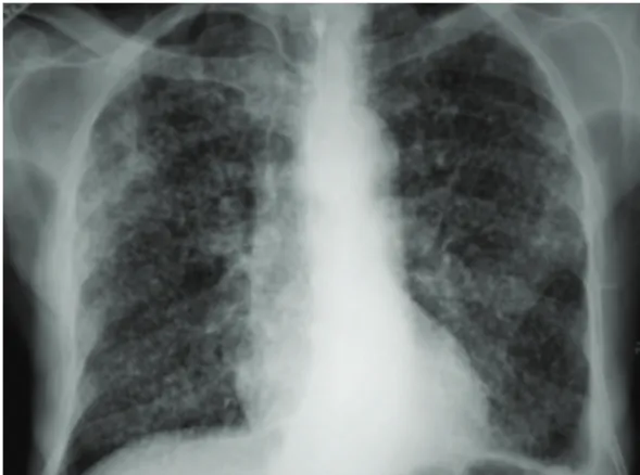
The most common radiological findings are reticular infiltrates and consolidation,(5,6,10,15,17) and cavitation can occur in 27-50% of cases. (6,10,16) Figures 1 to 4 show some of the main radiological patterns in this context.
The leading cause of ICU admission was respiratory failure, and Acute Physiology and Chronic Health Evaluation II (APACHE II) scores ranged from 13 to 23 in most of the studies. (5,6,10,15,17) Some authors evaluated the factors associated with the development of respiratory failure and the need for MV. Gram-negative pneumonia or sepsis, COPD, history of poor compliance with tuberculosis treatment, and cancer were predictors of respiratory failure.(9) In a series of 13 cases, 7 and 6 patients, respectively, had miliary/disseminated tuberculosis and tuberculous pneumonia requiring intensive care. Patients with miliary/disseminated tuberculosis were more likely to require MV than were those with tuberculous pneumonia (18.9% vs. 0.8%; p < 0.0001).(8)
Table 1 - Characteristics of the major studies included in the present review. Author Year Country/
number of centers
Type of study
Patients, n
Main results
Agarwal et al.(4) 1977 USA/1 Case series 16 5 deaths; survivors had severe restrictive lung
disease
Frame et al.(9) 1987 USA/1 Case series 43 An ARF incidence of 70% in hospitalized
patients with TB; an ICU mortality rate of 67%; a mortality rate of 81% in the patients with ARF
Levy et al.(7) 1987 USA/1 Cohort 15 An ARF incidence of 1.5% in hospitalized
patients with TB; time to treatment initiation: 3 ± 4 days; an in-hospital mortality rate of 33%; a post-discharge mortality rate of 47% Penner et al.(8) 1995 Canada/1 Cohort 13 A mortality rate of 69% in patients requiring
MV; patients with miliary TB were more likely to require MV than were those with tuberculous pneumonia (18.9 % vs. 0.8%) Zahar et al.(19) 2001 France/2 Retrospective
cohort
99 A TB/HIV co-infection rate of 38.4%; all patients had ARF; 50 patients (50.5%) required MV; 22 patients (22.2%) had ARDS; a 30-day mortality rate of 26.2%; factors associated with mortality: time from onset of symptoms to initiation of treatment > 1 month; number of failing organs; albumin > 20 g/L; large number of lobes involved, as seen on chest X-ray
Lee et al.(6) 2003 Taiwan/1 Retrospective
cohort
41 No HIV-positive patients; an in-hospital mortality rate of 65.9%; risk factors for mortality: consolidation on chest X-ray and multiple organ failure
Erbes et al.(10) 2006 Germany/1 Retrospective
cohort
58 22 patients (37.9%) required MV; an ICU mortality rate of 22.4%; factors associated with mortality: acute kidney injury; need for MV; chronic pancreatitis; sepsis; ARDS; and nosocomial pneumonia
Ryu et al.(5) 2007 South Korea/1 Retrospective
cohort
32 An in-hospital mortality rate of 59%; predictors of mortality: lungs destroyed by TB; APACHE II score ≥ 20; sepsis
Kim et al.(16) 2008 South Korea/1 Retrospective
cohort
90 An in-hospital mortality rate of 68.2% (pneumonia due to TB) and 58.3% (miliary TB); factors associated with mortality (in the group of patients with pneumonia due to TB): advanced age and shock unrelated to sepsis Lin et al.(17) 2009 Taiwan/1 Retrospective
cohort
59 An ICU mortality rate of 37.8%; factors associated with mortality: multiple organ failure syndrome and nosocomial pneumonia Silva et al.(15) 2010 Brazil/1 Retrospective
cohort
67 62 patients (92.5%) required MV; a TB/ HIV co-infection rate of 68.7%; an ICU mortality rate of 65.7%; factor associated with mortality: early ICU admission; protective factor: ventilator-associated pneumonia Lee et al.(35) 2011 South
Korea/10
Retrospective cohort
67 An ICU mortality rate of 58.2%; predictor of survival: SOFA score at the time of diagnosis of ARDS
The most common laboratory findings are anemia, leukopenia, leukocytosis, and hypoalbuminemia.(5,6,10,15) In a report of 6 cases of tuberculosis and acute respiratory failure, all of the patients were found to have anemia and hypoalbuminemia.(18)
Tuberculosis patients requiring intensive care can develop ARDS. The reported incidence of ARDS varies across studies: 12.1%(10); 13.4%(15); 28.1%(5); and over 60%.(6,16) In a case series of 15 hospitalized patients with tuberculosis and respiratory failure, it was found that, although the clinical and radiological characteristics were consistent with ARDS, the histopathological findings were consistent with tuberculous bronchopneumonia, with no evidence of ARDS. (7) In a study conducted in Brazil, the pulmonary histopathological changes found in autopsies of patients who died of acute respiratory failure
were reviewed, and the most common pattern associated with tuberculosis was diffuse alveolar damage.(11)
Difficulties in the diagnosis
Clinical characteristics and chest X-ray remain the main tools for the early diagnosis of active pulmonary tuberculosis. Mycobacterial culture takes 6-8 weeks. Therefore, the treatment of ICU patients can rarely be based on culture results. In addition, obtaining material for mycobacterial analysis can be difficult, especially in patients with extrapulmonary tuberculosis and in mechanically ventilated patients whose parameters preclude diagnostic procedures, such as bronchoscopy.
Although antituberculosis treatment is potentially toxic, it is recommended that patients admitted to an ICU with tuberculosis symptoms start receiving the medications before the results of diagnostic tests are available, given that delayed treatment initiation can result in death. In immunocompromised patients, the index of suspicion should be even higher.(19) Appropriate diagnostic investigation, as well as knowledge of the clinical and radiological presentations of severe tuberculosis, can contribute to earlier diagnosis and treatment initiation.
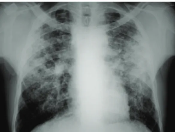
The time from onset of symptoms to initiation of antituberculosis treatment has been reported to be over 30 days in 28.8-34.0% of cases.(6,17) The time from admission to initiation of appropriate treatment was evaluated in only one study, which reported a mean of 4.3 days. In that retrospective study, the time from admission to initiation of treatment was shorter in patients with miliary tuberculosis than in those with tuberculous pneumonia (2.8 ± 2.5 days vs. 5.0 ± 7.0 days; p = 0.048).(16) There can be a delay in diagnosis and, consequently, in initiation of treatment because it is difficult to differentiate tuberculous pneumonia from severe bacterial pneumonia on X-rays. Considering this difficulty in distinguishing the two pathologies, one study evaluated the differences between ICU patients with severe pneumonia and tuberculosis and those with severe pneumonia without tuberculosis in terms of their clinical and radiological characteristics. Symptom duration longer than two weeks and the presence of micronodules or a cavitary pattern on chest X-ray were significantly associated with active pulmonary tuberculosis.(20) In addition, Figure 2 - Miliary pattern with an alveolar component.
a miliary pattern on chest X-ray can also be misinterpreted as congestive heart failure.(18)
In view of the aforementioned facts, the role of chest X-ray in the clinical diagnosis of pulmonary tuberculosis in ICU patients remains uncertain, and it is possible that chest X-ray does not contribute as much as expected. In a case-control study evaluating 89 patients with pulmonary tuberculosis and 89 controls, the most common radiological pattern was consolidation, which was found in 61 patients (6.5%). In the multivariate analysis, a history of pulmonary tuberculosis was associated with clinical suspicion of tuberculosis, although the radiological patterns were not associated with that suspicion.(21)
The introduction of new techniques, including early detection of the etiological agent by PCR, can aid in diagnosis and contribute to early initiation of treatment.(22) In addition, HRCT has been used in situations in which chest X-ray does not contribute to the diagnosis of active disease, such as in cases of minimal parenchymal changes and in the differentiation of old fibrotic lesions from those that are characteristic of bronchogenic dissemination.(22,23) Because HRCT allows the initiation of antituberculosis treatment before culture results are available, it is especially useful in patients with negative sputum smears.(24-26)
Treatment-related aspects
Appropriate antituberculosis treatment is an important factor that can affect patient outcome. Higher mortality is found among patients who do not receive optimal treatment including isoniazid and rifampin.(10) Oftentimes, ICU patients
with tuberculosis require alternative regimens (without isoniazid and rifampin) for maintenance therapy during recovery from hepatotoxicity. In addition, in critically ill patients, uncertain enteral absorption can occur.(27) Low serum concentrations of antituberculosis drugs have been associated with adverse outcomes, such as treatment failure, recurrence, acquired resistance, and death.(28-30) Furthermore, low serum albumin levels, which are common among ICU patients with tuberculosis, can impair drug absorption and are associated with reduced concentrations of rifampin and ethambutol.(31)
In the treatment of tuberculosis, corticosteroids are used as adjuvants, especially in extrapulmonary forms of the disease, such as meningeal and pericardial tuberculosis. Corticosteroids act by inhibiting the release of lymphokines and cytokines, which are responsible for constitutional symptoms and tissue damage. In addition, they allow antituberculosis drugs to penetrate granulomas, disrupting them.(32,33) Studies have suggested that corticosteroids can lead to faster resolution of pulmonary infiltrates and to cavity closure, especially in patients with more severe disease. (6,10,16,17) The beneficial effects of corticosteroids on the management of pulmonary tuberculosis with respiratory failure have been described in several case reports. However, such effects have yet to be evaluated in prospective studies, especially in randomized clinical trials. In general, corticosteroid use is considered for selected patients with severe forms of pulmonary tuberculosis, usually for those who develop ARDS.(6,10,16,17) There has been only one study(16) in which the mortality rate was found to be lower in patients with pulmonary
tuberculosis and respiratory failure who used corticosteroids (OR = 0.54; p = 0.011). However, mainly because of the retrospective nature of the study, the data presented do not allow us to conclude that corticosteroid use can be useful in patients with pulmonary tuberculosis.
Complications
In addition to the previously described development of ARDS, ICU patients with tuberculosis can develop other complications, such as ventilator-associated pneumonia (VAP), multiple organ failure, septic shock, acute kidney injury, disseminated intravascular coagulation, and digestive bleeding.(5,6,10,15-17) The incidence of these complications varies greatly across studies. The reported incidence of VAP is approximately 30% in most studies,(5,10,15) being 49.2% in one study. (17) The most commonly isolated pathogens are Pseudomonas aeruginosa, Acinetobacter baumannii, Acinetobacter spp., Staphylococcus aureus, and Stenotrophomonas maltophilia.(10,17)
Mortality and prognostic factors
In-hospital mortality of tuberculosis patients remains high, especially among those requiring ICU admission. Acute respiratory failure caused by tuberculosis and requiring MV has been associated with mortality rates ranging from 17.5% to 81.0%.(6,8-10,12,13,15)
Even after hospital discharge, mortality remains high. In a case series of 15 hospitalized patients with tuberculosis and respiratory failure, ICU mortality was 33% and 3-month post-discharge mortality was 47%.(7) In a retrospective cohort study,(15) with a 1-year follow-up period, post-discharge mortality was 17.4%, and the deaths occurred, on average, 3.7 months following discharge.
Some studies have reported factors that can contribute to mortality among critically ill tuberculosis patients. Disseminated disease, usually in the context of HIV infection, has been recognized as an important predictor of death. Other factors that can influence mortality rates include the presence of extensive fibrocavitary disease and consolidations on chest X-ray. Sepsis, ARDS, multiple organ failure, acute kidney injury, and nosocomial pneumonia also cause high mortality.(5,6,8,10) Likewise, a delay of more than 24 h in the initiation of treatment has been
reported to be associated with higher mortality (73.1% vs. 0%; p = 0.033).(17)
In a retrospective study over a 7-year period, 99 ICU patients with tuberculosis were evaluated and predictors of 30-day mortality were identified. Four factors were independently associated with mortality: time from onset of symptoms to initiation of treatment greater than 1 month (OR = 3.49; 95% CI: 1.20-10.20); number of failing organs (OR = 3.15; 95% CI: 1.76-5.76); large number of lung lobes involved, as seen on chest X-ray (OR = 1.83; 95% CI: 1.12-2.98); and serum albumin levels greater than 20 g/L (OR = 3.96; 95% CI: 1.04-15.10).(19) Serum levels of albumin and hemoglobin, reflecting the nutritional status, were found to be the best predictors of survival in a study of ICU patients with nonmiliary tuberculosis and respiratory failure.(34)
In the prognostic evaluation of such patients, APACHE II scores at ICU admission might underestimate the mortality rate among tuberculosis patients requiring MV. The median APACHE II score was found to be 16 in one study, a value that is usually associated with a mortality rate of 20-30% rather than with the 59% mortality rate reported in that study.(5) In two other studies,(15,17) the median APACHE II scores were 21.2 ± 6.5 and 22.8 ± 6.8, respectively, indicating a mortality rate of 30-40%. However, the mortality rates were found to be 67.8% and 65.7%, respectively. It is possible that events occurring during hospitalization, such as the development of VAP or other complications, contributed to the increased mortality rates.
In a multicenter study conducted in South Korea,(35) the APACHE II score was not a predictor of survival. In that study, the characteristics and outcomes of patients with ARDS caused by miliary tuberculosis were evaluated. A high in-hospital mortality and a high ICU mortality were found (61.2% and 58.2%, respectively), and Sequential Organ Failure Assessment score at the time of diagnosis of ARDS was found to be an important prognostic marker (OR = 0.809; 95% CI: 0.691-0.946; p = 0.008).
Biosafety
as isolation of suspected cases; b) environmental (or engineering) control measures—negative-pressure isolation rooms and use of high efficiency particulate air (HEPA) filters; and c) respiratory protection measures—use of N95 masks by the health care team.(22) Therefore, in the ICU setting, a high clinical suspicion is mandatory, with smear microscopy for AFB and culture of respiratory secretions in suspected cases. All suspected cases should be placed in individual negative-pressure rooms, and respiratory precautions should be implemented. Endotracheal suctioning without disconnection (closed system) should be used, and a bacterial filter should be placed in the expiratory line of the ventilator circuit.(36)
It is important that the recommendation for endotracheal intubation be emphasized. In suspected cases of tuberculosis, the procedure should be performed in a negative-pressure room with a ventilation rate of 6-12 air changes per hour. Vents that exhaust to the outside should be located away from windows/foot traffic or a HEPA filter system should be used. Health professionals should wear N95 masks or HEPA respirators while performing the intubation/ suctioning procedure, as well as while performing fiberoptic bronchoscopy.(36)
It is recommended that health professionals working in high-risk environments undergo tuberculin skin testing. Health professionals should be screened at the time of hiring, and periodic screening should be performed to identify those with tuberculin skin test conversion, i.e., with latent infection.(36,37) Those identified as such should be referred for treatment of latent infection in accordance with the consensus criteria.(22)
Final considerations
Tuberculosis patients requiring ICU admission or with respiratory failure have some peculiarities, especially regarding diagnosis and treatment. The diagnosis of tuberculosis in such cases is particularly complicated because of the possible misinterpretation of radiological findings, the difficulty in obtaining material for mycobacterial analysis, and the unavailability of culture results in most cases. Therefore, initiation of treatment is often delayed. In addition, the use of regimens without isoniazid and rifampin, as well as uncertain enteral absorption and low serum concentrations of antituberculosis drugs, can reduce the efficacy of treatment. For such patients, the prognosis
is generally poor and mortality rates are high. Measures aimed at promoting early diagnosis and treatment of tuberculosis in the public health care system, with special attention to patients at increased risk of developing respiratory failure and requiring intensive care, such as HIV-positive patients, can contribute to improving this situation.
References
1. World Health Organization [homepage on the Internet]. Geneva: World Health Organization. [cited 2011 Dec 1]. Global Tuberculosis Control. WHO Report 2011. Available from: www.who.int
2. Greenaway C, Menzies D, Fanning A, Grewal R, Yuan L, FitzGerald J, et al. Delay in diagnosis among hospitalized patients with active tuberculosis--predictors and outcomes. Am J Respir Crit Care Med. 2002;165(7):927-33. PMid:11934716.
3. Rao VK, Iademarco EP, Fraser VJ, Kollef MH. The impact of comorbidity on mortality following in-hospital diagnosis of tuberculosis. Chest. 1998;114(5):1244-52. PMid:9823996. http://dx.doi.org/10.1378/chest.114.5.1244
4. Agarwal MK, Muthuswamy PP, Banner AS, Shah RS, Addington WW. Respiratory failure in pulmonary tuberculosis. Chest. 1977;72(5):605-9. PMid:913139. http://dx.doi.org/10.1378/chest.72.5.605
5. Ryu YJ, Koh WJ, Kang EH, Suh GY, Chung MP, Kim H, et al. Prognostic factors in pulmonary tuberculosis requiring mechanical ventilation for acute respiratory failure. Respirology. 2007;12(3):406-11. PMid:17539846. http://dx.doi.org/10.1111/j.1440-1843.2006.01007.x 6. Lee PL, Jerng JS, Chang YL, Chen CF, Hsueh PR,
Yu CJ, et al. Patient mortality of active pulmonary tuberculosis requiring mechanical ventilation. Eur Respir J. 2003;22(1):141-7. PMid:12882464. http://dx.doi.org/ 10.1183/09031936.03.00038703
7. Levy H, Kallenbach JM, Feldman C, Thorburn JR, Abramowitz JA. Acute respiratory failure in active tuberculosis. Crit Care Med. 1987;15(3):221-5. PMid:3469061. http:// dx.doi.org/10.1097/00003246-198703000-00008 8. Penner C, Roberts D, Kunimoto D, Manfreda J, Long R.
Tuberculosis as a primary cause of respiratory failure requiring mechanical ventilation. Am J Respir Crit Care Med. 1995;151(3 Pt 1):867-72. PMid:7881684. 9. Frame RN, Johnson MC, Eichenhorn MS, Bower GC,
Popovich J Jr. Active tuberculosis in the medical intensive care unit: a 15-year retrospective analysis. Crit Care Med. 1987;15(11):1012-4. PMid:3677743. http://dx.doi. org/10.1097/00003246-198711000-00005
10. Erbes R, Oettel K, Raffenberg M, Mauch H, Schmidt-Ioanas M, Lode H. Characteristics and outcome of patients with active pulmonary tuberculosis requiring intensive care. Eur Respir J. 2006;27(6):1223-8. PMid:16481385. http:// dx.doi.org/10.1183/09031936.06.00088105
11. Soeiro Ade M, Parra ER, Canzian M, Farhat C, Capelozzi VL. Pulmonary histopathological alterations in patients with acute respiratory failure: an autopsy study. J Bras Pneumol. 2008;34(2):67-73. PMid:18345449. 12. Nogueira PA. Motivos e tempo de internação e o tipo
13. Ribeiro SA, Matsui TN. Hospitalização por tuberculose em hospital universitário. J Pneumol. 2003;29(1):9-14. http://dx.doi.org/10.1590/S0102-35862003000100004 14. Silva DR, Menegotto DM, Schulz LF, Gazzana MB,
Dalcin Pde T. Factors associated with mortality in hospitalized patients with newly diagnosed tuberculosis. Lung. 2010;188(1):33-41. http://dx.doi.org/10.1007/ s00408-009-9224-9
15. Silva DR, Menegotto DM, Schulz LF, Gazzana MB, Dalcin PT. Mortality among patients with tuberculosis requiring intensive care: a retrospective cohort study. BMC Infect Dis. 2010;10:54. PMid:20205952 PMCid:2843613. http:// dx.doi.org/10.1186/1471-2334-10-54
16. Kim YJ, Pack KM, Jeong E, Na JO, Oh YM, Lee SD, et al. Pulmonary tuberculosis with acute respiratory failure. Eur Respir J. 2008;32(6):1625-30. PMid:18614559. http://dx.doi.org/10.1183/09031936.00070907 17. Lin SM, Wang TY, Liu WT, Chang CC, Lin HC, Liu CY, et al.
Predictive factors for mortality among non-HIV-infected patients with pulmonary tuberculosis and respiratory failure. Int J Tuberc Lung Dis. 2009;13(3):335-40. PMid:19275793.
18. Heffner JE, Strange C, Sahn SA. The impact of respiratory failure on the diagnosis of tuberculosis. Arch Intern Med. 1988;148(5):1103-8. PMid:3130000. http://dx.doi. org/10.1001/archinte.1988.00380050107017 19. Zahar JR, Azoulay E, Klement E, De Lassence A, Lucet
JC, Regnier B, et al. Delayed treatment contributes to mortality in ICU patients with severe active pulmonary tuberculosis and acute respiratory failure. Intensive Care Med. 2001;27(3):513-20. PMid:11355119. http://dx.doi. org/10.1007/s001340000849
20. Hui C, Wu CL, Chan MC, Kuo IT, Chiang CD. Features of severe pneumonia in patients with undiagnosed pulmonary tuberculosis in an intensive care unit. J Formos Med Assoc. 2003;102(8):563-9. PMid:14569322.
21. Wu JY, Ku SC, Shu CC, Fan JY, Chen HY, Chen YC, et al. The role of chest radiography in the suspicion for and diagnosis of pulmonary tuberculosis in intensive care units. Int J Tuberc Lung Dis. 2009;13(11):1380-6. PMid:19861010.
22. Conde MB, Melo FA, Marques AM, Cardoso NC, Pinheiro VG, Dalcin Pde T, et al. III Brazilian Thoracic Association Guidelines on tuberculosis. J Bras Pneumol. 2009;35(10):1018-48. PMid:19918635. 23. Storla DG, Yimer S, Bjune GA. A systematic review of
delay in the diagnosis and treatment of tuberculosis. BMC Public Health. 2008;8:15. PMid:18194573 PMCid:2265684. http://dx.doi.org/10.1186/1471-2458-8-15
24. Im JG, Itoh H, Shim YS, Lee JH, Ahn J, Han MC, et al. Pulmonary tuberculosis: CT findings--early active disease and sequential change with antituberculous therapy. Radiology. 1993;186(3):653-60. PMid:8430169. 25. Oh YW, Kim YH, Lee NJ, Kim JH, Chung KB, Suh WH, et al.
High-resolution CT appearance of miliary tuberculosis. J
Comput Assist Tomogr. 1994;18(6):862-6. PMid:7962790. http://dx.doi.org/10.1097/00004728-199411000-00003 26. Hong SH, Im JG, Lee JS, Song JW, Lee HJ, Yeon KM.
High resolution CT findings of miliary tuberculosis. J Comput Assist Tomogr. 1998;22(2):220-4. PMid:9530383. http://dx.doi.org/10.1097/00004728-199803000-00011 27. Thompson JS. The intestinal response to critical illness.
Am J Gastroenterol. 1995;90(2):190-200. PMid:7847284. 28. Kimerling ME, Phillips P, Patterson P, Hall M, Robinson
CA, Dunlap NE. Low serum antimycobacterial drug levels in non-HIV-infected tuberculosis patients. Chest. 1998;113(5):1178-83. PMid:9596291. http:// dx.doi.org/10.1378/chest.113.5.1178
29. Mehta JB, Shantaveerapa H, Byrd RP Jr, Morton SE, Fountain F, Roy TM. Utility of rifampin blood levels in the treatment and follow-up of active pulmonary tuberculosis in patients who were slow to respond to routine directly observed therapy. Chest. 2001;120(5):1520-4. PMid:11713129. http:// dx.doi.org/10.1378/chest.120.5.1520
30. Weiner M, Benator D, Burman W, Peloquin CA, Khan A, Vernon A, et al. Association between acquired rifamycin resistance and the pharmacokinetics of rifabutin and isoniazid among patients with HIV and tuberculosis. Clin Infect Dis. 2005;40(10):1481-91. PMid:15844071. http://dx.doi.org/10.1086/429321
31. Tappero JW, Bradford WZ, Agerton TB, Hopewell P, Reingold AL, Lockman S, et al. Serum concentrations of antimycobacterial drugs in patients with pulmonary tuberculosis in Botswana. Clin Infect Dis. 2005;41(4):461-9. PMid:16028152. http://dx.doi.org/10.1086/431984 32. Muthuswamy P, Hu TC, Carasso B, Antonio M, Dandamudi
N. Prednisone as adjunctive therapy in the management of pulmonary tuberculosis. Report of 12 cases and review of the literature. Chest. 1995;107(6):1621-30. PMid:7781357. http://dx.doi.org/10.1378/chest.107.6.1621
33. Wallis RS. Reconsidering adjuvant immunotherapy for tuberculosis. Clin Infect Dis. 2005;41(2):201-8. PMid:15983916. http://dx.doi.org/10.1086/430914 34. Mehta JB, Fields CL, Byrd RP Jr, Roy TM. Nutritional status
and mortality in respiratory failure caused by tuberculosis. Tenn Med. 1996;89(10):369-71. PMid:8870488. 35. Lee K, Kim JH, Lee JH, Lee WY, Park MS, Kim JY, et al.
Acute respiratory distress syndrome caused by miliary tuberculosis: a multicentre survey in South Korea. Int J Tuberc Lung Dis. 2011;15(8):1099-103. PMid:21740675. http://dx.doi.org/10.5588/ijtld.10.0557
36. Jensen PA, Lambert LA, Iademarco MF, Ridzon R; CDC. Guidelines for preventing the transmission of Mycobacterium tuberculosis in health-care settings, 2005. MMWR Recomm Rep. 2005;54(RR-17):1-141.
About the authors
Denise Rossato Silva
Adjunct Professor of Pulmonology. Federal University of Rio Grande do Sul, Porto Alegre, Brazil.
Marcelo Basso Gazzana
Pulmonologist. Porto Alegre Hospital de Clínicas, Federal University of Rio Grande do Sul, Porto Alegre, Brazil.
Paulo de Tarso Roth Dalcin