ISSN 0102-695X DOI: 10.1590/S0102-695X2013005000055 Received 7 Jun 2013 Accepted 24 Jul 2013 Available online 20 Aug 2013 Revista Brasileira de Farmacognosia Brazilian Journal of Pharmacognosy 23(4): 651-661, Jul./Aug. 2013
Evaluation of antinociceptive and
anti-inl ammatory activities of extract and fractions
of
Eugenia jambolana
root bark and isolation
of phytoconstituents
Santanu Saha,
*,1Subrahmanyam E. V. S.,
2Chandrashekar
Kodangala,
3Shubhash C. Mandal,
4Shashidhara C. Shastry
51Department of Pharmacognosy, NGSM Institute of Pharmaceutical Sciences, Nitte
University, India,
2Department of Pharmaceutical Chemistry, Srinivas College of Pharmacy, Vallachil,
India,
3Department of Pharmacognosy, Manipal College of Pharmaceutical Sciences, Manipal
University, India,
4Department of Pharmaceutical Technology, Faculty of Engineering and Technology,
Jadavpur University, India,
5NGSM Institute of Pharmaceutical Sciences, Nitte University, India.
Abstract: Eugenia jambolana Lam., Myrtaceae, is a widely distributed and traditionally well known plant in India. The root bark of the plant was extracted with ethanol and then successively fractionated into petroleum ether fraction, chloroform fraction,
n-butanol fraction and methanol fraction. The extract and fractions of the plant material were evaluated for the antinociceptive activity by acetic acid-induced writhing test
and formalin-induced nociception test, and anti-inl ammatory activity was screened by
carrageenan-induced rat paw edema, cotton pellet induced granuloma formation and adjuvant induced arthritis in rat models. The test materials showed the antinociceptive and
anti-inl ammatory effect in dose dependent manner and the petroleum ether fraction was
found to be most potent among the test materials. At 400 mg/kg b.w., p.o. dose petroleum
ether fraction signii cantly inhibited 54.28% writhing response and 73.77% formalin induced nociception in mice. The fraction with same dose showed signii cant 79.31% inhibition of carrageenan-induced rat paw edema, 57.78% anti-proliferative effect and 77.93% inhibition of adjuvant induced arthritis. The bioactive petroleum ether fraction
was then subjected to column chromatography which led to isolate three compounds,
namely, β-sitosterol, stigmasterol and lupeol. These compounds were characterized and identii ed by IR, 1H NMR, 13C NMR and mass spectroscopy.
Keywords:
adjuvant induced arthritis anti-inflammatory antinociceptive formalin induced nociception
Eugenia jambolana
Myrtaceae
Introduction
Eugenia jambolana Lam., Myrtaceae, is a known Indian medicinal plant for traditional uses to treat different ailments. This large evergreen tree is distributed through out India and commonly known as ‘Jamun’. Root bark of the plant is pale brown at inner surface and from outside dark brown. Ayurveda and Unani system of medication have mentioned variety of therapeutic properties of the plant like astringent, carminative, diuretic, antidiabetic,
anthelmintic, antibacterial, analgesic, anti-inl ammatory,
antioxidant, as well as gastro protective agents (Kirtikar & Basu, 2006). Several phytoconstituents belonging to
category alkaloids, glycosides, l avonoids and volatile oil
has been reported from different parts of plant. Betulinic acid, ß-sitosterol, friedelin, epi-friedelanol and fatty acid ester of epi-friedelanol (eugenin) has been isolated from stem bark of E. jambolana (Sengupta & Das, 1965). Quercetin, myricetin, myricitrin, myricetin 3-O
different parts of this plant have also been reported earlier
(Ratsimamanga et al., 1972; Mukherjee et al., 1998;
Jagetia & Balinga, 2004; Sharma et al., 2006; Chaturvedi et al., 2007). Traditionally, barks of this plant are used
for treatment of inlammation (Nadkarni, 2009). Earlier studies reported the anti-inlammatory activity by stem
bark of Eugenia jambolana (Muruganandan et al., 2001).
As per our knowledge, there are no scientiic evidences available on antinociceptive and anti-inlammatory activity
of root bark. Hence, in this study we have evaluated the
antinociceptive and anti-inlammatory activity of ethanolic
extract and fractions of root bark of Eugenia jambolana
and isolated phytoconstituents from active fraction.
Materials and Methods
General methods
All the melting points were recorded in a Toshniwal melting point apparatus and were uncorrected. IR spectra of the compounds were recorded using the KBr pellet method
on a Perkin Elmer 700 IR spectrophotometer. NMR spectra
of the compound were taken on Bruker Avance II 400 NMR spectrometer in CDCl3, at 400 MHz for 1HNMR and
100.62 MHz for 13CNMR with tetramethylsilane (TMS) as internal standard. EIMS (Electron impact mass spectrum)
was taken on a Shimadzu LCMS-2010A. TLC was carried out using Silica gel 60 F254 plates (Merck). Column chromatography was carried out on neutral alumina of
70-300 mesh from S.D. ine chemicals Pvt. Ltd., Mumbai.
All the chemicals and reagents used were obtained in high
purity either from S.D. ine chemicals Pvt. Ltd; Bombay, India or E. Merck (India) Ltd., Mumbai.
Plant material
The root bark of Eugenia jambolana Lam., Myrtaceae, were collected from Derelakatte, Mangalore, during December 2009 and its botanical identity was
conirmed by Dr. Noeline J. Pinto Head of Botany
Department St. Agnes College, Mangalore. A voucher specimen (voucher no. 625e) has been deposited in NGSM Institute of Pharmaceutical Sciences, Derelakatte, Mangalore.
Extraction and fractionation of plant material
The shade dried powdered root barks (5 kg) were
extracted with ethanol (95%) by cold maceration method
for four times and the extract was concentrated by reduced pressure to yield 500 g of crude ethanolic extract. The ethanolic extract was suspended in distilled water (1:3, v/v) and successively partitioned with petroleum ether
(60-80 °C, 8x500 mL), chloroform (8x500 mL), n-butanol
(8x500 mL) and methanol (8x500 mL). The organic layers
were brought to dryness to yield petroleum ether (35 g), chloroform (50 g), n-butanol (55 g) and methanol (70 g) fractions.
Animals
Studies were carried out by using male Swiss
albino mice (18-25 g) and male albino Wistar rats (180-200
g). All animals were obtained from K.S. Hegde Medical Academy, Deralakatte, Mangalore. Animals were grouped and housed in polyacrylic cages and kept at ambient
temperature (25±2 ºC), 60±5% relative humidity and 12 h
light and dark cycle. They had been given standard pellet diet (Hindustan Lever Limited, Mumbai, India) and water
ad libitum throughout the course of the study. The study
protocols were approved by Institutional Animal Ethical Committee (KSHEMA /AEC/077/2008).
Chemicals and drugs
Carrageenan (Hi–Media Research Laboratories
Pvt. Ltd., Mumbai), tween 80 (S.D. ine Chemicals Pvt.
Ltd., Mumbai), Complete Freund’s adjuvant (FCA), morphine hydrochloride (Morphine) and indomethacin (Indo) (Sigma Aldrich St. Louis, USA). The ethanolic
extract (EJE) of root bark of E. jambolana and petroleum
ether fraction (EJPE), chloroform fraction (EJCE), n-butanol fraction (EJNB) and methanol fraction (EJME)
of ethanolic extract of root bark of E. jambolana were
used as a suspension in tween 80 (3 mL of 1% solution)
to screen biological activity. Indomethacin and morphine
hydrochloride were also used as a suspension in tween 80 (3 mL of 1% solution).
Acute toxicity study
Acute toxicity study was conducted to determine
the median lethal dose (LD50) of test materials EJE, EJPE, EJCE, EJNB and EJME, as described in previous study, in adult male albino Wistar rats and by following up and down procedure of OECD guideline no. 425 (OECD 425,
2001; Ranawat et al., 2010). Animals were administered by test material orally and observed at half hour intervals for 4 h, then after 24 h. Test materials were found to be safe up to 2000 mg/kg b. w., p.o. dose.
Selection of dose
The test materials (EJE, EJPE, EJCE, EJNB and EJME) did not exhibit any toxic effect up to 2000 mg/kg
Evaluation of antinociceptive and anti-inlammatory activities of extract and fractions of Eugenia jambolana root bark and isolation of
Santanu Saha et al.
Rev. Bras. Farmacogn. Braz. J. Pharmacogn. 23(4): Jul./Aug. 2013 of that one tenth dose, i.e. 200, 100 and 400 mg/kg b.w.,
p.o. doses, respectively. The antinociceptive and
anti-inlammatory activity studies were carried out by 100, 200 and 400mg/kg doses of all the test materials EJE, EJPE, EJCE, EJNB and EJME.
Antinociceptive activity studies in mice
Acetic acid-induced writhing in mice
Antinociceptive activity of drugs was evaluated
by the method described earlier (Collier et al., 1968). Male Swiss albino mice (18-25 g) were divided in eighteen
groups (n=10) and respectively treated orally with vehicle
(tween 80, 3 mL of 1% solution), indomethacin (Indo) 10
mg/kg b.w., p.o. and 100, 200 and 400 mg/kg b.w., p.o. of
EJE, EJPE, EJCE, EJNB and EJME at 1 h prior to acetic
acid injection. Morphine 1 mg/kg b.w. was administered intraperitoneally (i.p.) 30 min before the stimulus injection.
All the animals were injected with 0.6% v/v acetic acid
by intraperitoneal route and the numbers of writhe were counted over the period of 30 min. The percentage inhibition of number of writhings were calculated and compared with control group.
Formalin-induced nociception
This test was carried out as decribed by Hunskaar
& Hole (1987). In this experiment mice of respective
groups (n=10) were treated orally with indomethacin (Indo) 10 mg/kg b.w., p.o. and 100, 200 and 400 mg/kg b.w., p.o. of EJE, EJPE, EJCE, EJNB and EJME at 1 h prior to acetic acid injection. Animals of control group received
vehicle (tween 80, 3 mL of 1% solution). Morphine 10
mg/kg b.w. was administered intraperitoneally (i.p.).
After 1 h of drug treatment, 20 µL of 1% formalin (in 0.9% saline) were injected in dorsal surface of right hind
paw of all animals. The animals were placed immediately in transparent observation chambers to observe the time spent by animals in licking the injected paw during early phase (0-5 min) and late phase (20-30 min) after formalin injection.
Anti-inlammatory activity studies
Carrageenan induced rat paw edema
Acute anti-inlammatory activity of test materials (EJE, EJPE, EJCE, EJNB and EJME) were evaluated by
carrageenan induced rat paw edema study as described by
Winter et al. (1962). Paw edema was induced by injecting 0.1 mL of 1% (w/v) carrageenan suspension in 0.9%
(w/v) sterile saline into the plantar tissue of the left hind paw of albino wistar rats. Animals of respective groups
(n=8) were administered orally with vehicle (tween 80, 3
mL of 1% solution), indomethacin (Indo) 10 mg/kg b.w., p.o. and 100, 200 and 400 mg/kg b.w., p.o. of EJE, EJPE,
EJCE, EJNB and EJME at 1 h prior to the carrageenan
induced paw edema. The right paw served as a reference
to non-inlamed paw for comparison. Then every hour
relative increase in the paw volume was measured by plethysmograph for all groups up to 4 h after carrageenan injection. The percentage inhibition of edema volume by test materials and standard drug treated groups were compared with control group. The percentage inhibition of edema volume was calculated using the following formula (Suleyman et al., 1991)
Percentage inhibition = (1-Vt/Vc) x100
Where Vt and Vc are the relative changes in the edema of the test and control respectively.
Cotton pellets induced granuloma in rats
The cotton pellets induced granuloma formation in albino wistar rats were carried out by following the method described by D’ Arcy et al. (1960). All animals were anaesthetized with ether then shaved the fur and 10 mg of sterile cotton pellets were inserted, one in each
axilla. Test drugs (EJE, EJPE, EJCE, EJNB and EJME)
at 100, 200 and 400 mg/kg b.w., p.o. doses and standard drug (Indo) at 10 mg/kg b.w., p.o. dose were administered
orally to animals of respective groups (n=8) for seven days.
Animals of control groups received vehicle also for seven days. On eighth day animals were anaesthetized to remove cotton pellets surgically and made free from extraneous tissues. The moist pellets were weighed and then dried at
60 °C for 24 h, after that dried pellets were weighed again.
Increment in dry weight of pellets was taken as measure of granuloma formation. The percentage inhibition of weight was calculated and anti-proliferative effect of drug treated groups was compared with the control group.
Adjuvant induced arthritis in rats
Arthritis was induced by the injection of 0.1 mL of Freund’s Complete Adjuvant containing 1 mg/mL of heat killed Mycobacterium tuberculosis in parafin oil and mannide monooleate (Sigma Aldrich St. Louis, USA) into the subplantar region of right hind paw of rat on day 0 of
the experiment (Whittington & Green, 1970). On day 0, 2
h prior to induce arthritis all animals of respective groups
(n=8) were treated orally with indomethacin (Indo) 10 mg/
kg b.w., p.o. dose, 100, 200 and 400 mg/kg b.w., p.o. doses
of EJE, EJPE, EJCE, EJNB and EJME and control group
Statistical analysis
Values were expressed as mean±S.E.M. Statistical signiicance was determined by ANOVA,
followed by Dunnet’s t-test; values with p<0.05 and
p<0.01 were considered as statistically signiicant.
GraphPad Prism version 4.0, GraphPad Software Inc., was used for statistical analysis.
Isolation and characterization of constituents
Since EJPE (petroleum ether fraction), was
found to be most active fraction for antinociceptive and
anti-inlammatory activity, used further for isolation of
constituents. The petroleum ether fraction was dissolved in CHCl3 (30 mL) and adsorbed onto neutral alumina (30 g). After evaporation of the solvent it was loaded onto a silica gel column (150 g) prepared in petroleum ether
(60-80 ˚C). The column was eluted irst with petroleum ether (60-80 ˚C) followed by petroleum ether (60-80 ˚C):chloroform graded mixtures (95:5, 90:10, 80:20, 70:30, 60:40 and 50:50), then with chloroform and inally chloroform:methanol (95:5, 90:10, 80:20, 70:30, 60:40 and
50:50). The elution was monitored by TLC (Silica gel-G: visualization by vanillin-sulphuric acid reagent heated at
110 ºC). Each time 10 mL were collected in a test tube
and identical elutes (TLC monitored) were combined and concentrated to 10 mL and kept in a refrigerator. Three pure compounds was isolated respectively by petroleum ether
(60-80 °C):chloroform grade mixtures (90:10), petroleum ether (60-80°C):chloroform (80:20) and petroleum ether (60-80°C):chloroform (60:40).
Structure elucidation of the isolated compounds from petroleum ether fraction was carried out by IR, 1H and 13C NMR and mass spectrometry (EIMS). By comparing
the spectral data with the previously reported data these
compounds were identiied as β-sitosterol, stigmasterol and lupeol (Wenkert et al., 1978; Khalil & Ldler, 1980; Agarwal et al., 1985; Reynolds et al., 1986).
Results and Discussion
In the present study, root bark of Eugenia jambolana Lam., Myrtaceae, was extracted with ethanol and followed by successive fractionation of ethanolic
extract with petroleum ether (60-80 °C), chloroform, n-butanol and methanol. All the extract (EJE) and fractions (EJPE, EJCE, EJNB and EJME) were evaluated for antinociceptive and anti-inlammatory activity by in vivo studies. In acute toxicity study the ethanolic extract (EJE)
was found to be safe up to 2000 mg /kg b.w., p.o. dose. Thus, 100, 200 and 400 mg/kg b.w., p.o. doses were selected for all the test materials to carry out screening of biological activities. The studies to evaluate antinociceptive and
anti-inlammatory effects of root bark of Eugenia jambolana
were carried out for the irst time.
Acetic acid induced writhing test is a widely
employed visceral inlammatory pain model for
screening of peripherally acting analgesics. In the study, injecting acetic acid into the peritoneal cavity of animals augments the level of mediators like cytokine, histamine, serotonin, bradykinin etc., which are activating peripheral nociceptors to persuade writhing response, i.e. episodes of
stretching of abdomen (Gyires & Torna, 1984; Ikeda et al.,
2001; Lima et al., 2011). Furthermore, acetic acid induces prostaglandin biosynthesis which is also the possible
mechanism for the writhing response (Duarte et al., 1988).
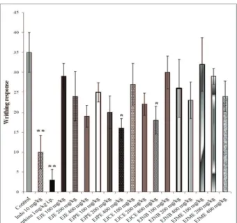
During the study, analgesic activity of drugs is measured by the ability of inhibition of writhing response. In this study, test drugs showed dose dependent response. As shown in Figure 1, 400 mg/kg b.w., p.o. dose of EJPE and
EJCE showed the signiicant (p<0.05) 54.28 and 48.57%
peripheral antinociceptive effect, respectively, and 400
mg/kg dose of EJPE was found to be most potent among
the test materials. Indo 10 mg/kg b.w., p.o. and morphine 1 mg/kg i.p. doses showed respectively 71.42% and 91.42%
signiicant (p<0.01) inhibition of writhing response. Hence, the result suggests that the possible mechanism for antinociceptive effect of test drug is the inhibition of the prostaglandin biosynthesis and the release of those
inlammatory mediators. The antinociceptive effect of
test materials were corroborated by formalin induced paw licking test. Formalin injection at mice paw stimulates the nociception effect by two distinct phases. In first phase, formalin directly acts on C-fibers which
cause acute nociceptive neurogenic pain. Where as the
late phase correspond inflammatory pain due to release of nociceptive mediators and persists for longer period (Tjolsen et al., 1992). The late phase is inhibited by peripherally acting drugs; where as, drugs act on opioid
system inhibit the first phase (Shibata et al., 1989). In
this study, Indo (10 mg/kg b.w., p.o.) showed significant
77.51% inhibition (p<0.01) of late phase (Figure 2).
Among the test materials EJE, EJPE and EJCE at 400
mg/kg b.w., p.o. dose inhibited significantly (p<0.01) the
late phase of nociception by 65.47, 73.77 and 69.87%, respectively, and EJPE at 400 mg/kg dose was found
to be most potent among the test materials. But the test materials did not show any significant inhibition of first phase of nociception. It has been seen that drugs which act primarily on central nervous system can inhibit both phases (Dubuisson & Dennis, 1977; Rosland et al., 1990). In this study, morphine 10 mg/kg i.p. dose showed the significant inhibition of early and late
Evaluation of antinociceptive and anti-inl ammatory activities of extract and fractions of Eugenia jambolana root bark and isolation of
Santanu Saha et al.
Rev. Bras. Farmacogn. Braz. J. Pharmacogn. 23(4): Jul./Aug. 2013
Figure 1. Effects of extract and fractions of Eugenia jambolana
root bark on acetic-acid induced writhing response in mice model. Mice were treated with vehicle (control), indomethacin (Indo) 10 mg/kg b.w., p.o., morphine 1 mg/kg b.w. intraperitoneally (i.p.), ethanolic extract (EJE), petroleum ether fraction (EJPE), chloroform fraction (EJCE), n-butanol fraction (EJNB) and methanol fraction (EJME) at 100, 200 and 400 mg/kg b.w., p.o. doses. Each value represents as mean±SEM. *p<0.05 and
**p<0.01 as compared with the control group (one-way ANOVA followed by Dunnet’s t-test).
Figure 2. Effects of extract and fractions of Eugenia jambolana
root bark on formalin induced nociception study in mice. Mice were treated with vehicle (control), indomethacin (Indo) 10 mg/ kg b.w., p.o., morphine (Morphi) 10 mg/kg b.w. intraperitoneally (i.p.), ethanolic extract (EJE), petroleum ether fraction (EJPE), chloroform fraction (EJCE), n-butanol fraction (EJNB) and methanol fraction (EJME) at 100, 200 and 400 mg/kg b.w., p.o. doses. Antinociceptive effects were observed in early phase
(0-5 min) and late phase (20-30 min). Each value represents as mean±SEM. *p<0.05 and **p<0.01 as compared with the control group (one-way ANOVA followed by Dunnet’s t-test).
The results of antinociceptive activity were
indicative for anti-inl ammatory properties of test drugs.
The hypothesis were evaluated by commonly used models
for screening of anti-inl ammatory activity of drugs, such as,
carrageenan-induced rat paw edema, cotton pellet induced granuloma formation and adjuvant induced arthritis in rat. The carrageenan induced rat paw edema is an experimental
model for acute inl ammation study and which is widely used to screen most of the anti-inl ammatory agents. The
carrageenan induced rat paw edema development is a biphasic response, where the initial stage is dependent on the release of signature mediators like histamine, kinin and bradykinin, and the last phase is attributed to the synthesis of prostaglandins (Vinegar et al., 1969; DiRosa et al., 1971).
Anti-inl ammatory drugs are regulating these mediators to reduce edema. Mostly, the anti-inl ammatory drugs are
found to be effective during late phase, as in this study indomethacin showed the inhibition of rat paw edema in third and fourth hour. As shown in Table 1, indomethacin
10 mg/kg showed signii cant 82.75% (p<0.01) inhibition
of rat paw edema at 4 h and 71.79% (p<0.05) at 3 h in the
present study. Like indomethacin, EJPE and EJCE 400 mg/
kg b.w., p.o. doses also respectively exhibited signii cant
79.31% (p<0.01) and 72.41% (p<0.01) inhibition of
rat paw edema at 4 h. During 3 h EJPE 400 mg/kg p.o.,
b.w. dose inhibited edema by 61.53% (p<0.05). Though,
EJE 400 mg/kg b.w., p.o. dose also showed signii cant 68.1% (p<0.05) inhibition at 4 h, EJPE 400 mg/kg dose
was found to be most potent among all the test materials (Figure 3), and this response may be due to the inhibition of biosynthesis of prostaglandins (Niemegeers et al., 1964).
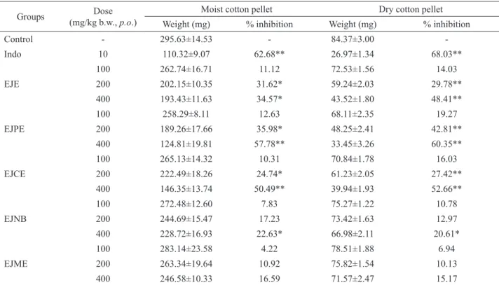
The cotton pellet induction in rat develops
granuloma as a sign of chronic inlammation by
accumulation of macrophages and lymphocytes around the foreign particles together with epitheloid and giant cells which are derived from macrophages (Swingle &
Shideman, 1972; Snedegard, 1985; Serhan & Savil, 2005).
The inhibition of granulomatous tissue formation in rat indicates the anti-proliferative effect of drugs, and which is measured by percentage inhibition of cotton pellet weight.
The wet weight of the pellet is inluenced by absorption of luid while the dry weight correlates with the amount of
granulomatous tissue formed (Spector, 1969). As shown in
Table 2, Indo 10 mg/kg signiicantly inhibited wet cotton pellet weight (62.68%, p<0.01). EJE, EJPE and EJCE
200 mg/kg b.w., p.o. doses respectively inhibited 31.62,
35.98 and 24.74% (p<0.05) weight of wet cotton pellet as
compared to control group (Figure 4). EJE and EJNB 400
mg/kg b.w., p.o. doses showed 34.57 and 22.63% (p<0.05) inhibition of weight of wet cotton pellet, respectively.
Where as, EJPE and EJCE 400 mg/kg b.w., p.o. doses
showed signiicant 57.78 and 50.49% (p<0.01) inhibition of wet cotton pellet weight, respectively. Indomethacin (10
mg/kg) also showed signiicant inhibition of the weight of dry cotton pellets (68.03%, p<0.01) (Table 2). EJPE 400
mg/kg b.w., p.o. dose showed signiicant 60.35% (p<0.01) inhibition of dry cotton pellets weight, which was closer
to the result showed by indomethacin (Figure 4). EJE and EJCE 400 mg/kg b.w., p.o. doses also signiicantly
inhibited 48.41 and 52.66% (p<0.01) weight of dry cotton
pellets, respectively. Hence, the indings suggested that the test drugs signiicantly (p<0.05 and p<0.01) inhibited the
proliferative phase of inlammation.
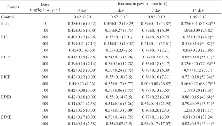
The FCA induced arthritis assessed by formation
of rat paw edema. The chronic inlammation induced by
adjuvant involved polyartritis in 14th day study (Ward
& Cloud, 1996). The arthritis was induced in this study by a sub-cutaneous injection of FCA, i.e. heat killed
Mycobacterium tuberculosis in parafin oil and mannide
monooleate, in the rat paw’s plantar surface. The arthritis was developed in the joints of hind limbs of rats by reduction of motor activity and increased paw diameter
was observed due to inlammation and edema (Calvino et al., 1987; Cain et al., 1997). The initial inlammatory
response was observed within hours, but more critical signs
of inlammation were developed with days and observed
till the 14th day of the study. EJPE and EJCE 400 mg/
kg b.w., p.o. doses on day 14th signiicantly inhibited rat paw swelling by 77.93 and 68.27% (p<0.01) respectively (Figure 5). Indo 10 mg/kg b.w., p.o. dose signiicantly inhibited the edema by 84.82% (p<0.05) on 14th day of study (Table 3). EJE 200 and 400 mg/kg b.w., p.o. doses on day 14th showed respectively 46.2 and 64.82% (p<0.05) inhibition of edema (Figure 5). EJPE, EJCE and EJNB
200mg/kg b.w., p.o. doses also showed signiicant (p<0.05)
55.17, 50.34 and 40.68% inhibition of adjuvant induced
rat paw edema, respectively (Figure 5). The test drugs Table 1. Anti-inlammatory activity of Eugenia jambolana root bark extracts in carrageenan induced rat paw edema model.
Groups Dose
(mg/kg b.w., p.o.)
Increase in paw volume (mL)
1 h 2 h 3 h 4 h
Control - 0.38±0.13 0.49±0.24 0.78±0.14 1.16±0.11
Indo 10 0.27±0.12 (28.94) 0.34±0.16 (30.61) 0.22±0.13 (71.79)* 0.20±0.17 (82.75)**
EJE
100 0.37±0.16 (2.63) 0.46±0.18 (6.12) 0.71±0.14 (8.97) 0.88±0.10 (24.13) 200 0.35±0.10 (7.89) 0.4±0.12 (18.36) 0.54±0.19 (30.76) 0.65±0.20 (43.96) 400 0.32±0.15 (15.78) 0.36±0.21 (26.53) 0.44±0.11 (43.58) 0.37±0.10 (68.10)*
EJPE
100 0.36±0.18 (5.26) 0.44±0.13 (10.20) 0.69±0.26 (11.53) 0.82±0.15 (29.31) 200 0.33±0.13 (13.15) 0.38±0.19 (22.44) 0.48±0.2 (38.46) 0.63±0.12 (45.68) 400 0.29±0.16 (23.68) 0.37±0.14 (24.48) 0.3±0.18 (61.53)* 0.24±0.11 (79.31)**
EJCE
100 0.37±0.14 (2.63) 0.45±0.18 (8.16) 0.70±0.23 (10.25) 0.86±0.17 (25.86) 200 0.34±0.18 (10.52) 0.42±0.12 (14.28) 0.51±0.16 (34.61) 0.69±0.33 (40.51) 400 0.32±0.17 (15.78) 0.39±0.23 (20.40) 0.41±0.08 (47.43) 0.32±0.20 (72.41)**
EJNB
100 0.37±0.25 (2.63) 0.47±0.12 (4.08) 0.73±0.1 (6.41) 0.91±0.20 (21.55) 200 0.36±0.14 (5.26) 0.45±0.16 (16.32) 0.63±0.22 (19.23) 0.79±0.15 (31.89) 400 0.34±0.16 (10.52) 0.42±0.11 (14.28) 0.59±0.14 (24.35) 0.70±0.06 (39.65)
EJME
100 0.37±0.11 (2.63) 0.48±0.13 (2.04) 0.74±0.15 (5.12) 0.95±0.10 (18.10) 200 0.36±0.14 (5.26) 0.48±0.17 (2.04) 0.67±0.11 (14.10) 0.87±0.14 (25.00) 400 0.35±0.19 (7.89) 0.43±0.12 (12.24) 0.64±0.15 (17.94) 0.74±0.13 (36.20)
Evaluation of antinociceptive and anti-inl ammatory activities of extract and fractions of Eugenia jambolana root bark and isolation of
Santanu Saha et al.
Rev. Bras. Farmacogn. Braz. J. Pharmacogn. 23(4): Jul./Aug. 2013
Table 2. Effects of Eugenia jambolana root bark extracts on cotton pellets induced granuloma formation in rats.
Groups Dose
(mg/kg b.w., p.o.)
Moist cotton pellet Dry cotton pellet
Weight (mg) % inhibition Weight (mg) % inhibition
Control - 295.63±14.53 - 84.37±3.00
-Indo 10 110.32±9.07 62.68** 26.97±1.34 68.03**
EJE
100 262.74±16.71 11.12 72.53±1.56 14.03
200 202.15±10.35 31.62* 59.24±2.03 29.78**
400 193.43±11.63 34.57* 43.52±1.80 48.41**
EJPE
100 258.29±8.11 12.63 68.11±2.35 19.27
200 189.26±17.66 35.98* 48.25±2.41 42.81**
400 124.81±19.81 57.78** 33.45±3.26 60.35**
EJCE
100 265.13±14.32 10.31 70.84±1.78 16.03
200 222.49±18.26 24.74* 61.23±2.05 27.42**
400 146.35±13.74 50.49** 39.94±1.93 52.66**
EJNB
100 272.48±12.60 7.83 75.27±1.22 10.78
200 244.69±15.47 17.23 73.42±1.63 12.97
400 228.72±16.93 22.63* 66.98±2.11 20.61*
EJME
100 283.14±23.58 4.22 78.51±1.88 6.94
200 263.34±19.64 10.92 75.82±1.54 10.13
400 246.58±10.33 16.59 71.57±2.47 15.17
All the result are expressed in term of mean±SEM. n=8 animals in each group; Statistical signii cance was determined by ANOVA, followed by Dunnet’s t-test. *p<0.05, **p<0.01, statistically signii cant.
Figure 4. Effects of extract and fractions of Eugenia jambolana root bark on cotton pellet induced granuloma in rat. Anti-proliferative effect of indomethacin (Indo) 10 mg/kg b.w., p.o., ethanolic extract (EJE), petroleum ether fraction (EJPE), chloroform fraction
(EJCE), n-butanol fraction (EJNB) and methanol fraction (EJME) at 100, 200 and 400 mg/kg b.w., p.o. doses were evaluated by
Table 3. Anti-inl ammatory activity of Eugenia jambolana root bark extracts in adjuvant induced arthritis in rats.
Groups Dose
(mg/kg b.w., p.o.)
Increase in paw volume (mL)
0 day 3 day 7 day 14 day
Control - 0.42±0.20 0.57±0.13 0.82±0.19 1.45±0.12
Indo 10 0.38±0.16 (9.52) 0.46±0.12 (19.29) 0.37±0.13 (54.87) 0.22±0.11 (84.82)**
EJE
100 0.42±0.15 (0.00) 0.56±0.27 (1.75) 0.77±0.14 (6.09) 1.09±0.09 (24.82) 200 0.40±0.12 (4.76) 0.53±0.1 (7.01) 0.74±0.19 (9.75) 0.78±0.15 (46.2)* 400 0.39±0.21 (7.14) 0.51±0.15 (10.52) 0.61±0.11 (25.61) 0.51±0.14 (64.82)*
EJPE
100 0.42±0.1 (0.00) 0.55±0.23 (3.5) 0.76±0.17 (7.31) 0.93±0.13 (35.86) 200 0.41±0.19 (2.38) 0.54±0.13 (5.26) 0.74±0.2 (9.75) 0.65±0.16 (55.17)* 400 0.39±0.14 (7.14) 0.5±0.16 (12.28) 0.56±0.18 (31.7) 0.32±0.10 (77.93)**
EJCE
100 0.42±0.15 (0.00) 0.56±0.24 (1.75) 0.77±0.13 (6.09) 0.97±0.12 (33.1) 200 0.42±0.12 (0.00) 0.55±0.18 (3.5) 0.76±0.16 (7.31) 0.72±0.18 (50.34)* 400 0.4±0.23 (4.76) 0.52±0.17 (8.77) 0.60±0.08 (26.82) 0.46±0.12 (68.27)**
EJNB
100 0.42±0.08 (0.00) 0.56±0.06 (1.75) 0.79±0.13 (3.65) 1.17±0.29 (19.31) 200 0.42±0.16 (0.00) 0.55±0.14 (3.5) 0.77±0.22 (6.09) 0.86±0.15 (40.68)* 400 0.41±0.11 (2.38) 0.54±0.16 (5.26) 0.64±0.14 (21.95) 0.79±0.09 (45.51)*
EJME
100 0.42±0.15 (0.00) 0.57±0.13 (0.00) 0.80±0.26 (2.43) 1.23±0.30 (15.17) 200 0.42±0.17 (0.00) 0.56±0.14 (1.75) 0.77±0.11 (6.09) 0.91±0.14 (37.24) 400 0.41±0.14 (2.38) 0.55±0.09 (3.5) 0.68±0.17 (17.07) 0.82±0.19 (43.44)*
All the result are expressed in term of mean±SEM.; n=8 animals in each group; number in parenthesis indicates percentage inhibition in increase in paw volume. Statistical signii cance was determined by ANOVA, followed by Dunnet’s t-test. *p<0.05, **p<0.01, statistically signii cant.
Figure 5. Effects of extract and fractions of Eugenia jambolana root bark on adjuvant induced arthritis in rat. The anti-arthritic activity of indomethacin (Indo) 10 mg/kg b.w., p.o., ethanolic extract (EJE), petroleum ether fraction (EJPE), chloroform fraction (EJCE), n-butanol fraction (EJNB) and methanol fraction (EJME) at 100, 200 and 400 mg/kg b.w., p.o. doses were evaluated by
Evaluation of antinociceptive and anti-inlammatory activities of extract and fractions of Eugenia jambolana root bark and isolation of
Santanu Saha et al.
Rev. Bras. Farmacogn. Braz. J. Pharmacogn. 23(4): Jul./Aug. 2013
showed the eficacy during the study and EJPE 400 mg/kg
b.w., p.o. dose showed the maximum reduction of rat paw edema among the other doses (Table 3) which was also comparable to the effect of standard drug. The inhibition
of rat paw edema was indicated the therapeutic eficacy of the test drug in chronic inlammatory state and the effect
was observed may be due to the inhibition of prostaglandin biosynthesis (Babu et al., 2009).
Therefore, these results suggested that the test
materials exhibited signiicant antinociceptive and anti-inlammatory effect and EJPE (400 mg/kg b.w., p.o. dose) was found to be most potent among all the test materials.
Hence, the current study has qualiied the traditional claim for anti-inlammatory effect by Eugenia jambolana.
Furthermore, the bioactive petroleum ether fraction of ethanolic extract of root bark of Eugenia jambolana (EJPE) was subjected to column chromatography which
led to isolate three pure compounds. These compounds were characterized by IR, 1H and 13C NMR and mass spectroscopy. These compounds were identiied as β-sitosterol, stigmasterol and lupeol by comparing with
previously reported spectroscopical data. Nevertheless,
by comparison with the previous reports the indings suggested that the antinociceptive and anti-inlammatory
activities of petroleum ether fraction of ethanolic extract of root bark of Eugenia jambolana may be due to the presence of these compounds (Garcia et al., 1999; Navarro et al., 2001; Geetha & Varalakshmi, 2001; Backhouse et
al., 2008). Therefore, the further studies on antinociceptive and anti-inlammatory activities need to be performed with
these isolated compounds.
Conclusion
The antinociceptive and anti-inlammatory
activity of ethanolic extract and different fractions of root bark of Eugenia jambolana were evaluated in the present study. The bioassay potential petroleum ether fraction was
led to isolate β-sitosterol, stigmasterol and lupeol. Hence, these indings suggested the signiicant antinociceptive and anti-inlammatory effects of root bark of Eugenia jambolana. However, the mechanisms underlying the
antinociceptive and anti-inlammatory effects of Eugenia
jambolana is still needed to be determined.
Acknowledgements
Authors are thankful to Nitte Gulabi Shetty Memorial Institute of Pharmaceutical Sciences for providing the necessary facilities and infrastructure for the study. Authors
are also grateful to Nitte University for the inancial
support.
Authors’ contributions
SS (PhD student) contributed in collecting
plant sample and identiication, confection of herbarium,
running the laboratory work, extraction of plant material and isolation of compound, biological studies, analysis of
the data and drafted the paper. EVSS guided and supervised
the research work and contributed to critical reading of the manuscript. CK contributed to chromatographic analysis, supervised the isolation of compounds and spectroscopical data analysis of compounds. SCM contributed to critical reading of the manuscript. SCS provided and made the availability of all the necessary facilities to carry out
the research work. All the authors have read the inal
manuscript and approved the submission.
References
Agarwal PK, Jain DC, Gupta RK, Thakur RS 1985. Carbon-13
NMR spectroscopy of steroidal sapogenins and steroidal saponins. Phytochemistry 24: 2476-2496.
Babu NP, Pandikumar P, Ignacimuthu S 2009. Anti-inlammatory
activity of Albizia lebbeck Benth., an ethnomedicinal
plant, in acute and chronic animal models of inlammation. J Ethnopharmacol 125: 356-360.
Backhouse N, Rosales L, Apablaza C, Goïty L, Erazo S,
Negrete R, Theodoluz C, Rodríguez J, Delporte C
2008. Analgesic, anti-inflammatory and antioxidant
properties of Buddleja globosa, Buddlejaceae. J Ethnopharmacol 116: 263-269.
Cain CK, Francis JM, Plone MA, Emerich DF, Lindner MD 1997.
Pain-related disability and effects of chronic morphine in the adjuvant-induced arthritis model of chronic pain.
Physiol Behav 62: 199-205.
Calvino B, Crepon-Bernard MO, Le Bars D 1987. Parallel clinical
and behavioral studies of adjuvant-induced arthritis in the rat: possible relationship with ‘chronic pain’. Behav Brain Res 24: 11-29.
Chaturvedi A, Kumar MM, Bhawani G, Chaturvedi H, Kumar
M, Goel RK 2007. Effect of ethanolic extract of Eugenia jambolana seeds on gastric ulceration and secretion in rats. Ind J Physiol Pharmacol 51: 131-140.
Collier HO, Dinneen LC, Johnson CA, Schneider C 1968. The
abdominal constriction response and its suppression by analgesic drugs in the mouse. Br J Pharmacol Chemother 32: 295-310.
D’Arcy PF, Howard EM, Muggleton PW, Townsend SB
1960. The antiinflammatory action of griseofulvin in experimental animals. J Pharm Pharmacol 12: 659-665.
DiRosa M, Giroud JP, Willoughby DA 1971. Studies on the mediators of the acute inlammatory response induced
in rats in different sites by carrageenan and turpentine. J Patho 104: 15-29.
sympathetic system in acetic acid-induced writhing in mice. Braz J Med Biol Res 21: 341-343.
Dubuisson D, Dennis SG 1977. The formalin test: a quantitative study of the analgesic effects of morphine, meperidine, and brain stem stimulation in rats and cats. Pain 4: 161-174.
Garcıa MD, Saenz MT, Gomez MA, Fernandez MA 1999. Topical antiinlammatory activity of phytosterols isolated from Eryngium foetidum on chronic and acute inlammation
models. Phytother Res 13: 78-80.
Geetha T, Varalakshmi P 2001. Anti-inlammatory activity of
lupeol and lupeol linoleate in rats. J Ethnopharmacol 76:
77-80.
Gyires K, Torna Z 1984. The use of the writhing test in mice
for screening different types of analgesics. Arch Int Pharmacodyn Ther 267: 131-140.
Hunskaar S, Hole K 1987. The formalin test in mice: dissociation between inlammatory and non-inlammatory pain. Pain 30: 103-114.
Ikeda Y, Ueno A, Naraba H, Oh-ishi S 2001. Involvement of vanilloid receptorVR1 and prostanoids in the acid-induced writhing responses of mice. Life Sci 69: 2911-2919.
Jagetia GC, Baliga MS 2004. The evaluation of nitric oxide scavenging activity of certain Indian medicinal plants in vitro: a preliminary study. J Med Food 7: 343-348.
Khalil MW, Ldler DR 1980. Sterols of scollop. III.
Characterization of some C-24 epimeric sterols by high resolution (220 MHz) nuclear magnetic resonance spectroscopy. Lipids 15: 69-73.
Kirtikar KR, Basu BD 2006. Indian Medicinal Plants, Vol. II.
Uttaranchal, India: International Book Distributors.
Lima GM, Bonim RR, Silva MR, Thomazzi SM, Santos MRV,
Quintans-Júnior LJ, Bonjardim LR, Araújo AAS 2011.
Assessment of antinociceptive and anti-inlammatory
activities of Porophyllum ruderale aqueous extract. Rev Bras Farmacogn 21: 486-490.
Mahmoud II, Marzouk MS, Moharram FA, El-Gindi MR, Hassan AM 2001. Acylated lavonol glycosides from Eugenia jambolana leaves. Phytochemistry 58: 1239-1244. Mukherjee PK, Saha K, Murugesan T, Mandal SC, Pal M, Saha
BP 1998. Screening of anti-diarrhoeal proile of some plant extracts of a speciic region of West Bengal, India. J Ethnopharmacol 60: 85-89.
Muruganandan S, Srinivasan K, Chandra S, Tandan SK, Lal
J, Raviprakash V 2001. Anti-inlammatory activity of Syzygium cumini bark. Fitoterapia 72: 369-375.
Nadkarni AK 2009. Dr. K. M. Nadkarni’s Indian Materia Medica, Vol. I. Bombay, India: Popular Prakashan.
Navarro A, De las Heras B, Villar A 2001. Anti-inlammatory and
immunomodulating properties of a sterol fraction from
Sideritis foetens Clem. Bio Pharm Bull 24: 470-473.
Niemegeers CJE, Verbruggen FJ, Janssen PAJ 1964. Effect of
various drugs on carrageenan-induced oedema in the rat hind paw. J Pharmacol 16: 810-816.
OECD 2001. OECD Guidelines for testing of chemicals. Organization for Economic Cooperation and
Development, Guideline 425, acute oral toxicity - Up and Down Procedure.
Ranawat L, Bhatt J, Patel J 2010. Hepatoprotective activity of ethanolic extracts of bark of Zanthoxylum armatum
DC in CCl4 induced hepatic damage in rats. J Ethnopharmacol 127: 777-780.
Ratsimamanga AR, Lefournier-Contensou C, Bibal-Prot P 1972. Comparative study of the effect of a principle extracted from Eugenia jambolana bark with NN-methyldimethyl biguanide and glybutamide on induced hypoglycemia in normal rats. C R Acad Sci Hebd Seances Acad Sci D 275: 913-915.
Reynolds WF, McLean S, Poplawski J, Enriquez RG, Escobar LI, Leon I 1986. Total assignment of 13C and 1H spectra of three isomeric triterpenoid derivatives by 2D NMR.
Tetrahedron 42: 3419-3428.
Rosland JH, Tjolsen A, Maehle B, Hole DK 1990. The formalin
test in mice: Effect of the formalin concentration. Pain 42: 235-242.
Sengupta P, Das PB 1965. Terpenoids and related compunds. Part IV, triterpenoids the stem bark of Eugenia jambolana
Lam. Ind Chem Soc 42: 255-258.
Serhan CN, Savil J 2005. Resolution of inlammation: the
beginning programs, the end. Nat Immunol 6: 1191-1197.
Sharma SB, Nasir A, Prabhu KM, Murthy PS 2006. Antihyperglycemic effect of the fruit-pulp of Eugenia jambolana in experimental diabetes mellitus. J Ethnopharmacol 104: 367-373.
Shibata M, Ahkubo T, Takahashi H, Inoki R 1989. Modiied
formalin test: characteristic biphasic pain response. Pain 38: 347-352.
Snedegard G 1985. Mediators of vascular permeability in inlammation. Pro Appl Microcir 7: 96-112.
Spector WG 1969. The granulomatous inlammatory exudates. Int Rev Exp Patho 8: 1-55.
Suleyman H, Demirezer LO, Kuruuzum A, Banoglu ZN, Gocer
F, Ozabakir G, Gepdiremen A 1991. Anti-inlammatory
effect of the aqueous extract from Rumex patientia L. roots. J Ethnopharmacol 65: 141-148.
Swingle KF, Shideman FE 1972. Phases of the inlammatory
response to subcutaneous implantation of a cotton pellet
and their modiication by certain anti-inlammatory
agents. J Pharmacol Exp Ther 183: 226-234.
Timbola AK, Szpoganicz B, Branco A, Monache FD, Pizzolatti
MG 2002. A new lavonol from leaves of Eugenia jambolana. Fitoterapia 73: 174-176.
Tjolsen A, Berge OG, Hunskaar S, Rosland JH, Hole K 1992. The formalin test: an evaluation of the method. Pain 51: 5-17.
Vinegar R, Schreiber W, Hugo R 1969. Biphasic development
Evaluation of antinociceptive and anti-inlammatory activities of extract and fractions of Eugenia jambolana root bark and isolation of
Santanu Saha et al.
Rev. Bras. Farmacogn. Braz. J. Pharmacogn. 23(4): Jul./Aug. 2013
Ward JR, Cloud RS 1996. Comparitive effect of antirhematic
drugs on adjuvant induced polyarthritis in rats. J Pharmacol Exp Ther 152: 116-121.
Wenkert E, Baddeley VG, Buritt IR, Morino LN 1978. C-13
nuclear magnetic resonance spectroscopy of naturally-occurring substances. Org Mag Res 11: 337-343.
Whittington H, Green AF 1970. Effects of azathioprine and
phenylbutazone in rat adjuvant arthritis. Br J Pharmacol 40: 167-168.
Winter CA, Risley EA, Nuss GW 1962. Carrageenan induced
edema in hind paw of the rat as an assay for anti
inlammatory drugs. Proc Soc Exp Bio Med 111: 544-547.
*Correspondence
Santanu Saha
Department of Pharmacognosy, NGSM Institute of Pharmaceutical Sciences, Nitte University
Paneer, Derelakatte, Mangalore 575 018, Karnataka, India