GRID COMPUTING IN THE OPTIMIZATION OF CONTENT-BASED
MEDICAL IMAGES RETRIEVAL*
Marcelo Costa Oliveira1
, Paulo Mazzoncini de Azevedo-Marques2
, Walfredo da Costa Cirne Filho3
OBJECTIVE: To utilize the grid computing technology to enable the utilization of a similarity measurement algorithm for content-based medical image retrieval. MATERIALS AND METHODS: The content-based im-ages retrieval technique is comprised of two sequential steps: texture analysis and similarity measurement algorithm. These steps have been adopted for head and knee images for evaluation of accuracy in the re-trieval of images of a single plane and acquisition sequence in a databank with 2,400 medical images. Ini-tially, texture analysis was utilized as a pre-selection resource to obtain a set of the 1,000 most similar images as compared with a reference image selected by a clinician. Then, these 1,000 images were processed uti-lizing a similarity measurement algorithm on a computational grid. RESULTS: The texture analysis has dem-onstrated low accuracy for sagittal knee images (0.54) and axial head images (0.40). Nevertheless, this technique has shown effectiveness as a filter, pre-selecting images to be evaluated by the similarity mea-surement algorithm. Content-based images retrieval with similarity meamea-surement algorithm applied on these pre-selected images has demonstrated satisfactory accuracy — 0.95 for sagittal knee images, and 0.92 for axial head images. The high computational cost of the similarity measurement algorithm was balanced by the utilization of grid computing. CONCLUSION: The approach combining texture analysis and similarity measurement algorithm for content-based images retrieval resulted in an accuracy of > 90%. Grid comput-ing has shown to be essential for the utilization of similarity measurement algorithm in the content-based images retrieval that otherwise would be limited to supercomputers.
Keywords: Content-based image retrieval; Texture analysis; Image registration; Grid computing.
Grades computacionais na otimização da recuperação de imagens médicas baseada em conteúdo. OBJETIVO: Utilizar o poder de processamento da tecnologia de grades computacionais para viabilizar a utiliza-ção do algoritmo de medida de similaridade na recuperautiliza-ção de imagens baseada em conteúdo. MATERIAIS E MÉTODOS: A técnica de recuperação de imagens baseada em conteúdo é composta de duas etapas se-qüenciais: análise de textura e algoritmo de medida de similaridade. Estas são aplicadas em imagens de joelho e cabeça, nas quais se avaliaram a eficiência em recuperar imagens do mesmo plano e a seqüência de aqui-sição em um banco de 2.400imagens médicas para testar a capacidade de recuperação de imagens baseada em conteúdo. A análise de textura foi utilizada inicialmente para pré-selecionar as 1.000 imagens mais se-melhantes a uma imagem de referência escolhida por um clínico. Essas 1.000 imagens foram processadas utilizando-se o algoritmo de medida de similaridade na grade computacional. RESULTADOS: A precisão encontrada na classificação por análise de textura foi de 0,54 para imagens sagitais de joelho e de 0,40 para imagens axiais de cabeça. A análise de textura foi útil como filtragem, pré-selecionando imagens a serem avaliadas pelo algoritmo de medida de similaridade. A recuperação de imagens baseada em conteúdo utili-zando o algoritmo de medida de similaridade aplicado nas imagens pré-selecionadas por análise de textura resultou em precisão de 0,95 para as imagens sagitais de joelho e de 0,92 para as imagens axiais de cabeça. O alto custo computacional do algoritmo de medida de similaridade foi amortizado pela grade computacio-nal. CONCLUSÃO: A utilização da abordagem mista das técnicas de análise de textura e algoritmo de medida de similaridade no processo de recuperação de imagens baseada em conteúdo resultou em eficiência acima de 90%. A grade computacional é indispensável para utilização do algoritmo de medida de similaridade na recuperação de imagens baseada em conteúdo, que de outra forma seria limitado a supercomputadores. Unitermos: Recuperação de imagens baseada em conteúdo; Análise de textura; Registro de imagens; Gra-des computacionais.
Abstract
Resumo
* Study developed in the Laboratório de Sistemas Distribuí-dos do Departamento de Sistemas e Computação da Universi-dade Federal de Campina Grande (UFCG), Campina Grande, PB, Brazil.
1. PhD of Medicine in the field of Medical Information Tech-nology, Master in Physics applied to Medicine, Bachelor in Com-puting Sciences, Professor at Universidade Federal de Alagoas, Campus Arapiraca, Arapiraca, AL, Brazil.
2. Electronic Engineer, PhD in Applied Physics, Lecturer at Centro de Ciências das Imagens e Física Médica da Faculdade de Me-dicina de Ribeirão Preto da Universidade de São Paulo (CCIFM/ FMRP-USP), Ribeirão Preto, SP, Brazil.
INTRODUCTION
The amount of data generated in hospi-tals and medical centers grows at increas-ing rates. The yearly production of data gen-erated by diagnostic imaging in important radiological centers achieves two terabytes, as a result of acquisition and storage of data 3. PhD in Computing Sciences, Lecturer at Department of
Sys-tems and Computation, Coordinator for Laboratório de Sistemas Distribuídos da Universidade Federal de Campina Grande (UFCG), Campina Grande, PB, Brazil.
Mailing address: Dr. Marcelo Costa Oliveira. Universidade Fe-deral de Alagoas, Campus Arapiraca, Coordenação de Ciência da Computação. Rodovia AL 115, km 6,5, Sementeira. Ara-piraca, AL, Brazil. E-mail: [email protected]
regarding patients in consequence of the increasing importance and utilization of imaging diagnosis. There is a need for in-telligent and safe indexation and storage of a huge amount of data, considering that they play an essential role in clinical diag-nosis(1,2).
The increasing use of computer-aided diagnosis (CAD) applications is related to the rapid development of medical algo-rithms. The CAD objective is to improve the diagnostic accuracy, as well as the con-sistency in diagnostic images interpretation by means of a diagnostic response sug-gested by a computer(3). However, some recognized CAD applications have not been utilized in the clinical routine yet be-cause of their high computational cost, lim-iting their utilization to those centers that have high capacity computers(4).
Difficulties in applying these CAD al-gorithms in the clinical routine and the current limits on images storage, process-ing, search and retrieval in large data banks have led companies and research institu-tions to find new soluinstitu-tions for the accom-plishment of these tasks(5,6).
Grid computing
The grid computing (GC) technology is the most recent and promising tool in the field of distributed computing. In summary, distributed computing consists of a collec-tion of independent computers presenting to the user as a single and consistent sys-tem. This technology allows integration between remotely distributed and con-nected by means of long distance networks. With this integration capacity, a virtual or cooperative computing system is created to resolve problems regarding mass data stor-age and access, as well as regarding appli-cations running with high computational cost(6,7).
The GC technology offers a single en-vironment for data sharing, storage and processing. Additionally, it allows the medical community to utilize a single dis-tributed database capable of providing re-sources, data and knowledge sharing. These capabilities allow a greater interac-tion among medical clinics, offering new opportunities for small clinics and research laboratories with poor computational re-sources(7).
Besides, GC offers a flexible, scalable and less vulnerable infrastructure, and, therefore more reliable and capable of guar-anteeing a safe access to any installed ap-plication. Differently from distributed com-puting and the methodology of groups of terminals or workstations connected to a server or group of servers (clusters technol-ogy), the GC resources present administra-tive autonomy and system heterogeneity. These two features allow a higher scala-bility and robustness to applications. How-ever, these same features require that the computational grid components are com-pliant with standards aiming at an open scalability and sharing of computational resources(8).
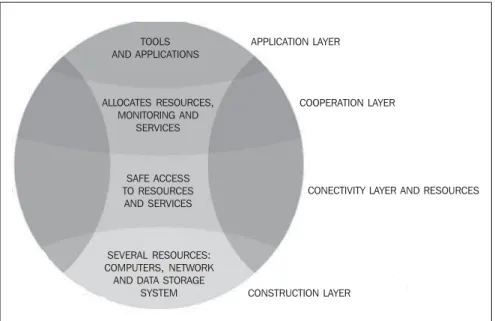
Foster & Kesselman(9) have presented a proposal of GC architecture and compo-nents. The grid formal architecture com-prises four layers (Figure 1). The contion layer is the lowest level of the struc-ture and represents the physical resources and devices that users want to share and access (computers, network, file systems, catalogs, softwares and digital instru-ments). Just above the construction layer, is the resources and connectivity layer, re-sponsible for communication and authen-tication required for resources exchange, user validation, monitoring and control over resources sharing. The third layer, or cooperation layer, holds the protocols and performs the services responsible for the
resources exchange (resources discovery and allocation, monitoring and diagnosis of services functionality, data replication, and policies regulating users’ privileges for accessing the grid resources). The user ap-plication layer is at the top of the structure and is responsible for invoking all the other layers.
There is a great number of GC-related projects described in the literature (for ex-ample: Globus(10), Legion(11), Condor(12) and OurGrid(13)), based on different tech-nologies, and aimed at determined areas and purposes like applications for data stor-age and processing, Web portals and infra-structure services for interinstitutional col-laboration(14,15).
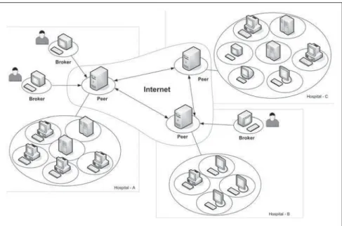
The baseline utilization of GC by the user is accomplished by means of a soft-ware interface that allows the user to com-municate with the data processing center of the computational grid known as broker. The broker can find the resources required for the tasks execution. After finishing the task execution, the broker returns the ap-plication result to the user(16). Figure 2 dem-onstrates the baseline functioning of the OurGrid project.
Content-based medical images retrieval
Among the several CAD techniques, content-based images retrieval (CBIR) are the systems that most benefit from the GC technology due to their features and
re-Figure 1. Computational grids architecture.
CAMADA DE CONSTRUÇÃO
CAMADA DE CONECTIVIDADE E RECURSOS
CAMADA DE COOPERAÇÃO CAMADA DE APLICAÇÃO
TOOLS AND APPLICATIONS
ALLOCATES RESOURCES, MONITORING AND
SERVICES
SAFE ACCESS TO RESOURCES
AND SERVICES
SEVERAL RESOURCES: COMPUTERS, NETWORK AND DATA STORAGE
SYSTEM
APPLICATION LAYER
COOPERATION LAYER
CONECTIVITY LAYER AND RESOURCES
(pixels) for each gray-scale intensity. Gray-scale distribution presents ambiguity (where different images may generate the same summation), and for this reason is not effective for the whole CBIR; however, considering its simplicity and low compu-tational cost it can and should be utilized as a initial filter for other more complex and costly methods.
Texture-based features are related to the quantification of image intensity variation and scale. In the literature, one of the most frequently utilized methods for extracting texture attributes is the co-occurrence ma-trix(24). Haralick et al.(26) have defined the texture attributes that can be obtained from the co-occurrence matrix with texture dis-crimination purposes. Approximately 20 statistical functions are proposed in the lit-erature for acquisition of information from the co-occurrence matrix(27). Some of the most significant functions producing a sat-isfactory textures classification are: en-tropy, inertia, energy, shade, inverse differ-ence moment, promenance, correlation and variance(27–33).
Shape-based image retrieval is one of the most complex issues to be approached by CBIR systems, considering the com-plexity of the method for automatic seg-mentation of medical images. After the segmentation, the structures are described by their shape characteristics, including quirements: processing intensity and
com-plexity and great amount of stored im-ages(17).
Through CBIR, and based on a refer-ence image, it is possible to find similar images included in one or several image banks utilizing inherent attributes. In the clinical decision-making process, CBIR presents great advantages, and is capable of retrieving images of a same modality, ana-tomical region and with the same structural alterations caused by certain diseases. Therefore, CBIR has awakened the medi-cal community interest because of its ca-pacity to retrieve already diagnosed images to compare with an image being studied, allowing the specialist to confirm his/her diagnostic hypothesis(18). Although part of this information may be shown on the medical images letterhead, this textual la-beling may present a high rate of error, with case reports of up to 16%(19). A great num-ber of scientific papers emphasize the need for adopting alternative methods of access-ing data manually inserted into the medi-cal images letterheads(20–23).
Besides the techniques of clinical deci-sion-making support, research and teach-ing benefit from CBIR systems. In educa-tion, CBIR aids both teachers and students in utilizing educational image banks and visual analysis of results. Besides evalua-tion based on diagnosis and anatomical region, analysis of visually similar cases, although with different diagnosis, result in an improvement of the educational qual-ity(24).
Content-based images retrieval is one of the computational vision techniques more intensely studied in the last tem years, and is based on three classes of visual charac-teristics: color, texture and shape(25). These attributes allow the development of robust computational tools capable of character-izing images by their own contents, adding advantages to the images identification based only on textual descriptors that con-stitute the traditional classification of medi-cal images files(23).
The gray-scale distribution is the sim-plest feature to be characterized. Character-ization is performed by comparison be-tween gray-scale histograms utilizing the summation of absolute or quadratic differ-ences on the number of image elements
Figure 2. The user enters the CG through the broker installed in his/her PC. The broker can find resources, and request local or remote computers to accomplish user’s tasks. The peer is responsible for the man-agement of the local network hardware for the interaction with remote machines.
information on rotation, translation and scale(34).
Another CBIR technique described in the literature is the images registration(5,17). This technique calculates a rigid 2D coor-dinates transformation including rotation, translation and scale, searching the maxi-mum matching between two images or between two image volumes. The rigid transformation is based on the minimiza-tion of the quadratic error or sum of square differences between the structures contour utilizing similarity measurement algorithms between two images intensities (35,36).
The present study presents a singular approach to systems of content-based medical images retrieval, utilizing texture attributes and the computational power of the recent GC technology applied to the similarity measurement algorithm based on the sum of square differences.
MATERIALS AND METHODS
acqui-sition planes, with gray levels ranging from 4,096 to 65,536.
The system comprises two CBIR mod-ules. The first module utilizes second or-der texture analysis parameters (co-occur-rence matrix) to classify the most similar images according to this technique. In the second module, the similarity measurement algorithm is applied on the images selected in the first module. Because of the high computational cost of the similarity mea-surement algorithm, the second module is processed on the OurGrid computational grid that is a cooperative, open and free-access network. OurGrid, currently, hooks together approximately 500 machines.
The user/GC interface is performed by means of MyGrid 3.2 (OurGrid; Campina Grande, PB, Brazil) that is the OurGrid broker, capable of selecting the computa-tional resources to be utilized in the appli-cation execution, besides releasing the user from the GC complexity, so the user utilizes de grid as if it was a single computer(13).
All of the database images have an as-sociated characteristic vector obtained from the gray levels co-occurrence matrix and its attributes. The co-occurrence matrix followed orientation at 0°, 45°, 90° and 135° and distances between images ele-ments (pixels) = 1. Texture attributes uti-lized were: energy, entropy, inverse differ-ence moment, shadow, inertia, promenance, correlation and variance. The utilization of eight texture attributes and four angular orientations resulted in a 32-dimension characteristic vector.
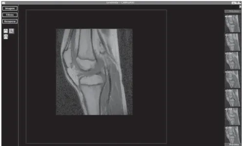
The system offers a graphic interface (Figure 3) allowing the specialist to select a DICOM (digital imaging and communi-cation in medicine) reference image at the beginning of the first module. At the end of the module, the images are classified according the lower value of the Euclidean distance between the characteristic vectors of the reference image and the database images.
The second module utilizes the 1,000 most similar images according to the first module. This module also requires that the specialist define the number of tasks for the similarity measurement algorithm process-ing e distribution on the GC. That is to say, which is the application “granularity”. The granularity is related to the amount of
im-ages to be processed by the similarity mea-surement algorithm on each GC machine. The similarity measurement algorithm uti-lizes similar transformations and linear in-terpolation aiming at the mapping of the homologue points between two images.
RESULTS
The results of the present study origi-nated from the selection of images of two anatomical regions — knee and head — in an images bank. The knee studies included 20 sagittal, T1-weighted images, and head studies included 40 axial, T2-weighted images. The experiments were repeated for three times, with slices different from the described studies. The images were consid-ered as correct when the application re-turned images from the same plane and sequence of the reference images.
The first module classified the most similar images according to texture at-tributes. The mean processing time in the first module was 2.3 minutes, and was ob-tained by the calculation of the Euclidean distance between the characteristic vector of each of the 2,400 images, and the char-acteristic vector of the reference image. The algorithms were processed by the local computer utilizing a 2.8 GHz Pentium 4 processor with 1 Gbyte memory.
Results were evaluated with “precision” and “recall” parameters which are typically utilized for evaluating systems of content-based images retrieval and information
re-trieval. “Recall” means the ratio of relevant images over the number of images re-trieved in the query. On the other hand, “precision” is the ratio of retrieved images that are relevant for reference(38).
Figure 4 shows the results of the execu-tion of the first module with mean values of precision-recall curves of the Euclidean distance between characteristic vectors of the reference images in relation to the im-ages of the database. This result allowed the evaluation of the CBIR effectiveness utilizing the texture in the classification of the most similar images for the second module. Although the mean precision ob-tained in the experiments is 0.54 (sagittal knee), and 0.40 (axial head), it is sufficient for filtering the images to be submitted to the second module. In the second module, the images are processed with the similar-ity measurement algorithm on the compu-tational grid. CBIR with the similarity measurement algorithm resulted in a satis-factory precision for both anatomical re-gions — 0.95 (sagittal knee) and 0.92 (axial head) —, according to the mean precision-recall curves between the reference images and those classified by the first module (Figure 5).
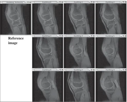
Figure 6 shows the classification of the most similar images after the application execution. For space reasons, only nine of the most similar images are shown.
The high computational cost of the similarity measurement algorithm was bal-anced by the utilization of the computation
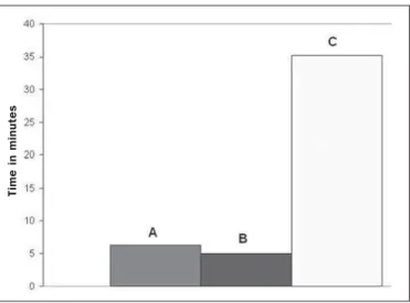
grid of the OurGrid system. On average, the processing time of the similarity measure-ment algorithm applied to the experimeasure-ments utilizing 50 processor of the grid was re-duced by 116.97 minutes for knee images and 95.15 minutes for head images in re-lation to processing times obtained in the local computer (Figure 7).
In the present study, the application was divided into 20 tasks comprised of 50 im-ages each. Imim-ages were compressed before being sent to the computational grid, and the mean size of the files with 50 images
was 4 Mbytes. Images were sent to the computational grid with a single identifi-cation file specifying the number of the image and the respective task.
On average, the compressed images were sent to the computational grid ma-chines is 22.2 seconds, and the mean pro-cessing time for the 50 images by each computer of the grid was 11.45 seconds. The images send-time was short because the greatest part of the tasks was executed on computers connected to the local net-work.
Also, OurGrid allowed that the librar-ies required for the application execution were stored in remote computers avoiding the necessity of re-sending data.
The mean time for experiments has also been analyzed, changing the application granularity among 10, 20, and 50 images/ task (Figure 8). The use of the smallest grain, i.e., 50 images /task, implied a greater total amount of images/task. So, a higher number of computers of the computational grid were requested because of the increase in the quantity of tasks to be processed.
Figure 6. Application result utilizing sagittal knee image and the most similar images acquired with the application.
The necessity of allocating 50 machines for executing the application implied the distribution of tasks for being processed out of the local network. So, the total ap-plication time was affected by the time of data transmission to remote computers.
Nevertheless, decomposing the applica-tion into larger tasks (tem tasks in total), i.e., larger grain, implied the requisition of less computers and transmission of greater files with higher processing time/machine. Therefore, a fixed and intermediate num-ber of 20 tasks were adopted.
DISCUSSION
The CG technology has shown to be a promising tool in the processing and stor-age of great data volumes. However, more benefits should be expected, according to Liu et al.(7), who have utilized the GC ar-chitecture to make medical images backup copies in several PACS (picture archiving and communication system).
Figure 5. Curves regarding the application efficacy in content-based im-ages retrieval.
Figure 4. Curves regarding the execution of the first module utilized for images filtering.
Sagittal knee Axial head Sagittal knee Axial head
The present study has adopted a mixed approach of CBIR techniques to classify similar images of different planes and ana-tomical regions utilizing the high CG pro-cessing capacity. The system has utilized CBIR techniques based on texture analy-sis and similarity measurement algorithm. The texture analysis approximates the human visual perception and has been uti-lized in many systems as an aid to the clini-cal diagnosis(39,40). The mean texture analy-sis accuracy — 0.54 for knee images, and 0.40 for head images -, despite being rela-tively low, was effective as an initial filter for the second module. A possible solution to increase the efficiency of this filtering would be the development of methods to detect motion artifacts, since texture infor-mation may be missed when rotation, trans-lation and scaling are involved in the im-ages processing(29).
The utilization of the similarity mea-surement algorithm of the sum of the square differences applied to the second module presented a quite satisfactory mean accuracy — 0.95 for knee, and 0.92 for head. The algorithm could retrieve similar images of different anatomical regions and planes. The majority of studies in the litera-ture are restricted to a determined anatomi-cal region, modality or diagnostic proce-dure, only utilizing characteristic vec-tors(41). However, because of their high computational cost, the utilization of simi-larity measurement algorithms executed in a single computer becomes unfeasible in
computer-aided diagnosis. The GC tech-nology enables the utilization of the simi-larity measurement technique because of the capability of parallel data processing in the several computers connected to the computational grid.
Although the computational grid uti-lized in the present study is constituted by approximately 500 computers spread over more than 20 locations, the ten- and twenty-task experiments were processed in the lo-cal network machines without affecting the application execution time. However, the 50-task experiments required processing out of the local network, so they were af-fected by the high costs of data transmis-sion. In these cases, the cost-benefit ratio between processing time and data-trans-mission should be evaluated.
The utilization of GC in medical appli-cations is still at its beginning; however this is a promising technology and significant developments in IT applied to the health care field can be expected in the near fu-ture.
Aiming at improving the results of the present study, two new components are presently in development: similarity mea-surement based on cross-correlation, and automatic segmentation of brain structures. The cross-correlation algorithm will allow the search in different modalities to mini-mize a limitation of the sum of square dif-ferences. Another limitation of this algo-rithm is the high sensitivity to small amounts of pixels with great differences in
intensity between two images, like in cases of contrast injection(35). The automatic seg-mentation algorithm will restrict the image retrieval to determined structures, allowing more specific queries than those performed in comparison with complete images. An integrate utilization of different methods could result in a more accurate differentia-tion between images(42).
Acknowledgments
The authors thank Projeto GridVida, Laboratório de Sistemas Distribuídos da Universidade Federal de Campina Grande (UFCG), and Centro de Ciências das Ima-gens e Física Médica da Faculdade de Me-dicina de Ribeirão Preto da Universidade de São Paulo (CCIFM/FMRP-USP).
REFERENCES
1. Montagnat J, Breton V, Magnin IE. Using tech-nologies to face medical image analysis chal-lenges. Proceedings of the IEEE CCGrid03 2003, Tokyo, Japan.
2. Montagnat J, Breton V, Magnin IE. Partitioning medical image databases for content-based que-ries on a Grid. Methods Inform Med 2005;44: 154–160.
3. Azevedo-Marques PM. Diagnóstico auxiliado por computador na radiologia. Radiol Bras 2001;34: 285–293.
4. HealthGrid, HealthGrid White Paper. [Acessado em: 10/10/2006]. Disponível em: http://www. heathgrid.org
5. Montagnat J, Bellet F, Benoit-Catin H, et al. Medi-cal images simulation, storage, and processing on the European DataGrid testbed. J Grid Comput 2004;2:387–400.
6. Breton V, Blanchet C, Legré Y, Maigne L, Monta-gnat J. Grid technology for biomedical applica-tions. Lecture Notes in Computed Science 2005; 204–218.
Figure 7. Mean processing times. A: Axial head, local. B: Axial head, grid.
C: Sagittal knee, local. D: Sagittal knee, grid.
T
im
e
i
n
m
in
u
te
s
Figure 8. Comparison between mean times utilizing different granularities in the sagittal knee images processing, 10 tasks (A), 20 tasks (B) and 50 tasks (C).
T
im
e
i
n
m
in
u
te
7. Liu BJ, Zhou MZ, Documet J. Utilizing data Grid architecture for the backup and recovery of clini-cal image data. Comput Med Imaging Graph 2005;29:95–102.
8. Foster I, Kesselman C, Tuecke S. The anatomy of the Grid: enabling scalable virtual organizations. International Journal of High Performance Com-puting Applications 2001;15:200–222. 9. Foster I, Kesselman C. The Grid 2: blueprint for
a new computing infrastructure. San Francisco, CA: Morgan Kaufmann Publishers, 2004. 10. Foster I, Kesselman C. Globus: a metacomputing
infrastructure toolkit. International Journal of Supercomputing Applications 1997;11:115–128. 11. Grimshaw AS, Wulf WA. The legion vision of a worldwide virtual computer. Communications of the ACM 1997;40:39–45.
12. Condor. [Acessado em: 13/9/2006]. Disponível em: http://www.cs.wisc.edu/condor
13. Cirne W, Brasileiro F, Andrade N, et al. Labs of the World, Unite!!! UFCG/DSC Technical Report 07/2005;1–12.
14. de Roure D, Baker M, Jennings NR, Shadbolt N. The evolution of the Grid. In: Berman F, Fox G, Hey AJG, editors. Grid computing – making the global infrastructure a reality. New York, NY: Wiley, 2003;65–100.
15. Foster I. The Grid: computing without bounds. Scientific American April 2003,228:80–85. 16. Grid Café. [Acessado em: 29/8/2006].
Disponí-vel em: http://gridcafe.web.cern.ch/gridcafe 17. Montagnat J, Duque H, Pierson JM, Breton V,
Brunie L, Magnin IE. Medical image content-based queries using the Grid. Proceedings of the First European HealthGrid Conference 2004, Lyon, France.
18. Rahman M, Wang T, Desai B. Medical image retrieval and registration: towards computer as-sisted diagnostic approach. In: IDEAS Workshop on Medical Information Systems: The Digital Hospital (IDEAS-DH’04), 2004. Washington, DC: IEEE Computer Society, 2004;78–89. 19. Güld MO, Kohnen M, Keysers D. Quality of
DICOM header information for image
categori-zation. Proceedings of the International Sympo-sium on Medical Imaging 2002, San Diego, CA. 20. Tagare HD, Jaffe C, Duncan J. Medical image databases: a content-based retrieval approach. J Am Med Inform Assoc 1997;4:184–198. 21. Traina Júnior C, Traina AJM, Santos RR, Senzako
EJ. A support system for content-based medical image retrieval in object oriented databases. J Med Syst 1997;21:339–352.
22. Rosset A, Ratib O, Valle J. Integration of a mul-timedia teaching and reference database in a PACS environment. RadioGraphics 2002;22: 1567–1577.
23. Petrakis EGM. Content-based retrieval of medi-cal images. Int J Comput Res 2002;11:171–182. 24. Müller H, Michoux N, Bandon D, Geissbuhler A. A review of content-based image retrieval systems in medical applications – clinical benefits and fu-ture directions. Int J Med Inform 2004;73:1–23. 25. Azevedo-Marques P, Honda MH, Rodrigues JAH, et al. Recuperação de imagem baseada em con-teúdo: uso de atributos de textura para caracteri-zação de microcalcificações mamográficas. Radiol Bras 2002;35:93–98.
26. Haralick RM, Shanmuga K, Dinstein I. Textural features for image classification. IEEE Trans Syst Man Cybern 1973;SMC3:610–621.
27. Walker RF, Jackway P, Longstaff ID. Improving co-occurrence matrix feature discrimination. In: Proc DICTA ’95, 3rd Conference on Digital Im-age Computing: Techniques and Application 1995;643–648.
28. McLean GF. Vector quantization for texture clas-sification. IEEE Trans Syst Cybern 1993;23:637– 644.
29. Freeborough PA, Fox NC. MR image texture analysis applied to the diagnosis and tracking of Alzheimer’s disease. IEEE Trans Med Imaging 1998;17:475–479.
30. Mathias JM, Tofts PS, Losseff NA. Texture analy-sis of spinal cord pathology in multiple scleroanaly-sis. Magn Reson Med 1999;42:929–935. 31. Materka A, Strzelecki M. Texture analysis
meth-ods – a review. In: COST B11 Report. Lodz,
Po-land: Technical University of Lodz, Institute of Electronics, 1998.
32. Konak ES. A content-based image retrieval sys-tem for texture and color queries. (M.Sc. degree thesis). Ankara, Turkey: Department of Computer Engineering and Institute of Engineering and Science, Bilkent University, 2002.
33. Sharma M, Singh S. Evaluation of texture meth-ods for image analysis. 7th Australian and New Zealand Intelligent Information System Confer-ence. Perth, Australia, 2001;117–121. 34. Veltkamp RC, Hagedoorn M. State-of-the-art in
shape matching. In: Lew M, editor. Principles of visual information retrieval. London: Springer-Verlag, 2000;87–119.
35. Hajnal JV, Hill DLG, Hawkes DJ. Medical image registration. In: Neuman MR, editor. Biomedical engineering. Boca Raton, FL: CRC Press, 2001. 36. Yoo TS. Insight into images: principles and prac-tice for segmentation, registration, and image analysis. Wellesley, MA: AK Peters, 2004. 37. InsightToolkit. [Acessado em: 8/10/2006].
Dis-ponível em: http://www.itk.org
38. Bueno JM. Suporte à recuperação de imagens médicas baseada em conteúdo através de histogra-mas métricos. São Carlos, SP: Instituto de Ciên-cias Matemáticas e de Computação, Universida-de Universida-de São Paulo, 2002.
39. Shyu CR, Bradley CE, Kak AC, Kosaka A, Aisen AM, Broderick LS. ASSERT: a physician-in-the-loop content-based retrieval system for HRCT image databases. Computer Vision and Image Understanding 1999;75:111–132.
40. Kuo WJ, Chang RF, Lee CC, Moon WK, Chen DR. Retrieval technique for the diagnosis of solid breast tumors on sonogram. Ultrasound Med Biol 2002;28:903–909.
41. Lehmann TM, Güld MO, Thies O, et al. Content-based image retrieval in medical applications. Methods Inform Med 2004;43:354–361. 42. Traina AJM, Traina C, Bueno JM, Chino FJT,