Spontaneous complete regression of cerebral
arteriovenous malformation
Regressão completa espontânea de malformação arteriovenosa cerebral
Lucas Alverne Freitas de Albuquerque
1, Jander Moreira Monteiro
2, Marcos Dellaretti
1,3,
Gerival Vieira Junior
1, Atos Alves de Sousa
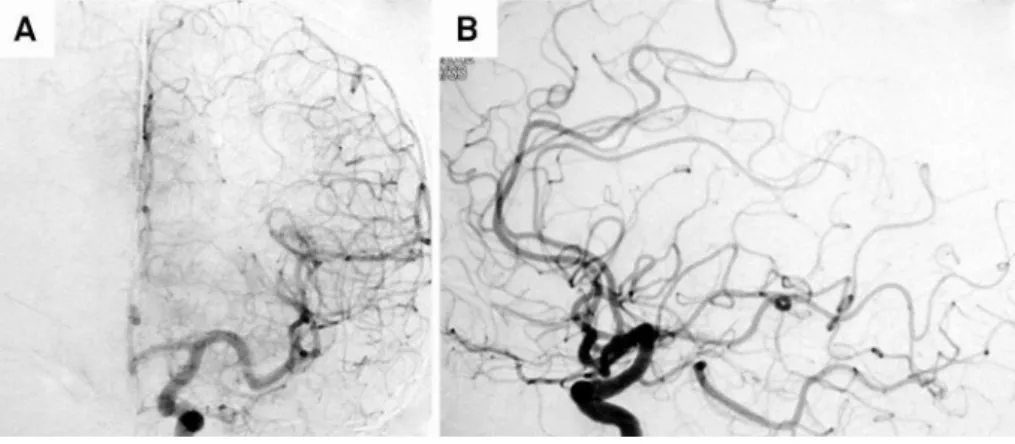
1,3A 55 year-old man started with headaches in 2005. He
underwent MRI that showed left frontal arteriovenous
mal-formations (AVM) without evidence of hemorrhage. An
arteriography revealed AVM supplied by the left anterior
cerebral artery (Figures 1A and 1B).
In 2010 the patient had an episode of cerebral
hemorrhage confirmed with CT and MRI (Figures 2A,
2B and 2C). Referred to our service for treatment in
2012, when he was made a new arteriography that
revealed complete spontaneous regression of the AVM
1,2(Figure 3).
Complete spontaneous regression of the AVM is
extre-mely rare, with only few cases in literature and an estimated
prevalence of 0.8 to 1.3%
1,2,3.
1Departamento de Neurocirurgia, Santa Casa de Belo Horizonte, Belo Horizonte MG, Brazil;
2Escola Bahiana de Medicina e Saúde Pública, Salvador BA, Brazil;
3Faculdade de Ciências Médicas de Minais Gerais, Belo Horizonte MG, Brazil.
Correspondence:Lucas Alverne Freitas de Albuquerque; Rua Padre Rolim, 492 / ap.101; 30130-090 Belo Horizonte MG, Brasil; E-mail: [email protected]
Conflict of interest:There is no conflict of interest to declare.
Received 16 May 2014; Received in final form 05 August 2014; Accepted 25 August 2014.
Figure 2.
(A) CT scan shows an intracerebral hematoma in the left frontal lobe; (B) Axial without gadolinium T1-weighted MR image
suggests a hematoma. The tubular structures representing the AVM are seen as signal voids; (C) Sagittal gadolinium-enhanced
T1-weighted MR image suggests a hematoma. The tubular structures representing the AVM are seen as high signal intensity.
Figure 1.
(A) Early-phase left internal carotid artery angiogram showed an AVM supplied by the left anterior cerebral artery;
(B) Delayed-phase left internal carotid artery angiogram shows an early-draining single vein which drains into the sagittal sinus.
DOI:10.1590/0004-282X20140158
IMAGES IN NEUROLOGY
References
1. Abdulrauf SI, Malik GM, Awad IA. Spontaneous angiographic obliteration of cerebral arteriovenous malformations. Neurosurgery.1999;44(2):280-7. http://dx.doi.org/10.1097/00006123-199902000-00021
2. Buis DR, Berg R, Lycklama G, Worp HB, Dirven CM, Vandertop WP. Spontaneous regression of brain arteriovenous malformations. A
clinical study and a systematic review of the literature. J Neurol. 2004;251(11):1375-82. http://dx.doi.org/10.1007/s00415-004-0548-3
3. Patel MC, Hodgson TJ, Kemeny AA, Forster DM. Spontaneous obliteration ofpial arteriovenous malformations: a review of 27 cases. Am J Neuroradiol. 2001;22(3):531-6.