Ricardo António Neves Moreira
Dissertation presented to obtain the Ph.D degree
in Biology
Instituto de Tecnologia Química e Biológica
Universidade Nova de Lisboa
Oeiras, December, 2011
Financial Support from Fundação para a Ciência e Tecnologia (FCT) – Ph.D: grant - SFRH/BD/36027/2007.
Work performed at:
Control of Gene Expression Laboratory Instituto de Tecnologia Química e Biológica Av. da República (EAN)
Supervisor :
Professora Doutora Cecília Maria Pais de Faria de Andrade Arraiano – Investigadora Coordenadora, Instituto de Tecnologia Química e Biológica, Universidade Nova de Lisboa.
(Head of the Laboratory of Control of Gene Expression, where the work of this Dissertation was performed)
Examiners
:
Professora Doutora Paloma López Garcia - Head of Department of Molecular Microbiology & Infection Biology, Centro de Investigaciones Biológicas, Madrid (Principal Examiner).
Professora Doutora Leonilde Morais Moreira – Assistant Professor, Instituto Superior Técnico, Universidade Técnica de Lisboa (Principal Examiner).
Professora Doutora Maria Teresa Crespo – Principal Investigator, Instituto de Biologia Experimental e Tecnológica, Oeiras.
"Magic is just science we don't understand yet."
I would like to thank all the people that directly and indirectly contributed and supported my work, and without whom this PhD dissertation would not have been possible:
Instituto de Tecnologia Química e Biológica (ITQB) - the Institution that made
this work possible, by providing the facilities and resources. I would like to thank the support of two former ITQB directors Professor Miguel Teixeira,Professor
José Martinho Simões andthe present Director Professor Luís Paulo Rebelo for providing the conditions for the presentation of this dissertation.
Fundação para a Ciência e Tecnologia (FCT) for the financial support crucial for the accomplishment of this work.
Professor Cecília Arraiano for receiving me in her laboratory and accepting to be
my PhD supervisor. Thank you for your support, for your enthusiasm, inspiration and endless capacity to see always the bright side, even in hard and more challenging moments. I also thank you for giving me so many and different
opportunities to expand my scientific knowledge participating and presenting my work in several meetings, both in Portugal and abroad.
To all past and present colleagues and friends in the lab for making it such an enjoyable place to be and work at.
Ana – My favorite dance floor partner. Thank you for all the positive energy you
transmit, for all your support, ideas and helpful discussions. Thank you for always welcome me even when it was for the most “silly” question.
Filipa – for your friendship, all the funny moments, conversations and also
Andreia Aires – for rescuing me countless times during our so called
“emergency” happenings and to help me when I needed.
Cátia Bárria– for making me laugh with the amazing capacity of telling jokes that
everyone thinks are real.
Clémentine Dressaire – For all the help, all the knowledge shared and for
contributing so much to my work even when far away.
Inês Guinote– for all the long conversations and the lessons about… well… so
many things. “Theoretically…” will be the word I will always remember.
Inês Silva – for the enjoyable movies and TV series discussions and all the
encouragement during my experimental adventures in the RNA world.
Joana Pissara – for being our audio daily news and for always have an explanation (just like an encyclopedia) when doubt arises.
Zé–for the “laughing out loud” moments at lunch time.
Margarida Matos – for the non-stop good mood. Thanks for your kindness and
even for teaching me that I could avoid contaminations in the LB stock by making an aliquot in a falcon tube.
Margarida Saramago – to make me giggle with her daily youtube selection
movies.
Michal – for sharing different points of view and ideas work wise.
Patrick – for all the knowledge transferred, all the technics you taught me and all
the amazing brainstorming afternoons. Thank you for all your help, support and trust on me.
Rute– for helping when needed and for always be present in the breakfast team
for some socializing morning moments.
Sandra – for all the work tips you gave me and the helpful discussions when
necessary. For make me laugh so much with your singing skills, mainly with
Susana – for the helpful discussions and shared experiences having always a
good example (in her dozen's lab books) and making things more practical. Vânia– For the helpful bioinformatics tips.
To all my colleagues in the other labs in the Institute, specially to the 2008 class of the ITQB PhD course. Thank you for having participated, in one way or another,
in this work, by exchanging experiences and friendly words during coffee breaks and dinner parties.
To all my friends – specially to Alexander, Rita, Zé, Nuno, Ana, Damião and
Soraia, for all the comprehension, friendship, support, fun and socializing moments. Thank you for your comments, suggestions and remarks.
To all my family, Mum, Dad, Brother and Sisters, for their eternal support and
unconditional love.
To my Mum and Dad - for your love, strength, for being present and always curious about what I was doing in the lab.
My Brother and Sisters - for being the greatest and call me the family mad
scientist – I like it! Thank you for your love and support.
Thank you all!
Table of Contents
ABSTRACT ... XV
RESUMO ... XXI
LIST OF PUBLICATIONS ...XXIX
DISSERTATION OUTLINE ...XXXIII
ABBREVIATIONS ... XXXIX
CHAPTER 1... 1
CHAPTER 2... 29
CHAPTER 3... 57
CHAPTER 4... 81
CHAPTER 5... 109
CHAPTER 6... 145
The cellular concentration of a given RNA is the result of the balance
between its synthesis and degradation. Both DNA transcription and RNA decay
control the final levels of each protein in the cell. BolA is an Escherichia coli (E. coli)
protein, which induces changes in cell morphology when present in high levels.
BolA expression is regulated by two different promoters, a sigma 70 (σ70) promoter responsible for the basal levels of this gene in exponential phase and a
sigma S (σS) gearbox promoter important in stress situations and stationary phase of bacterial growth.
The first objective of this PhD work was to further characterize the
expression of the bolA gene. Based on bioinformatic analysis, we have identified
the H-NS protein as a putative transcriptional regulator of BolA. H-NS is a
relatively small protein, abundant in bacterial cells and is often compared to
eukaryotic histones due to its high affinity for DNA. In order to clarify the
possible role of H-NS in BolA transcription, we have constructed an hns E. coli mutant. This mutant was compared to the wild type regarding the levels of bolA
mRNA transcript and in vitro DNA-protein interaction studies were performed. These experiments allowed us to demonstrate that H-NS is able to down-regulate
the levels of bolA mRNA in exponential phase and bind to the bolA promoter region. In addition, the DNA-protein interaction studies revealed that H-NS has a
special affinity to the curved bolA promoter region encompassing both bolA1p and bolA2p promoters.
In the second part of this doctoral project, the aim was to study the
specific role of BolA as a transcription factor and characterize its role in cell
starvation. Previous studies have revealed the influence of BolA in the
transcription of different genes involved in cell wall synthesis, such as the mRNA
levels of the penicillin binding protein 5 (PBP5) and penicillin binding protein 6
(PBP6). We studied the effect of BolA overexpression in growing cells and
showed that this protein can inhibit the cell elongation mechanism. Furthermore,
by RT-PCR and dot-blot experiments, we demonstrated that this inhibition is
caused by a reduction of mreB mRNA levels. Protein levels were also studied, and
the effect observed at the mRNA level was reflected in the amount of MreB
protein. For the first time BolA has been shown to bind DNA and directly
regulate the levels of MreB. MreB filaments are crucial for the bacterial cell
cytoskeleton and are essential for the maintenance of a cellular rod shape. The
inhibitor effect of BolA on MreB levels might be enough to prevent filament
polymerization. Thus, BolA induced morphology is involved in a complex
pathway that comprises PBP5, PBP6 and MreB regulation.
The conclusions obtained until this point were quite important for the
regulation of a gene with such pleiotropic effects in cell shape maintenance and
its functional characterization. To pursue our task of understanding the role of
BolA as a new transcription factor, we performed microarrays to study the global
effect of BolA in E. coli transcription regulation. Our results displayed a great variety of genes affected by the presence of BolA in the cell. These genes are
related not just with cell morphology but also with cell metabolism, cell motility
and stress response. Among the stress response genes, sigma E (σE) was the unique polymerase subunit to be significantly affected at the mRNA level by
BolA. In E. coli one of the key pathways involved in maintaining cell envelope integrity during stress and normal growth is controlled by σE. The regulation of this sigma factor by BolA was assayed by northern blot and confirmed. It is
coli small RNAs (sRNAs) that control different outer membrane porins (OMPs) during the envelope stress response. In order to establish a relationship between
the BolA properties to alter the OmpC/OmpF ratio, we analyzed the possibility of
OMP-related sRNAs being affected. Our results showed several sRNAs varying
with the BolA presence during the exponential growth of cells. From the three
sRNAs regulated by σE, RybB and CyaR were both overexpressed when the cells grow in the presence of elevated BolA. Moreover, the levels of six other sRNAs
were also affected, either negatively or positively. The mechanism of regulation is
still not clear. However, preliminary experiments showed that apparently, BolA is
not influencing sRNAs stability, and probably the regulation is at the
transcription level.
Although transcription is quite important to determine steady-state levels
of a given mRNA, post-transcriptional control is critical in the regulation of gene
expression. To finalize this doctoral study I also dedicated some time performing
studies on the post-transcriptional regulation of gene expression. RNases are the
enzymes that intervene in the processing, degradation and quality control of all
types of RNAs. RNase R is a processive 3’-5’ exoribonuclease that belongs to the RNase II family of enzymes that is expressed in high amounts when cells are
faced with a stress challenge. This protein has been implicated in the virulence
mechanisms of different pathogenic organisms. In Streptococcus pneumoniae (S. pneumoniae), there is a unique homologue of the RNase II family of enzymes that was shown to be a RNase R-like protein. We challenged cells growing in different
temperatures, and we observed that RNase R responds to cold shock, increasing
quality control mechanism targets the degradation of mRNAs without a stop
codon, their respective abnormal proteins and rescues the stalled ribosomes. In
collaboration with Doctor Paloma López (Centro de Investigaciones Biológicas,
Madrid) and Doctor Mónica Amblar (Centro Nacional de Microbiología, Instituto
de Salud Carlos III, Madrid) we have constructed S. penumoniae RNase R and SmpB mutants and studied the possible inter-regulation of these two partners.
Our results showed that RNase R protein levels are dependent on the SmpB and
vice-versa. Furthermore, the smpB mRNA levels were significantly higher in the rnr mutant strain, indicating a possible role of this ribonuclease in the degradation of smpB transcript.
The work of this dissertation further characterized the role of E. coli BolA protein in terms of its complex network of regulation and on its influence in cell
morphology and envelope maintenance. Moreover, the characterization of RNase
R and its possible impact in the trans-translation process can be applied to better
A concentração celular de RNA é o resultado de um balanço entre a sua
transcrição e a sua degradação. Estes dois processos permitem controlar o nível
de proteína final na célula. A proteína BolA de Escherichia coli (E. coli) induz
modificações na morfologia ceuluar quando presente em grandes quantidades. A
nível transcripcional, a expressão do seu gene é regulada por dois promotores. A
expressão basal deste gene na fase exponencial de crescimento é controlada pela
proteina sigma 70 (σ70). Em situações de stress ou em fase estacionária do crescimento a proteína sigma S (σS) é responsável pelos seus níveis de expressão a partir de um promotor “gearbox”.
O primeiro objectivo deste trabalho foi a continuação da caracterização da
expressão do gene bolA. Com base em análises bioinformáticas, identificámos um
possível regulador da expressão do gene bolA, a proteína H-NS. Esta é uma
proteína relativamente pequena, abundante nas células bacterianas e
frequentemente comparada com as histonas de células eucariotas devido à sua
grande afinidade para moléculas de DNA. De forma a estudar o possível efeito da
proteína H-NS na transcrição do gene bolA em E. coli, construímos um mutante
desta proteína e comparámos os níveis de RNA mensageiro do gene bolA entre
este mutante e a correspondente estirpe selvagem. Adicionalmente foram
também efectuados estudos in vitro de forma a determinar a interacção
DNA-proteína. Os resultados obtidos permitiram demonstrar que em fase exponencial
a proteína H-NS se liga à região promotora do gene bolA reduzindo a sua
expressão. Além do mais, as interacções DNA-proteína revelaram que a proteína
H-NS tem uma grande afinidade para a região reguladora a montante do gene
bolA que engloba os dois promotores responsáveis pela sua transcrição, região
Na segunda parte deste trabalho, o objectivo foi estudar a função da
proteína BolA como factor de transcrição. Em condições de crescimento não
favoráveis, como por exemplo fase estacionária, a proteína BolA é essencial para
manter a morfologia das células. Estudos anteriores mostraram ainda a influência
desta proteína na expressão de vários genes cujos produtos estão envolvidos na
síntese da parede celular, tais como os genes dacA e dacC que codificam para as
proteínas PBP5 e PBP6. Neste contexto, estudámos o efeito da sobre-expressão da
proteína BolA durante o crescimento celular e mostrámos que esta proteína inibe
o mecanismo de alongamento celular. Adicionalmente, utilizando RT-PCR e RNA
dot-blot, demonstrámos que esta inibição é causada pela redução dos níveis de
RNA do gene mreB. Os níveis celulares da proteína MreB foram também
analisados e o efeito observado ao nível do RNA reflecte-se também nos níveis de
proteína detectada. Foi demonstrado pela primeira vez que a proteína BolA tem a
capacidade de se ligar ao DNA e acima de tudo, que é um regulador da
transcrição do gene mreB. A proteína MreB é caracterizada por se polimerizar em
forma de hélice distribuida ao longo da célula, sendo uma proteína essencial para
a manutenção da forma de bacilo. O efeito inibidor da proteína BolA nos níveis
da proteína MreB podem ser assim suficientes para a prevenção da formação dos
filamentos. Concluiu-se então que as alterações morfológicas causadas pela
proteína BolA deverão envolver uma complexa rede que compreende as
proteínas PBP5, PBP6 e MreB.
As conclusões obtidas até este ponto foram de elevada relevância para a
compreensão da função de uma proteína com variados efeitos na morfologia
celular. De forma a prosseguir os estudos relativos à função da proteína BolA
como factor de transcrição, realizámos estudos de transcriptómica para avaliar o
seu efeito global na célula. Os resultados obtidos foram surpreendentes e
genes. Esses genes não estão somente relacionados com morfologia celular, mas
também com o metabolismo, a motilidade e a resposta a stress. De todos os
factores sigma envolvidos na resposta a stress, a proteína sigma E (σE) foi a única
que se observou estar significativamente afectada a nível transcricional pela
presença da proteína BolA. É sabido que em E. coli uma das vias mais importantes
na manutanção da integridade da parede celular é controlada por σE. Os efeitos
observados a nível da regulação da expressão desta sub-unidade da polimerase
foram analisados e confirmados por northern blot. Adicionalmente, sabe-se que a
proteína σE está envolvida na regulação da expressão de pelo menos três
pequenos RNAs (sRNAs) que estão envolvidos no controlo da expressão de
diferentes proteínas da membrana externa (OMPs) em situações de stress
relacionado com a membrana celular. De forma a estabelecer uma relação entre a
proteína BolA e as alterações no rácio OmpC/OmpF anteriormente observadas,
analisou-se a possibilidade dos sRNAs relacionados com a expressão das OMPs
estarem a ser afectados a nível transcripcional. Os resultados obtidos mostraram
que na presença da proteína BolA em fase exponencial, há oito sRNAs distintos
regulados a nível transcripcional. Dos três que se sabem ser regulados pela
proteína σE, RybB e CyaR foram aqueles que se observou apresentarem maior variação na presença de uma elevada concentração de BolA. O mecanismo de
regulação pelo qual a proteína BolA afecta a expressão dos sRNAs é
desconhecido. Contudo, resultados preliminares mostram que a proteína BolA
não parece afectar a estabilidade dos sRNAs e que provavelmente estará a ter um
papel fundamental no controlo da transcrição dos mesmos.
Embora o controlo da transcrição seja muito importante para os níveis de
mRNA na célula, o controlo pós-transcripcional desempenha também um papel
enzimas que intervêm no controlo da degradação, processamento e controlo de
qualidade de todos os tipos de RNAs na célula. A proteína RNase R, uma
ribonuclease da família da proteína Rnase II, está caracterizada como sendo capaz
de degradar RNAs estruturados no sentido 3’-5’ sendo expressa em maior quantidade situações de stress. Esta proteína está ainda envolvida em
mecanismos de virulência de diferentes agentes patogénicos. Em Streptococcus
pneumoniae (S. pneumoniae), foi recentemente descrita uma proteína homóloga
estruturalmente e bioquimicamente muito semelhante à RNase R de E. coli. De
forma a prosseguir com os estudos efectuados sobre esta enzima, diferentes
culturas de células de S. pneumoniae foram sujeitas a diferentes temperaturas
observando-se, tal como em E. coli, um aumento dos níveis de mRNA que
codifica esta enzima, assim como os níveis da expressão da proteína.
Adicionalmente, por análise bioinformática do genoma de S. pneumoniae,
observou-se que neste organismo, a proteína RNase R está localizada a montante
do gene smpB e a sua extremidade 3’ sobrepõem-se com a extremidade 5’ desse
mesmo gene. Sendo que estes dois genes codificam para duas proteínas importantes envolvidas no mecanismo de controlo de qualidade “ trans-translation”, o possível significado biológico da sobreposição destes dois genes foi alvo de estudo. O mecanismo “trans-translation” está envolvido na degradação de mRNAs abrerrantes, como por exemplo, mRNAs que não possuem codão
stop, e libertação dos respectivos ribossomas que ficam impedidos de continuar a
tradução dos mRNAs. Em colaboração com a Dra. Paloma López (Centro de
Investigaciones Biológicas, Madrid) e Dra. Mónica Amblar (Centro Nacional de
Microbiologia, Instituto de Salud Carlos III, Madrid), construímos mutantes das
proteínas RNase R e SmpB de S. pneumoniae e estudámos a possível
inter-regulação destes dois parceiros. Os resultados obtidos mostraram que os níveis
da proteína RNase R são dependentes da proteína SmpB e vice-versa.
significativamente mais elevados numa estirpe mutante do gene rnr, indicando
uma possível função desta ribonuclease na degradação do transcrito smpB.
O trabalho descrito nesta dissertação contribuíu para a caracterização da
proteína BolA de E. coli relativamente à sua complexa rede de regulação,
respectiva influência na morfologia celular e manutenção da homeostase da
membrana celular. Alvo deste estudo foi também a caracterização e análise do
envolvimento da RNase R de S. pneumoniae na regulação da expressão do gene
smpB, que codifica para uma proteína importante no mecanismo “trans
Moreira, R.N., C. Dressaire, and C.M. Arraiano. 2011. Multiple target regulation by BolA: The impact on stress related small non-coding RNAs in Escherichia coli. Manuscript in preparation
Guinote I.B., R.N. Moreira, P. Freire, C.M. Arraiano. 2011 Characterization of the BolA homologue IbaG: a new gene involved in acid resistance. Submitted to J Microbiol Biotechnol
Moreira R.N.*, S. Domingues*, S.C. Viegas, C.M. Arraiano. 2011 Criss-cross regulation of RNase R and SmpB in Streptococcus pneumoniae. Submitted to J. Mol. Biol.*- Authors contributed equally
Guinote, I.B., R.N. Moreira, P. Freire, M. Vicente, C.M. Arraiano. 2011 Gram-negative cell wall regulation and BolA mediated protection against stresses. World J Microbiol Biotechnol. Accepted
Moreira, R.N., C. Dressaire, S. Domingues, and C.M. Arraiano. 2011. A new target for an old regulator: H-NS represses transcription of bolA morphogene by direct binding to both promoters. Biochem Biophys Res Commun. 411:50-55
Arraiano, C.M., J.M. Andrade, S. Domingues, I.B. Guinote, M. Malecki, R.G. Matos, R.N. Moreira, V. Pobre, F.P. Reis, M. Saramago, I.J. Silva, and S.C. Viegas. 2010. The critical role of RNA processing and degradation in the control of gene expression. FEMS Microbiol Rev. 34:883-923
This Dissertation is divided into six chapters.
Chapter one consists of a general introduction on transcription and
post-transcriptional regulation of gene expression. A particular emphasis is given to
BolA, small non-coding RNAs and RNase R, the main focus of this Dissertation.
Chapter two consists of an article published in the Biochemical and Biophysical
Research Communications in which the author of this dissertation played a major
contribution. A mutant of hns was constructed and the influence of H-NS protein
in bolA transcription was studied, leading to important conclusions regarding the
regulation of this E. coli morphogene.
In chapter three the role of BolA as a transcription factor and the characterization
of its role in the cell division and cell shape maintenance were analysed. We
studied the effect of BolA overexpression in growing cells and showed that this
protein acts as a transcription factor and inhibits cell elongation mechanism. This
chapter is presented as a manuscript that was published in the Journal of Molecular
Biology.
Chapter four is centred in the newly found characteristic of BolA as a
transcription factor. We performed microarrays to study the global effect of BolA
in E. coli transcription regulation. We showed that the stress sigma factor E and
outer membrane proteins related sRNAs are affected by BolA in exponential
Chapter five focuses on the post-transcriptional regulation of gene expression in
the human pathogen bacteria Streptococcus pneumoniae. The main focus of this
chapter was to study the expression of RNase R in cold-shock and its involvement
in the trans-translation system. This chapter was submitted to Journal of Molecular
Biology.
To finalize, Chapter six is the final discussion that brings together all the
Aadenine
Amp ampicillin
ATP adenosine triphosphate
bp base pair
BSA bovin serum albumine
B. subtillis Bacillus subtillis
°C degree Celsius
C citosine
CDS cold shock domain
Cm chloramphenicol
cpm counts per minute
deletion
Da dalton
DTT dithiothreitol
dATP 2’-deoxyadenosine 5’-triphosphate DNA deoxyribonucleic acid
DNase deoxyribonuclease
dsDNA double stranded DNA
dsRNA double stranded RNA
E. coli Escherichia coli
EDTA ethylenediaminetetraacetic acid
EF-Tu elongation factor TU
EMSA electrophoretic mobility shift assay
G guanine
g relative centrifugal force
GFP green fluorescence protein
Glu glutamate
h hour
His histidine
H-NS histone-like nucleoid structuring protein
HU heat unstable protein
IHF integration host factor
IPTG IsoPropyl--D-thiogalactopyranoside Kan kanamycin
Kb kilobase
KD dissociation constant
kDa kilodalton
L liter
LB luria- bertani broth
Log logarithm
M molar/ molarity (mol/L)
mg milligram
g microgram l microliter M micromolar
Mg magensuim
ml milliliter
min minute
mM milliMolar
mmol millimole
Mol mole
Mr molecular mass
mRNA messenger RNA
ng nanogram
MW molecular weight
nM nanoMolar
nmol nanomole
nt nucleotide
OD optical density
Oligo oligonucleotide
32P phosphorus 32 radionucleotide
PAA polyacrylamide
PAGE polyacrylamide gel electrophoresis
PAP I Poly(A) Polymerase I
PBP penicillin binding protein
PBS phosphate-buffered saline buffer
PCR polymerase chain reaction
pmol picomol
PNPase polynucleotide phosphorylase
Poly(A) polyadenylate
psi pressure unit
RBS ribosome binding site
rcf relative centrifugal force
RNA ribonucleic acid
RNase ribonuclease
rpm rotations per minute
rRNA ribosomal RNA
RT reverse transcriptase
RT-PCR reverse transcriptase polymerase chain
reaction
s second
SDS sodium dodecyl sulfate
SOC SOB ("Super Optimal Broth") with glucose
S. pneumoniae Streptococcus pneumoniae
sRNA small RNA
SSC sodium cholride/ sodium citrate
ssDNA single stranded DNA
ssRNA single stranded RNA
StpA suppressor of td- phenotype A
T tymine
Tet tetracycline
Thy thymine
tmRNA transfer messenger RNA
Tris trishydroxymethylaminomethane
(2-Amino-2-(hydroxymethyl)propane-1,3-diol)
tRNA transfer RNA
U uracil
UTP uracil triphosphate
UV ultraviolet radiation
V volt
vol volume
v/v volume/volume
wt wild-type
Chapter 1
Introduction
Part of this chapter was based on:
Guinote, I.B., R.N. Moreira, P. Freire, M. Vicente, C.M. Arraiano. 2011 Gram-negative cell wall regulation and BolA mediated protection against stresses. World J Microbiol Biotechnol. Accepted
and
Introduction ... 5
Transcriptional Regulation ... 6
TRANSCRIPTION ACTIVATORS ... 8
TRANSCRIPTION REPRESSORS ... 9
FOLDED CHROMOSOMES AND TRANSCRIPTION ... 10
BOLA MORPHOGENE AND TRANSCRIPTIONAL REGULATION IN E. COLI ... 11 BOLA REGULATORY NETWORK ... 12
Post-Transcriptional Regulation ... 14
SMALL REGULATORY RNAS ... 15
MRNA DEGRADATION IN THE GRAM-NEGATIVE BACTERIA ... 17
MRNA DEGRADATION IN THE GRAM-POSITIVE BACTERIA ... 18 RNASE R AND THE POST-TRANSCRIPTIONAL REGULATION ... 20
Aim of this Dissertation ... 22
Introduction
Environmental changes induce adaptive cellular responses that can lead to
alterations in the genetic expression and physiology of the cell. These
modifications allow the cell to survive and adapt to the new environment.
Regulation of transcription allows controlling the concentration of an RNA
molecule during its synthesis. Gene products are conserved among different
species of bacteria and the extraordinary variety of bacterial life results from
differences in the relative amounts of these products and in the timing of their
expression. Regulation can occur at every step on the pathway to gene expression
(Browning and Busby, 2004). mRNAs vary greatly in stability and alterations in
mRNA decay have a very high impact in cellular processes. The cellular
concentration of a given RNA is the result of the balance between its synthesis
and degradation. Both transcription and decay control the levels of each protein
in the cell. Regulating gene expression at the messenger level is of utmost
importance to give adaptability in the context of the small genome size found in
prokaryotes where transcription and translation are coupled.
Transcriptional Regulation
The central component in transcriptional regulation in bacteria is the
multi-subunit DNA-dependent RNA polymerase, which is responsible for all
transcription (FIG. 2) (Browning and Busby, 2004; Ebright, 2000; Murakami et al.,
2002). In bacteria, RNA polymerase exists in two different states. One form,
known as the core enzyme, can catalyse RNA synthesis but is unable to bind to
the promoters in the DNA sequence. The second form of RNA polymerase, the
holoenzyme, is capable of both RNA synthesis and promoter recognition.
The RNA polymerase holoenzyme contains an additional subunit, σ, giving the complex three main functions: to ensure the recognition of specific promoter
sequences; to position the RNA polymerase holoenzyme at a target promoter; and
to facilitate unwinding of the DNA duplex near the transcript start site (Wosten,
1998). Seven sigma factors have been identified in E. coli (Gross et al., 1998;
Helmann and Chamberlin, 1988; Ishihama, 1997) and each σ subunit is required by RNA polymerase in order to transcribe specific set of genes.
Promoters control the transcription of all genes. Transcription initiation
requires the interaction of RNA polymerase with promoter DNA and the
formation of an open complex, in which the duplex DNA around the transcript
start-point is unwound (FIG. 2b) (deHaseth et al., 1998). Promoters recognized by
sigma contain two well-characterized elements, the -10 and -35 promoter
sequences, which are located 10 and 35 base pairs upstream of the transcription
start site. These elements specify the initial binding of RNA polymerase to a
promoter, but the relative contribution of each element differs from promoter to
promoter. As the role of these promoter elements seems to be primarily to dock
the RNA polymerase, deficiencies in one element can be compensated by another.
The amount of free RNA polymerase that is available in the cell is limited
(Ishihama, 2000). Additionally, the supply of σ factors is limited, so there is intense competition between different promoters for RNA polymerase
holoenzyme (Ishihama, 2000; Maeda et al., 2000). Five distinct molecular
mechanisms seem to ensure the prudent distribution of RNA polymerase
between competing promoters. These involve promoter DNA sequences, σ factors, small ligands, transcription factors and the folded bacterial chromosome
structure. Some promoters are active in the absence of additional factors and
when the genes under their control are not required, they are silenced by
transcription repressors (Martinez-Antonio and Collado-Vides, 2003). The E. coli
genome contains more than 300 genes that encode proteins that are predicted to
bind to promoters, and to either up- or downregulate transcription (Madan Babu
Transcription activators
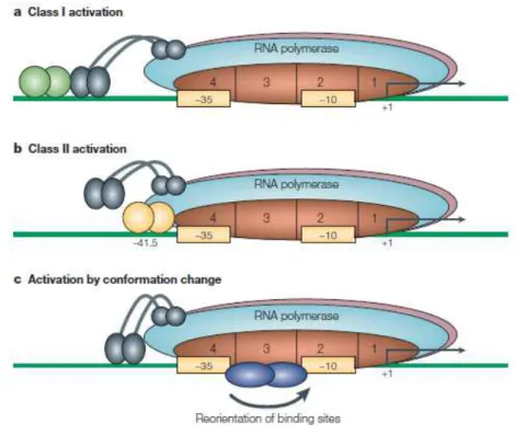
Three general mechanisms are described for transcription activation (FIG.
3). In Class I (FIG. 3a), the activator binds to a target that is located upstream of
the promoter –35 element and recruits RNA polymerase to the promoter by directly interacting with the RNA polymerase αCTD. In Class II activation (FIG. 3b), the activator binds to a target that overlaps the promoter –35 element and
contacts domain 4 of the RNA polymerase σ subunit (Dove et al., 2003). This
Fig. 3. Many activators function as dimers, and are shown as dimers here. Interacting proteins are shown adjacent to each other. (a) Class I activation. The activator is bound to an upstream site and contacts the αCTD of RNA polymerase recruiting thepolymerase to the promoter. (b) Class II activation. The activator binds to a target that is adjacent to the promoter –35 element, and the bound activator interacts with domain 4 of σ70. (c) Activation by conformation changes. The
contact also results in recruitment of RNA polymerase to the promoter, but other
steps in initiation can also be affected. The third mechanism for simple activation
is found in cases where the activator alters the conformation to enable the
interaction of RNA polymerase with the promoter -10 and/or –35 elements. This requires the activator to bind at, or very near to, the promoter elements (FIG 3c).
Transcription repressors
Repressor proteins reduce transcription initiation at target promoters (FIG.
4). Steric hindrance of RNA polymerase binding to promoter DNA is probably
the simplest mechanism of repression (FIG. 4a). The repressor binding site is
located in, or close to, the core promoter elements. However, in some cases, the
repressor might not prevent binding of RNA polymerase to the promoter, but
instead might interfere with post-recruitment steps in transcription initiation
(Muller-Hill, 1998). At other promoters, multiple repressor molecules bind to
promoter-distal sites, and repression might be caused by DNA looping, which
shuts off transcription initiation in the looped domain (FIG. 4b). Finally, complex
cases have been found where the repressor functions as an anti-activator (FIG.
4c).
Folded chromosomes and transcription
In addition to RNA polymerase and transcription factors, the chromosome of
bacteria cells is bound by a battery of proteins involved in DNA repair,
replication, protection, and folding. The folded E. coli chromosome is called the nucleoid and proteins involved in folding the chromosome are known as
nucleoid-associated proteins. In E. coli, panoply of proteins are involved in this compaction, including Fis, IHF, H-NS and HU, StpA (an H-NS homologue) and
Dps. These so-called nucleoid proteins are abundant in the cell, although the
concentrations of some fluctuate sharply depending on the growth conditions.
The binding of these nucleoid proteins to DNA, and the resulting folding of the
bacterial chromosome, must affect the distribution of RNA polymerase between
promoters. The effects of these proteins have been unravelled on a case-by-case
basis at individual promoters. H-NS (Histone-like protein) protein is one of the
most studied nucleoid associated proteins and was seen as a protein that can
completely silence gene expression by forming extended nucleoprotein structures
(Jordi and Higgins, 2000; Petersen et al., 2002; Schnetz, 1995). Interestingly, in E. coli, it has been found that many locations where H-NS is bound to the DNA also contain RNA polymerase (Grainger et al., 2006; Oshima et al., 2006) perhaps
transcription regulation by H-NS. Nucleoid associated factors can also work
together to influence transcription, causing activation or repression, depending
on the context of their binding sites (McLeod and Johnson, 2001).
Overall, transcription is a very important mechanism in the cell and being
a very complex process it needs to be tightly regulated. Part of this thesis is
focused on the transcriptional studies of bolA gene. The first part will give emphasis to the transcriptional regulation of this E. coli morphogene. In a second part, the hypothesis of BolA protein acting in the cell as a transcriptional factor is
analysed.
bolA
morphogene and transcriptional regulation in
E. coli
The gene bolA was first described as a stationary phase gene (Aldea et al., 1989; Aldea et al., 1990). The expression of bolA is growth phase-regulated and controlled by the sigma S (σS) (Lange and Hengge-Aronis, 1991). However, bolA
has also been established as a general stress response gene induced during
exponential phase in response to several stresses (Santos et al., 1999). The
overexpression of bolA leads to substantial changes in the cell and the bacterial bacilli transform into spheres. This effect of bolA on cell morphology is mediated by the induction of penicillin binding protein 5 (PBP5) and penicillin binding
protein 6 (PBP6) D,D-carboxypeptidases (Aldea et al., 1988; Guinote et al., 2011;
Santos et al., 2002). Escherichia coli BolA protein contains one defined BolA/YrbA domain with potential for DNA-binding and regulatory activity. It was already
shown that BolA can bind to the dacA and dacC promoter regions and upregulate these genes, hence expanding its potential as a transcriptional regulator, with
permeability (Freire et al., 2006b). As a result BolA constitutes a privileged target
to study molecular mechanisms of adaptation of Escherichia coli when facing adverse growth conditions.
bolA
regulatory network
bolA encodes a protein with a predicted molecular weight of 13,5 KDa and is transcribed in a clockwise direction in the E. coli chromosome. This gene has two different upstream promoters, a P1 promoter, under the control of σS and an
upstream P2 promoter, controlled by σ70. (FIG. 5) Moreover, bolA mRNA
transcribed by P2 is always detected along bacterial growth, in low amounts,
being classified as a weak and constitutive promoter (Aldea et al., 1989; Lange
and Hengge-Aronis, 1991). Nevertheless, transcript originated by P1 promoter is
primarily detected when cells are already in late-exponential phase of growth,
entrance to the stationary phase or upon stress conditions with a concomitant
decrease of bolA P2 levels (Aldea et al., 1989). This observation gave rise to a
promoter occlusion hypothesis stating that the physical interaction of the trans-
Fig. 5. bolA transcriptional and post-transcriptional regulation. At the transcriptional level, bolA
-cription machinery by σS at the P1 promoter might interfere with the ability of
RNA polymerase to either interact or proceed from P2. P1 promoter is growth
phase and growth rate regulated and contains a gearbox element characteristic of
several other structural genes. Additionally, it was also observed that P1,
similarly to σS, is down regulated by cAMP levels (Lange and Hengge-Aronis,
1991). At transcriptional level, Ribonuclease III (RNase III) has an important role
in the expression of the bolA gene (Freire et al., 2006a; Santos et al., 1997). Also polyadenilation, which inversely correlates with bacterial growth rate (Jasiecki
and Wegrzyn, 2003), reduces RssB-mediated ClpXP σS proteolysis, increasing
rpoS protein levels, thus contributing for the transcription of σS dependent genes
like bolA (Santos et al., 2006). Additionally, Yamamoto and co-workers, saw in vitro that bolAp1 is negatively regulated by phosphor-OmpR (Yamamoto, 2000).
At post-transcriptional level, under carbon starvation, it was shown that
RNase III is also involved in the bolAp1 stability (Freire et al., 2006a). The bolAp1
mRNA is induced nine-fold in a WT strain while in an rnc mutant strain is four-fold induced, showing a decrease in bolAp1 RNA in the absence of RNase III. Taking together this data, RNase III was classified as a contributor to the stability
of bolAp1 mRNA, thus correlating with its role in rpoS RNA.
As it can be observed in the example given above, bolA transcriptional regulation is a complex network where different proteins or transcriptional
factors are involved. The expression of a gene depends not only on the rate of
synthesis of its RNA in the cell (transcription) but also on its rate of decay.
Although transcription is quite important to determine steady-state levels,
Post-Transcriptional Regulation
RNA degradation plays a fundamental role in all biological processes, since
the fast turnover of mRNA permits rapid changes in the pattern of gene
expression. mRNAs vary greatly in stability, and alterations in mRNA decay have
a very high impact in cellular processes. The balance between mRNA degradation
and mRNA synthesis determines the intracellular levels of individual mRNAs in
the cells. There are two very well studied and major post-transcriptional groups
of regulators in bacteria: small regulatory RNAs (sRNAs) and Ribonucleases
(RNases).
Regulatory RNAs can modulate transcription, translation, mRNA stability,
and DNA maintenance or silencing. This panoply of events occur through a
variety of mechanisms, such as changes in RNA conformation, protein binding,
base pairing with other RNAs, and interactions with DNA (Waters and Storz,
2009). sRNAs have been identified in a wide range of bacteria. The major families
of sRNAs include antisense RNAs, synthesized from the strand complementary
to the mRNA they regulate, trans encoded sRNAs that act by limited
complementarity with their targets, and sRNAs that regulate proteins by binding
to and affecting protein activity (Gottesman and Storz, 2010).
sRNAs have been extensively studied over the last years because of their
high importance in the post-transcriptional regulation of bacterial gene
expression.
In the third part of this dissertation, we will study the direct/indirect role of BolA
Small regulatory RNAs
sRNAs from enterobacterial species are usually of ~ 50 to 200 nucleotides,
often expressed under specific growth, stress or virulence conditions (Papenfort
and Vogel, 2009). Whilst some sRNAs modulate the activity of proteins
(Gottesman and Storz, 2010), most of the characterized sRNAs regulate gene
expression by base pairing with mRNAs and those can be split into two different
classes: the ones having perfect base pairing with their target RNA and those with
more limited complementarity. For the first class, one of their functions is to allow
the directed cleavage of the mRNA encoded on the opposite strand. However, the
most prevalent role for antisense sRNAs in bacteria has been the repression of
genes that encode toxic proteins, functionally working as a toxin/anti-toxin
system (Fozo et al., 2008).
The second class of base pairing sRNAs is the trans-encoded sRNAs, which
share only limited complementarity with their target mRNAs and are normally
induced under stress conditions. These regulators modulate the translation
and/or stability of target mRNAs mainly in a negative manner (Aiba, 2007). The
contact between the sRNA and its target mRNA usually leads to repression of
protein levels through translational inhibition and/or mRNA degradation.
However, sRNAs can also activate expression of their target mRNAs through an
anti-antisense mechanism whereby base pairing of the sRNA disrupts an
inhibitory secondary structure (Prevost et al., 2007). The RNA chaperone Hfq is,
in many cases, required for trans-encoded sRNA-mediated regulation, most
probably by actively remodeling the interacting RNAs to melt secondary
structures or by indirectly increase the local RNA concentrations by bringing
together sRNAs and target mRNAs (Valentin-Hansen et al., 2004).
Trans-encoded sRNA typically has multiple target mRNAs (Fig. 6)
given stress condition, this biological particularity means that a single sRNA can
globally modulate a particular physiological response, in much the same manner
as a transcription factor, but at the post-transcriptional level. For example, it is
intriguing that a disproportionate number of trans-encoded sRNAs regulate outer
membrane proteins (MicA, MicC, MicF, RybB, CyaR, OmrA and OmrB) or
transporters (SgrS, RydC, GcvB).
RNases are the enzymes that intervene in the processing, degradation and
quality control of all types of RNAs, including the sRNAs and targets duplex. A
limited number of RNases can exert a determinant level of control acting as a
global regulatory network, monitoring and adapting the RNA levels to the cell
needs. Different enzymes are involved in the RNA degradation mechanisms. Fig. 6. Different targets of sRNAs in E. coli, Salmonella and S. aureus. sRNAs are shown in
There are other accessory enzymes that can also intervene in these processes like
helicases, polymerases (PAP) and RNA binding proteins.
mRNA degradation in the Gram-negative bacteria
In prokaryotes there are three possible pathways by which the mRNA
molecules are degraded (FIG. 7). The more common model for RNA decay in E. coli (the model organism) usually begins with an endonucleolytic cleavage at one or more internal sites on the RNA molecule by the action of an endoribonuclease
(either by RNase E, RNase G or by RNase III) (Arraiano et al., 2010; Carpousis et
al., 2009). After the endonucleolytic cleavages, the transcripts are available for a
direct exoribonucleolytic digestion to oligo- and mononucleotides by
exoribonucleases such as RNase II, RNase R and/or PNPase (Coburn and Mackie,
1998). An alternative path for the RNA degradation relies on the
endoribonuclease RNase E cleavage followed by polyadenylation by the action of
the Poly(A) polymerase (PAP I). The addition of polyA tails to the 3´end of the
mRNA molecules will “help” the activity of exoribonucleases, since these residues are the preferred substrate of some exoribonucleases. Furthermore,
polyadenylation facilitates decay by providing a single-stranded platform for the
3’-exoribonucleases (Coburn and Mackie, 1998). The later mechanism described is of particular importance in the absence of endonucleolytic cleavages. In order to
proceed with the degradation of mRNA molecules, polyadenylation is of extreme
relevance for the removal of secondary structures. In this case mRNA decay can
be achieved by successive cycles of polyadenylation followed by
exoribonucleolytic cleavage (Regnier and Arraiano, 2000). The final step in the
degradation pathway is the degradation of oligoribonucleotides by a
oligoribonuclease of the short mRNA resulting from previews steps (Ghosh and
mRNA degradation in the Gram-positive bacteria
In the gram-positive model bacteria B. subtilis, RNase E is not present to start the mRNA decay pathway. RNase J1 seems to take over this function (FIG. 8)
(Arraiano et al., 2010). Not so long ago, the exoribonucleolytic activity was
believed to be just in the 3´to 5´direction. However, very recently this dogma was
broken, since it was observed that, there is a 5’to 3’ exoribonucleolytic activity in the maturation of 16S ribosomal RNA (Mathy et al., 2007). RNase J1 has been
mRNA turnover. It can be associated with RNase J2 or in a single unit form. For
the initiation of endonuclease cleavage, RNase J1 either binds to the 5’ end or
directly to the internal site of the mRNA. The upstream product is rapidly
degraded by the 3’–5’ exonuclease activity of PNPase. Furthermore, the downstream RNA fragment with the 5’-monophosphate end can be a target of
new RNase J1 endonuclease cleavage or processive 5’–3’ exonucleolytic decay from the 5’ end (Bechhofer, 2009). It was also shown that RNase J1 requires a single-stranded 5’ end with AU-rich regions to allow the exoribonucleolytic activity (Mathy et al., 2007). Another endonuclease sensitive to the 5’ end phosphorylation state of the substrate was recently discovered. RNase Y is
dependent riboswitches (Shahbabian et al., 2009), which controls the expression
of 11 transcriptional units (Henkin, 2008; Winkler and Breaker, 2005). The enzyme
has a major function in the initiation of mRNA degradation in this organism,
affecting mRNA stability >30% in an RNase J1/J2 double-mutant strain.
RNases are mostly responsible for the post-transcriptional control of
RNA in bacteria. They are in charge not just of the mRNA degradation but are
also involved in the quality control of the mRNA in the cells and processing of
specific RNA molecules during their maturation. In many cases, these enzymes
are also related with virulence. In the fourth part of this dissertation, we will
study the role of Streptococcus pneumoniae RNase R, the unique hydrolytic ribonuclease described until now in this human pathogen.
RNase R and the post-transcriptional regulation
RNase R encoded by the rnr gene (previously vacB) is a 3’–5’ hydrolytic exoribonuclease from the RNase II family of exoribonucleases (Cheng and
Deutscher, 2002; Vincent and Deutscher, 2006). In E. coli, the rnr gene is in an operon and its transcription is driven from a putative σ70 promoter upstream of
nsrR (Cairrao et al., 2003; Cheng et al., 1998). RNase R is a processive and sequence-independent enzyme, with a wide impact on RNA metabolism (Cairrao
et al., 2003; Cheng and Deutscher, 2005; Andrade et al., 2006; Andrade et al., 2009;
Purusharth et al., 2007). It is unique among the RNA-degradative exonucleases
present in E. coli as it can easily degrade highly structured RNAs (Awano et al., 2010; Cheng and Deutscher, 2002; Cheng and Deutscher, 2003). This enzyme was
seen to be involved in the control of gene expression (Andrade et al., 2006).
gene expression that could not be replaced by any of the other exoribonucleases.
The activity of RNase R is modulated according to the growth conditions of the
cell and responds to environmental stimuli. RNase R levels are increased under
several stresses, namely in cold shock, and the stationary phase of growth
(Andrade et al., 2006; Cairrao et al., 2003). This protein is not essential for growth
at optimal temperature; however, it is important for growth and viability at low
temperatures (Charpentier et al., 2008). RNase R-like enzymes are widespread in
most sequenced genomes. Even though most knowledge on this protein came
from the work in E. coli, RNase R in other bacteria has been identified. Remarkably, RNase R has also been implicated in the establishment of virulence
in a growing number of pathogens being involved in the modulation of the
expression of virulence in a number of different pathogenic organisms (Cheng et
al., 1998; Erova et al., 2008; Tobe et al., 1992; Tsao et al., 2009). In Streptococcus pneumoniae, there is a unique homologue of the RNase II family of enzymes that was shown to be an RNase R-like protein (Domingues et al., 2009). Proteins
isolated from virulent and non-virulent S. pneumonia strains are different with respect to their activity and RNA affinity (Domingues et al., 2009). In the
gram-positive model oganisms B. subtilis, RNase R was suggested not to play a critical role in RNA degradation; however, it may play a role in mRNA turnover when
polyadenylation at the 3’ end occurs (Oussenko et al., 2005). Moreover, B. subtilis
RNase R was shown to be important for the quality control of tRNAs
(Campos-Guillen et al., 2010). Overall, RNase R-deficient bacteria have been shown to be
less virulent than the wild-type parental strains. However, how this is achieved is
still not completely clear. This is probably related to critical RNA degradation
pathways. The fact that RNase R was found to be the key in the degradation of
sRNAs, namely the virulence regulator SsrA/tmRNA, is probably linked to its
data suggest that bacterial RNase R may be attractive as a potential therapeutic
agent, but clearly more studies are required.
Aim of this Dissertation
In this Dissertation we wanted to focus on both transcriptional and
post-transcriptional regulation of gene expression.
In a first part we have looked for possible regulators of bolA. We searched bioinformatically for putative candidates and H-NS was a possible regulator of
bolA. By mRNA levels studies and different protein-DNA interaction techniques, we investigated if H-NS was regulating bolA and we have characterized its mode of action.
In a second part we aimed at the study of bolA gene as a transcription factor. It has been shown before that bolA was involved in the modulation of the mRNA levels of certain genes related with cell wall synthesis, cell wall structure and cell
division. We have analyzed the impact of bolA overexpression and its absence in the mreB gene that codes for MreB protein that forms the bacterial “cytoskeleton”.
We were also interested in studying the possible role of BolA protein in the
modulation of non-coding RNAs, namely those transcribed by sigma E. In this
part of the work we have studied how a transcription factor can have a role in
post-transcriptional regulation by sRNAs.
Continuing our study on post-transcriptional control we have investigated
the expression of a ribonuclease involved in RNA degradation in the pathogenic
bacterium Streptococcus pneumoniae. We constructed an RNase R mutant (the unique hydrolytic exoribonuclease described in this organism) and characterized
In summary, the theme of this thesis was focused on BolA and RNase R, two
proteins that respectively affect transcription and post-transcriptional
mechanisms in the cell. Every step on the path to understanding how gene
expression is regulated is of major importance in any organism. The pleiotropic
effects of bolA in cell morphology and cell division triggered our curiosity to study the regulation of this gene and understand how it affects such important
mechanisms in the cell. Furthermore, deciphering the role of the until know
unique RNase II family of enzymes in Streptococcus pneumoniae, the RNase R, was also of interest due to the involvement of this important post-transcriptional
regulator in mRNA decay and virulence factors expression in this bacterium.
References
Aiba, H. 2007. Mechanism of RNA silencing by Hfq-binding small RNAs. Curr Opin Microbiol. 10:134-139.
Aldea, M., T. Garrido, C. Hernandez-Chico, M. Vicente, and S.R. Kushner. 1989. Induction of a growth-phase-dependent promoter triggers transcription of bolA, an Escherichia coli morphogene. EMBO J. 8:3923-3931.
Aldea, M., T. Garrido, J. Pla, and M. Vicente. 1990. Division genes in Escherichia coli are expressed coordinately to cell septum requirements by gearbox promoters. EMBO J. 9:3787-3794.
Aldea, M., C. Hernandez-Chico, A.G. de la Campa, S.R. Kushner, and M. Vicente. 1988. Identification, cloning, and expression of bolA, an ftsZ-dependent morphogene of Escherichia coli. J Bacteriol. 170:5169-5176.
Andrade, J.M., F. Cairrao, and C.M. Arraiano. 2006. RNase R affects gene expression in stationary phase: regulation of ompA. Mol Microbiol. 60:219-228.
Andrade, J.M., V. Pobre, I.J. Silva, S. Domingues, and C.M. Arraiano. 2009. The role of 3'-5' exoribonucleases in RNA degradation. Prog Mol Biol Transl Sci. 85:187-229.
Awano, N., V. Rajagopal, M. Arbing, S. Patel, J. Hunt, M. Inouye, and S. Phadtare. 2010. Escherichia coli RNase R has dual activities, helicase and RNase. J Bacteriol. 192:1344-1352.
Bechhofer, D.H. 2009. Messenger RNA decay and maturation in Bacillus subtilis.
Prog Mol Biol Transl Sci. 85:231-273.
Browning, D.F., and S.J. Busby. 2004. The regulation of bacterial transcription initiation. Nat Rev Microbiol. 2:57-65.
Cairrao, F., A. Cruz, H. Mori, and C.M. Arraiano. 2003. Cold shock induction of RNase R and its role in the maturation of the quality control mediator SsrA/tmRNA. Mol Microbiol. 50:1349-1360.
Campos-Guillen, J., J.L. Arvizu-Gomez, G.H. Jones, and G. Olmedo-Alvarez. 2010. Characterization of tRNA(Cys) processing in a conditional Bacillus subtilis CCase mutant reveals the participation of RNase R in its quality control. Microbiology. 156:2102-2111.
Carballido-Lopez, R. 2006. The bacterial actin-like cytoskeleton. Microbiol Mol Biol Rev. 70:888-909.
Carpousis, A.J., B.F. Luisi, and K.J. McDowall. 2009. Endonucleolytic initiation of mRNA decay in Escherichia coli. Prog Mol Biol Transl Sci. 85:91-135. Charpentier, X., S.P. Faucher, S. Kalachikov, and H.A. Shuman. 2008. Loss of
RNase R induces competence development in Legionella pneumophila. J Bacteriol. 190:8126-8136.
Cheng, Z.F., and M.P. Deutscher. 2002. Purification and characterization of the Escherichia coli exoribonuclease RNase R. Comparison with RNase II. J Biol Chem. 277:21624-21629.
Cheng, Z.F., and M.P. Deutscher. 2003. Quality control of ribosomal RNA mediated by polynucleotide phosphorylase and RNase R. Proc Natl Acad Sci U S A. 100:6388-6393.
Cheng, Z.F., and M.P. Deutscher. 2005. An important role for RNase R in mRNA decay. Mol Cell. 17:313-318.
Cheng, Z.F., Y. Zuo, Z. Li, K.E. Rudd, and M.P. Deutscher. 1998. The vacB gene required for virulence in Shigella flexneri and Escherichia coli encodes the exoribonuclease RNase R. J Biol Chem. 273:14077-14080.
Coburn, G.A., and G.A. Mackie. 1998. Reconstitution of the degradation of the mRNA for ribosomal protein S20 with purified enzymes. J Mol Biol. 279:1061-1074.
deHaseth, P.L., M.L. Zupancic, and M.T. Record, Jr. 1998. RNA polymerase-promoter interactions: the comings and goings of RNA polymerase. J Bacteriol. 180:3019-3025.
exoribonucleases from the human pathogens Salmonella typhimurium and Streptococcus pneumoniae. Biochemistry. 48:11848-11857.
Dove, S.L., S.A. Darst, and A. Hochschild. 2003. Region 4 of sigma as a target for transcription regulation. Mol Microbiol. 48:863-874.
Ebright, R.H. 2000. RNA polymerase: structural similarities between bacterial RNA polymerase and eukaryotic RNA polymerase II. J Mol Biol. 304:687-698.
Erova, T.E., V.G. Kosykh, A.A. Fadl, J. Sha, A.J. Horneman, and A.K. Chopra. 2008. Cold shock exoribonuclease R (VacB) is involved in Aeromonas hydrophila pathogenesis. J Bacteriol. 190:3467-3474.
Fozo, E.M., M.R. Hemm, and G. Storz. 2008. Small toxic proteins and the antisense RNAs that repress them. Microbiol Mol Biol Rev. 72:579-589, Table of Contents.
Freire, P., J.D. Amaral, J.M. Santos, and C.M. Arraiano. 2006a. Adaptation to carbon starvation: RNase III ensures normal expression levels of bolAp1 mRNA and sigma(S). Biochimie. 88:341-346.
Freire, P., R.N. Moreira, and C.M. Arraiano. 2009. BolA inhibits cell elongation and regulates MreB expression levels. J Mol Biol. 385:1345-1351.
Freire, P., H.L. Vieira, A.R. Furtado, M.A. de Pedro, and C.M. Arraiano. 2006b. Effect of the morphogene bolA on the permeability of the Escherichia coli outer membrane. FEMS Microbiol Lett. 260:106-111.
Ghosh, S., and M.P. Deutscher. 1999. Oligoribonuclease is an essential component of the mRNA decay pathway. Proc Natl Acad Sci U S A. 96:4372-4377. Gottesman, S., and G. Storz. 2010. Bacterial Small RNA Regulators: Versatile
Roles and Rapidly Evolving Variations. Cold Spring Harb Perspect Biol. Grainger, D.C., D. Hurd, M.D. Goldberg, and S.J. Busby. 2006. Association of
nucleoid proteins with coding and non-coding segments of the Escherichia coli genome. Nucleic Acids Res. 34:4642-4652.
Gross, C.A., C. Chan, A. Dombroski, T. Gruber, M. Sharp, J. Tupy, and B. Young. 1998. The functional and regulatory roles of sigma factors in transcription. Cold Spring Harb Symp Quant Biol. 63:141-155.
Guinote, I.B., R.G. Matos, P. Freire, and C.M. Arraiano. 2011. BolA affects cell growth, and binds to the promoters of penicillin-binding proteins 5 and 6 and regulates their expression. J Microbiol Biotechnol. 21:243-251.
Helmann, J.D., and M.J. Chamberlin. 1988. Structure and function of bacterial sigma factors. Annu Rev Biochem. 57:839-872.
Henkin, T.M. 2008. Riboswitch RNAs: using RNA to sense cellular metabolism.
Genes Dev. 22:3383-3390.
Ishihama, A. 1997. Adaptation of gene expression in stationary phase bacteria.