Brazilian Journal of Microbiology (2009) 40: 933-942 ISSN 1517-8382
MECHANISM OF STERIGMATOCYSTIN BIOSYNTHESIS REGULATION BY PH IN ASPERGILLUS NIDULANS
Francisco Delgado-Virgen, Doralinda Guzman-de-Peña*
Departamento de Biotecnología y Bioquímica. Centro de Investigación y de Estudios Avanzados del IPN Campus Guanajuato.
Km. 9.6 Libramiento Norte Irapuato-León. Irapuato, Guanajuato. 36821, México.
Submitted: February 23, 2009; Returned to authors for corrections: April 26, 2009; Approved: May 15, 2009.
ABSTRACT
External pH constitutes one of the most important environmental factors that control growth, metabolism
and differentiation in microorganisms, including fungi. We have analyzed the effect of external pH on
sterigmatocystin biosynthesis in Aspergillus nidulans. It was observed in repeated experiments that
alkaline pH, in opposition to acid pH, increased sterigmatocystin production and the transcript levels of
aflR, the master gene that regulates expression of the sterigmatocystin cluster in A. nidulans.
It is known that pH effects in fungi operate mostly through the Pal/Pac signaling pathway, originally
described in Aspergillus nidulans. Accordingly, we studied the role of this signaling pathway in ST
biosynthesis. It was observed that aflR transcript levels were increased in the “alkalinity mimicking”
mutant pacCc14 and were minimal in the “acidity mimicking” mutant palA1. No sterigmatocystin was
produced by palA1 or pacC- mutants at neither acid or alkaline pH of incubation. Finally, fluG and flbA,
genes known to regulate both conidiation and sterigmatocystin synthesis upstream in the regulatory
cascade, were up-regulated at alkaline pH.
Key words: Aspergillus nidulans; pH; sterigmatocystin; pacC
INTRODUCTION
Mycotoxins are a group of toxic secondary metabolites
produced by fungi, mainly from the genera Aspergillus,
Penicillium and Fusarium (22). They are frequently found
contaminating a wide variety of food and feed, representing a
serious threat to human and animal health and causing severe
economic losses (8). Aflatoxins (AF) are among the most
studied mycotoxins. Aflatoxins are produced mainly by
Aspergillus flavus and A. parasiticus. Aflatoxin B1 is not only
toxic but also teratogenic and mutagenic; being the most
potent carcinogenic compound found in nature (15).
Studies using the model organism A. nidulans, which
produces the AF precursor sterigmatocystin (ST), have led to
a better understanding of the biosynthetic steps involved in
AF production (4). The fact that the mycotoxin gene clusters
from A. nidulans and A. parasiticus have high homology (25)
has also been helpful in the study of AF/ST synthesis and
regulation.
Among the most important physiological determinants
that regulate AF/ST production in Aspergilli are the carbon
and nitrogen sources, and the pH of the medium. Simple
carbohydrates, such as glucose and sucrose, favor AF
biosynthesis in A. parasiticus and ST biosynthesis in A.
Delgado-Virgen, F. et al.
nidulans (19). It is also known that while ammonium favors
AF biosynthesis in A. parasiticus, it inhibits ST production in
A. nidulans (13). However, studies on the effect of pH on
mycotoxin synthesis have often produced complex and at
times contradictory results; e.g. data in the literature have
reported either a positive (9, 11, 18) or no effect (5) of acid
pH in aflatoxin biosynthesis by A. flavus and/or A.
parasiticus, but no information exists on the mechanism
involved (for a discussion see (7)). It has also been reported
that ST biosynthesis was stimulated by acid pH in A.
nidulans (18).
Regulation of fungal metabolism by pH is mostly
effected through the Pal/Pac signaling pathway, described for
the first time in A. nidulans (6). PacC is a transcription factor
activated at alkaline pH by the products of six pal genes that
sense external pH and transduces the activating signal. PacC
activation may lead to repression of genes expressed
preferentially under acidic growth conditions, and
up-regulation of genes transcribed preferentially under alkaline
growth conditions, including pacC itself (23). Loss of
function mutations in any of the pal genes or pacC, lead to an
“acidity mimicking” phenotype; i.e., display a pattern of gene
expression similar to that of the wild-type strains grown
under acidic conditions, independently of ambient pH. In
contrast, pacC constitutive mutants show an “alkalinity
mimicking” phenotype, displaying a gene expression pattern
similar to that of the wild-type strains grown under alkaline
conditions, irrespective of ambient pH (see (20, 21) for
helpful reviews). A pacC null mutant from A. nidulans
displays an “acidity mimicking” phenotype, and exhibits
poor growth and almost null conidiation, leading to the
hypothesis that PacC might also be involved in development
(23). Interestingly, Guzman-de-Peña and Ruiz-Herrera (12)
found a correlation between AF biosynthesis and sporulation
in A. parasiticus; and between both sexual and asexual
sporulation and ST production in A. nidulans (13), thus
linking mycotoxin synthesis and differentiation in Aspergilli.
Further on, several reports that proposed a model
G-protein/cAMP/PKA signaling pathway that connected
growth, conidiation and ST production in A. nidulans have
been published (3, 14).
Taking into consideration all these data, we proceeded to
analyze the regulation of ST production by ambient pH in A.
nidulans, and whether the Pal/Pac signaling pathway was or
not involved in this process.
MATERIALS AND METHODS
Strains and Growth Media and Conditions
The strains used for these studies were Aspergillus
nidulans FGSC26 (biA1, veA1), A. nidulans MAD002 (biA1,
veA1), A. nidulans MAD134 (biA1, vea1, palA1, wA3), A.
nidulans MAD135 [biA1,veA1, pacCc14 (5-492)] and
MAD812 (pantoB100, veA1, pacC+/-209). A. nidulans
FGSC26 and A. nidulans MAD002 are both good ST
producers. A. nidulans MAD134 is a mutant strain unable to
process the inactive form of PacC, and therefore mimicks
growth in acidic conditions regardless of the pH of the
medium (“acidity mimicking” phenotype). On the other
hand, the A. nidulans mutant strain MAD135 produces only a
pH-independent activated form of PacC, mimicking growth
in alkaline conditions, also regardless of the pH of the
medium (“alkalinity mimicking” phenotype); and strain
MAD812 has a stringent loss of function mutation. All MAD
strains were kindly donated by Miguel A. Peñalva (Centro de
Investigaciones Biológicas, Madrid, Spain).
All strains were kept at 4 °C as silica stocks. For conidia
production, A. nidulans strains FGSC26, MAD002,
MAD135, and MAD812 aliquots from the stocks were
transferred to solid Kafer medium (16) with sodium nitrate as
the sole nitrogen source and supplemented with 5% sucrose
as carbon source, and grown for 5 days at 37 °C. A. nidulans
MAD134 was grown at 28 °C on solid Kafer medium with
ammonium tartrate and 5% sucrose (acidic growth
conditions) in order to avoid extragenic suppressors of the
Sterigmatocystin Biosynthesis Regulation in A. nidulans
Spore suspensions
Conidia were harvested by adding 10 ml of a Triton
X-100 solution (0.01%) to each Petri dish, counted with a
hemocytometer and adjusted to the desired inoculum
concentration with sterile distilled water. Before inoculation
conidia suspensions were kept overnight at 4 °C to ensure an
even germination.
Growth conditions
Conidia were inoculated into 100 x 15 mm Petri dishes
containing 50 ml of liquid (1x10⁷) or solid (1x10⁶) Kafer
medium (see Results) supplemented with 5% sucrose and 70
mM sodium nitrate as carbon and nitrogen sources
respectively, and incubated under static conditions at 37 °C
for variable periods of time. In some experiments, pH of the
media was adjusted with 100 mM citrate buffer.
At the end of the incubation period, in liquid cultures the
medium was separated from the mycelium by filtration.
Mycelia were transferred to a Petri dish, washed with 10 ml
of distilled water containing 0.01% Triton X-100, and gently
shaken to release the spores. This procedure was repeated
twice. Mycelia were then dried at 75 °C for 24 h, stored at
room temperature in desiccators with silica gel as drying
agent for another 24 h, and weighed. The residual growth
medium was processed immediately or stored at 4 °C until
used. When mycelia were used for RNA extraction, they
were collected by vacuum filtration, frozen with liquid
nitrogen and stored at -70 °C until used. When solid medium
was used, after spore recovery ST was extracted as described
below, agar was melted in a microwave oven, mycelia were
recovered with the use of tweezers, washed with hot water,
dried and weighed as above.
ST quantitation
ST was extracted from the mixture of culture medium,
dry mycelium (see above) and conidia, with 50 ml acetone
for 30 min, followed by 50 ml chloroform by further 30 min
(17). The organic phase was separated, filtered through
anhydrous sodium sulfate and evaporated in a fume hood in a
water boiling bath. The residue was resuspended in 500 µL
HPLC grade methanol and filtered through C-18 columns
(Alltech).
Analysis and quantitation were performed by HPLC as
previously described (13). The detection limit was 3 ng ST in
20 µl samples.
RT-PCR analysis of the expression of genes encoding proteins involved in growth and ST synthesis
Total RNA extraction was performed using TriZOL
(Invitrogen). RNA purification was achieved using RNeasy
mini Kit (Qiagen). RNA integrity was confirmed by
electrophoresis in a 1.5% agarose gel in TAE 1X. The
expression levels of the genes fluG, flbA, laeA, pacC and
aflR, were evaluated semi quantitatively by RT-PCR.
First-strand cDNA was obtained using 1 µg of total RNA, adding 1
µl Oligo dT (Invitrogen), 1 µl dNTP mix (10 µM each) and
enough RNase-free water to adjust to 12 µl, heating the mix
at 65 °C for 15 min and transferring it immediately to ice. 4
µl of 5x RT Buffer, 2 µl of DTT 0.1 M and 1 µl RNase OUT
(Invitrogen) were added to each reaction, gently mixed and
incubated at 42 °C for 2 min. After that, 1 µl Superscript II
Reverse Transcriptase (Invitrogen) was added to each
reaction for a final volume of 20 µl. RT conditions were: an
initial incubation at 42 °C for 50 min, followed by 15 min at
70 °C. The cDNA derived from 0.1 µg of total RNA was
used as a template. PCR conditions were as follows: After an
initial incubation at 94 °C for 3 min, 28 cycles of 94 °C for
45 s, 54 °C for 1 min and 72 °C for 1 min were performed,
followed by a final incubation at 72 °C for 7 min.
The PCR primers for each gene were as follows:
5’-ACCCTAATGTTTATTTGGAT-3’ (forward) and
5’-TGGATAGGTCTGGTATAAGG-3’ (reverse) for fluG; 5’-,
TCCCTCAAATTCTCTCAATCGAACCGG-3’ (forward)
and 5’-GTAGAATGACAGGTTTTCTTCGCAGA-3’
(reverse) for flbA; 5’-GGTGACGATTTGTATAGTCC-3’
(forward) and 5’-CTCTTCATGAAACTGGTTTC-3’
(reverse) for laeA; 5’-GACTGACGGTATGACTTCTG-3’
Delgado-Virgen, F. et al.
(reverse) for pacC; 5’-GCCATCCTGTCTCCGAATAC-3’
(forward) and 5’-CGAACCTCTACGACTGTCTTG-3’
(reverse) for aflR; and
5’-CCAAGGCCAACCGCGAGAAGATGAC-3 (forward) and
5’-AGGGTACATGGTGGTGCCGCCAGAC-3’ (reverse)
for γ-actin (control gene). Transcription levels were
normalized by comparing the UV absorption intensity of the
bands to that of γ-actin using ImageJ software
(http://rsb.info.nih.gov/ij/).
Statistical analyses
Each experiment was repeated three to five times, and
values presented in this paper are means of these replicates.
Differences between means were evaluated by Tukey’s Test
(p=0.05) using SAS (version 6.12; SAS Institute, Cary, NC,
USA). Coefficients of determination (r2) and regression data
are included in the corresponding results.
RESULTS
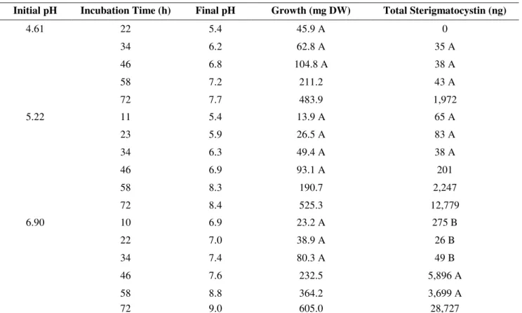
Relationship between initial pH, growth and ST production
A. nidulans MAD002 (wild-type strain) was grown in
Kafer medium at three different initial pH values for 72 h.
Growth, conidiation and ST synthesis were evaluated
approximately every 12 h. When initial pH was 4.61, it was
observed that only basal levels of ST were produced until 58
hours of incubation, ST synthesis being activated after this
time (Table 1). Activation of ST synthesis occurred earlier
when the initial pH value was raised (58 h at initial pH= 5.22,
and 46 h at pH= 6.90) (Table 1). As it may be seen in this
table, time of activation of ST synthesis occurred when pH
reached a value above 7.5. The amount of ST produced and
mycelial growth at 72 h of incubation were also linearly
increased as initial pH value of the medium increased (r2 =
0.977 and r2 = 0.993, respectively).
Table 1. Rate of growth and ST synthesis in A. nidulans MAD002 (wild-type).
Initial pH Incubation Time (h) Final pH Growth (mg DW) Total Sterigmatocystin (ng)
22 5.4 45.9 A 0
34 6.2 62.8 A 35 A
46 6.8 104.8 A 38 A
58 7.2 211.2 43 A
4.61
72 7.7 483.9 1,972
11 5.4 13.9 A 65 A
23 5.9 26.5 A 83 A
34 6.3 49.4 A 38 A
46 6.9 93.1 A 201
58 8.3 190.7 2,247
5.22
72 8.4 525.3 12,779
10 6.9 23.2 A 275 B
22 7.0 38.9 A 26 B
34 7.4 80.3 A 49 B
46 7.6 232.5 5,896 A
58 8.8 364.2 3,699 A
6.90
72 9.0 605.0 28,727
Sterigmatocystin Biosynthesis Regulation in A. nidulans
Transcription of regulatory genes through time of incubation
The previous experiment suggested a correlation
between pH and ST production. Taking into consideration
that both phenomena are regulated in Aspergilli by known
pathways (6, 25), we decided to investigate the
correlation existing between the drift in external pH
during incubation, and the expression of the
corresponding regulatory genes. A. nidulans MAD002
was incubated in Kafer medium pH = 6.60 for 83 h.
Mycelia were collected at different intervals, and levels of
transcripts of the regulatory gene aflR was evaluated (Fig.
1). aflR expression levels increased through time linearly
(r2 = 0.922) as pH of the medium was increased, raising
the hypothesis that alkaline pH promotes ST biosynthesis.
Figure 1. Effect of pH on the transcription of the regulatory gene aflR in the wild-type strain MAD002 through incubation time. Cultures were incubated in large Petri dishes containing liquid pH= 6.5 unbuffered Kafer medium under static conditions
at 37 °C. At intervals, sets of dishes were recovered, RNA was extracted and transcription levels were measured by
semi-quantitative RT-PCR. γ-actin was used as an endogenous reference. The amount of each mRNA was normalized to the amount
of γ-actin mRNA in each sample. Data are means ± SD (n= 3). Time of incubation and actual pH of the different samples are
indicated.
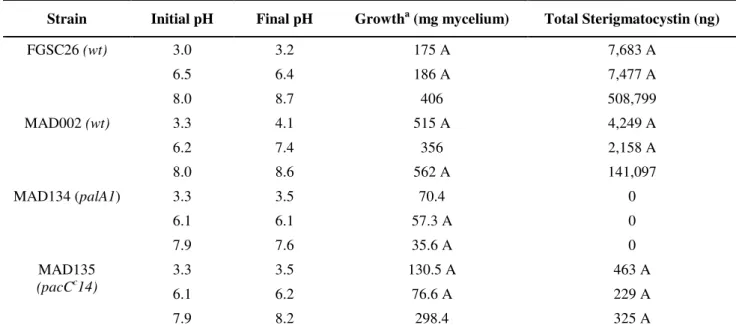
Determination of the optimum pH for growth and ST production in the wild-type and mutant strains affected in the Pal/Pac pathway
A. nidulans mutants affected in the Pal/Pac pathway were
included in these experiments because of the general
observation on the role of this pathway in the regulation of
different phenomena by pH in Aspergilli. All strains were
grown in Kafer (liquid or solid) medium containing 100 mM
citrate buffer at different pH values. It was observed that
mycelial growth of wild-type strains (FGSC26 and MAD002)
was higher at alkaline pH. ST levels were about 30-70-fold
higher at alkaline pH as compared to acidic or neutral
conditions in both strains. In contrast to these results, the A.
nidulans “acidity mimicking” mutant MAD134 (palA1) grew
poorly at these three different pH values compared to the
wild-type strains grown at similar pH values and time. 0
0.2 0.4 0.6 0.8 1 1.2 1.4 1.6 1.8 2
24 h 35 h 48 h 59 h 72 h 83 h
aflR
7.0 7.0 7.3 7.7 8.2 8.3
Actual pH Incubation time
Delgado-Virgen, F. et al.
Interestingly, this mutant was unable to produce ST at any of
the pH values tested. On the other hand, the MAD135
(pacCc14) mutant produced ST regardless of the pH of the
medium. Nevertheless, as previously reported (18), its levels
of ST production were well below those from the wild-type
strains. These results are shown in Table 2. Finally, contrary
to the behavior of strain MAD135, a pacC negative mutant
(MAD812) was unable to synthesize ST at neither acid nor
alkaline pH (Table 3).
Table 2. Growth and ST synthesis in A. nidulans strains grown in buffered medium.
Strain Initial pH Final pH Growtha (mg mycelium) Total Sterigmatocystin (ng)
3.0 3.2 175 A 7,683 A
6.5 6.4 186 A 7,477 A
FGSC26 (wt)
8.0 8.7 406 508,799
3.3 4.1 515 A 4,249 A
6.2 7.4 356 2,158 A
MAD002 (wt)
8.0 8.6 562 A 141,097
3.3 3.5 70.4 0
6.1 6.1 57.3 A 0
MAD134 (palA1)
7.9 7.6 35.6 A 0
3.3 3.5 130.5 A 463 A
6.1 6.2 76.6 A 229 A
MAD135 (pacCc14)
7.9 8.2 298.4 325 A
Cultures were obtained in buffered (100 mM citrate) liquid Kafer medium incubated under static conditions, and growth and ST formation were measured as described in Materials and methods. Statistical analysis of samples (n=5) proceeded by the Tukey test (p=0.05). Statistical significance of the differences in the same experimental block (same strain and medium) were treated as described for Table 1.
Table 3. Growth and ST synthesis of A. nidulans MAD002 and MAD812 strains grown in solid buffered medium.
Strain Initial pH Final pH Growth (mg¹) Total ST
3.1 4.4 726.9 A 29,076
MAD002 (wt)
7.8 8.9 747.0 A 2’530,700
3.1 4.0 707.3 0
MAD812 (pacC+/209)
7.8 8.3 260.6 0
Cultures were obtained in buffered (100 mM citrate) solid Kafer medium, and growth and ST formation were measured as described in Materials and methods. Statistical analysis of samples (n=5) proceeded by the Tukey test (p=0.05). Statistical significance of the differences in the same experimental block (same strain and medium) were treated as described for Table 1.
Determination of the expression of regulatory genes in wild-type and mutants affected in the Pal/Pac pathway
In order to analyze the effect of pH on the expression of
the regulatory genes (pacC and aflR) in wild-type and Pal/Pac
affected mutants, we used buffered medium of different pH
values as described above. It was observed that expression
levels of pacC and aflR in strain FGSC26 increased linearly
as pH of the medium increased (r2 = 0.981 and r2 = 0.997,
respectively). Expression levels of pacC in strain MAD134
Sterigmatocystin Biosynthesis Regulation in A. nidulans
levels were also basal, in agreement with the absence of ST
production at any pH value. Transcript levels from strain
MAD135 (pacCc14) contrasted with those of strain MAD134
(palA1). Levels of pacC transcript in pacCc14 were twice
higher than those from palA1 mutant, and its aflR transcript
levels were high regardless of the pH of the medium,
correlating with ST production in this strain (these results are
shown in Fig. 2).
Figure 2. Effect of pH on the transcription of pacC and aflR genes in wild-type and mutant strains of A. nidulans grown at three different pH values. Culture conditions proceeded as described for Fig. 1, but medium was added of 100 mM citrate
buffer at the indicated pH values, and incubation lasted for 72 h. Gene transcription levels were analyzed as described for Fig.
1. Data are means ± SD (n= 3). a) wild-type strain FGSC26; b) “acidity mimicking” mutant MAD134 (palA1); c) “alkalinity
mimicking” mutant MAD135 (pacCc14). Initial and final pH values of the different samples are indicated.
0 0.2 0.4 0.6 0.8 1 1.2 1.4 1.6 1.8 2
3.0 6.5 8.0
pacC
aflR
(r2= 0.981) (r2= 0.997)
3.2 6.4 8.7
Initial Final
a
0 0.2 0.4 0.6 0.8 1 1.2 1.4 1.6 1.8 2
3.3 6.1 7.9
pacC aflR
Initial
Final pH 3.5 6.1 7.6
b
Initial pH
Final pH 3.5 6.2 8.2
0 0.2 0.4 0.6 0.8 1 1.2 1.4 1.6 1.8 2
3.3 6.1 7.9
pacC aflR
Delgado-Virgen, F. et al.
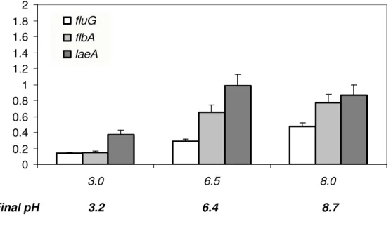
Effect of pH on the regulatory mechanism of ST synthesis We further proceeded to determine the probable level at
which external pH affects ST production. It has been
suggested that fluG and flbA encoded proteins have a positive
role in ST production (17). In this model, laeA transcript
product also activates ST formation (3). Transcript levels of
these genes were evaluated in FGSC26 and MAD134 (palA1)
strains grown at three different pH values in citrate buffered
medium for 72 h (Fig. 3). It was noted that fluG (r2= 0.998 in
wild-type and r2= 0.971 in MAD134 strains) and flbA (r2=
0.889 and r2= 0.976, respectively) were preferentially
expressed at alkaline pH in both strains. On the other hand
transcript levels of laeA were higher at neutral and alkaline
pH in the wild-type strain (Fig. 3), but in the MAD134
(palA1) strain an opposite trend in expression was observed,
decreasing with an increase in pH (data not shown).
Figure 3. Effect of pH on the transcription of fluG, flbA and laeA genes in the wild-type strain FGSC26 grown at three different pH values. Culture conditions proceeded as described for Fig. 2, and transcription levels were analyzed as described
for Fig. 1. Data are means ± SD (n= 3).
DISCUSSION
The formation of natural products resulting from the
secondary metabolism of plants and microorganisms is
subjected to different environmental cues, such as
temperature, humidity, light, pH, nutrient source, etc.
Aflatoxins (AF), probably the most important and studied
mycotoxins, do not escape to this rule. Accordingly, it has
been described that temperature and nitrogen source affect
AF production in A. parasiticus and ST synthesis in A.
nidulans, but in different ways (11). It was demonstrated that
nitrate as the sole nitrogen source promoted ST synthesis in
A. nidulans, while ammonium promoted AF production in A.
parasiticus. It was also observed that AF production by A.
parasiticus was best at 27 °C, while ST was maximally
produced by A. nidulans when incubated at 37 °C (11).
pH is also an important factor in the production of
aflatoxins. Acid pH has been described to stimulate AF
synthesis in A. parasiticus (10, 18). According to the results
described in this paper, we may conclude that pH of the
medium does have an effect on ST synthesis in A. nidulans,
with alkaline pH promoting mycotoxin production. These
data are in contradiction with a previous communication
reporting stimulation of ST biosynthesis at acid pH (18). The
0 0.2 0.4 0.6 0.8 1 1.2 1.4 1.6 1.8 2
3.0 6.5 8.0
fluG flbA laeA
reasons for this discrepancy are difficult to pin point.
Possibly, conditions of growth, time of incubation and the
determination only of ST present in the mycelium (contrary
to our protocols) may be partially responsible for this
behavior.
Among our data that support the conclusion that it is
alkaline pH the one that promotes ST production, we may
cite the following: 1) the repeated observation that higher
levels of ST are produced at alkaline pH in the wild-type
strains; 2) the observation that expression levels of aflR (the
transcriptional regulator of the gene cluster encoding proteins
involved in ST synthesis (24)) in the wild-type strain of A.
nidulans were high under alkaline conditions, and very low at
acidic pH; 3) the observation that an “acidity mimicking”
palA mutant did not produce ST at any pH value; 4) the
observation that a PacC constitutive “alkalinity mimicking”
mutant produced similar levels of ST at all the pH values
tested, although as reported (18), at lower levels than the
wild-type strain; 5) the fact that a pacC negative mutant did
not form ST at any pH value. These last results also meet the
criteria for recognizing gene regulation by pH through the
Pal/Pac signaling pathway (1); and finally 6) it is also
important to point out that the regulation of other genes
involved in the common control of ST biosynthesis (laeA,
fluG and flbA) are in agreement with a positive regulation by
alkaline pH.
It is accepted that LaeA is a global regulator of
secondary metabolism in A. nidulans (2). Levels of
transcription of laeA gene were higher at neutral to alkaline
growth conditions in the wild-type strain.
fluG and flbA are known to be involved in a
G-protein/cAMP/PKA signaling pathway that connects ST
synthesis and development in A. nidulans (3, 14). The
observation that transcription of both genes was stimulated
by alkaline pH of the medium agrees with the general
observation that sterigmatocystin production is stimulated at
alkaline pH.
In summary, based on our results, we may suggest that
alkaline pH regulates the signaling pathway that controls ST
Sterigmatocystin Biosynthesis Regulation in A. nidulans
synthesis in A. nidulans at the level of aflR by the Pal/Pac
signaling pathway.
ACKNOWLEDGMENTS
We wish to thank Prof. Miguel A. Peñalva for providing
us all of the A. nidulans MAD strains, and to Prof. José Ruiz
Herrera for his helpful comments and suggestions to this
manuscript. We acknowledge technical support to Gloria
Laura Anguiano-Ruvalcaba and Yolanda Rodriguez-Aza.
FDV Ph.D fellowship (181787), was supported by
CONACYT, México
REFERENCES
1. Arst, H.N. Jr; Peñalva, M.A. (2003). Recognizing gene regulation by ambient pH. Fungal Genet. Biol, 40, 1-3.
2. Bok, J.W.; Keller, N.P. (2004). LaeA, a regulator of secondary metabolism in Aspergillus spp. Eukaryotic cell 3, 527-535.
3. Brodhagen, M.; Keller, N.P. (2006). Signaling pathways connecting mycotoxin production and sporulation. Mol. Plant Pathol. 7, 285-301. 4. Brown, D.W.; Yu, J.H.; Kelkar, H.S.; Fernandes, M.; Nesbitt, T.C.;
Keller, N.P.; Adams, T.H.; Leonard, T.J. (1996). Twenty-five coregulated transcripts define a sterigmatocystin gene cluster in
Aspergillus nidulans. Proc. Natl. Acad. Sci. USA 93, 1418–1422. 5. Buchanan, R.L.; Ayres, J.C. (1975). Effect of initial pH on aflatoxin
production. Appl Microbiol. 30, 1050-1051.
6. Caddick, M.X.; Brownlee, A.G.; Arst, H.N.Jr. (1986). Regulation of gene expression by pH of the growth medium in Aspergillus nidulans. Mol. Gen. Genet. 203, 346-353.
7. Calvo, A.M.; Wilson, R.A.; Bok, J.W.; Keller, N.P. (2002). Relationship between secondary metabolism and fungal development.
Microbiol. Mol. Biol. Rev. 66, 447-459.
8. Cardwell, K.F.; Desjardins, A.; Henry, H.S.; Munkvold, G., Robens, J. (2001). Mycotoxins: The cost of achieving food security and food
quality. APSnet.org. Available at:
http://apsnet.org/online/feature/mycotoxin/top.html. Accessed 13 Feb
2009.
9. Cotty, P.J. (1988). Aflatoxin and sclerotial production by Aspergillus flavus: Influence of pH. Phytopathology 78, 1250-1253.
10. Detroy, R.W.; Hesseltine, C.W. (1969). Net synthesis of 14C-labeled lipids and aflatoxins in resting cells of Aspergillus parasiticus. Dev. Ind. Microbiol. 10, 127–133.
Delgado-Virgen, F. et al.
in Aspergillus parasiticus and A. nidulans. Appl. Environ. Microbiol. 64, 2275-2277.
12. Guzman-de-Peña, D.; Ruiz-Herrera, J. (1997). Relationship between aflatoxin biosynthesis and sporulation in Aspergillus parasiticus.
Fungal Gen. Biol. 21, 198-205.
13. Guzman-de-Peña, D.; Aguirre, J.; Ruiz-Herrrera, J. (1998). Corrrelation between the regulation of sterigmatocystin biosynthesis and asexual and sexual sporulation in Emericella nidulans. Antonie Van Leeuwenhock. 73, 199-205.
14. Hicks, J.K.; Yu, J.H.; Keller, N.P.; Adams, T.H. (1997). Aspergillus
sporulation and mycotoxin production both require inactivation of the FadA Ga protein-dependent signaling pathway. EMBO J. 16, 4916-4923.
15. International Agency for Research on Cancer (IARC). (2002). Aflatoxins (Naturally occurring mixtures). Monogr. Eval. Carcinog. Risks. Hum. 82. Available at http://monographs.iarc.fr. Accessed 13 Feb 2009.
16. Kafer, E. (1977). Meiotic and mitotic recombination in Aspergillus and its chromosomal aberrations. Adv. Genet. 19, 131-133.
17. Keller, N.P.; Kantz, N.J.; Adams, T.H. (1994). Aspergillus nidulans veA is required for production of the mycotoxin sterigmatocystin. Appl. Environ. Microbiol. 60, 1444-1450.
18. Keller, N.P.; Nesbitt, C.; Sarr, B.; Phillips, T.D.; Burow, G.B. (1997). pH regulation of Sterigmatocystin and Aflatoxin Biosynthesis in
Aspergillus spp. Phytopathology. 87, 643-648.
19. Payne, G.A.; Brown, M.P. (1998). Genetics and physiology of aflatoxin biosynthesis. Annu. Rev. Phytopathol. 36, 329-362.
20. Peñalva, M.A.; Arst, H.N. Jr. (2002). Regulation of gene expresión by ambient pH in filamentous fungi and yeasts. Microbiol. Mol. Biol. Rev.
66, 426-446.
21. Peñalva, M.A.; Arst, H.N.Jr. (2004). Recent advances in the characterization of ambient pH regulation of gene expression in filamentous fungi and yeasts. Ann. Rev. Microbiol. 58, 425-451. 22. Task Force Report. (2003). Fungal growth and mycotoxin development
by major mycotoxigenic fungi. In Mycotoxins: Risks in plant, animal, and human systems, pp. 129-135. Edited by the Council for Agricultural Science and Technology. CAST, Ames, Iowa, USA. 23. Tilburn, J.; Sarkar, S.; Widdick, D.A.; Espeso, E.A.; Orejas, M.;
Mungroso, J.; Peñalva, M.A.; Arst, H.N.Jr. (1995). The Aspergillus
PacC zinc finger transcription factor mediates regulation of both acid- and alkaline-expressed genes by ambient pH. EMBO J. 14, 779-790. 24. Woloshuk, C.P.; Foutz, K.R.; Brewer, J.F.; Bathnagar, D.; Cleveland,
T.E.; Payne, G.A. (1994). Molecular characterization of aflR, a regulatory locus for aflatoxin biosynthesis. Appl. Environ. Microbiol. 60, 2408-2414.
25. Yu, J.; Chang, P.K.; Erlich, K.C.; Cary, J.W.; Bhatnagar, D.;