INTRODUCTION
Plants synthesize toxic chemicals in large amounts, primarily as a defense against bacterial, fungal, insect and other animal predators. Various toxicological studies have assessed the mutagenic, teratogenic and carcinogenic prop-erties of some of these chemicals (Ames, 1983; Konstan-topoulou et al., 1992; Varanda et al., 1997). In many cases, the active compounds have been shown to be sesqui- and diterpenes (Gilbert et al., 1970).
The sesquiterpene lactones (SL) from plants com-prise a group of substances with a variety of biological ef-fects. These compounds are terpenoids and are characteris-tic of the Asteraceae but may also occur in other angiosperm plant families as well as in plants of the genus Hepatica; their principal structural characteristic is the presence of α,β -unsaturated γ-lactone (Rodriguez et al., 1976). SL possess antibacterial, antifungal, antitumor, antiprotozoal, antihel-minthic, schistosomicidal, cytotoxic and analgesic activi-ties (Picman, 1986). Glaucolide B (Figure 1) is an SL that is active against the embryos and adults of Biomphalaria glabrata snails, causing approximately 70 and 90% mortal-ity, respectively, within 24 h; this compound has an analge-sic action in mice and shows strong antimicrobial activity against Bacillus cereus (Alarcon et al., 1990).
Based on these data, we have examined the clastogenic and cytotoxic activities of glaucolide B in tem-porary cultures of human lymphocytes and in bone mar-row cells of BALB/c mice.
MATERIAL AND METHODS
Glaucolide B preparation
Glaucolide B extracted from Vernonia eremophila Mart. (family Asteraceae) (Alarcon et al., 1990) was dis-solved in dimethylsulfoxide (DMSO, Merck) and diluted in RPMI-1640 medium (Sigma) to the desired concentra-tions for the in vitro assays. For tests in vivo, the lactone was suspended in a solution of powdered milk at a con-centration of 320 mg/kg body weight (b.w.).
Assays with human peripheral blood lymphocytes
Human peripheral blood lymphocytes were ob-tained from six volunteers (three women and three men) and analyzed for chromosomal aberrations. For the analy-sis of analy-sister chromatid exchanges (SCE) and proliferation index (PI), lymphocytes were obtained from three non-smoking, healthy volunteers (two women and one man) aged 18-21 years. Metaphase preparations were obtained as described by Moorhead et al. (1960). The lymphocytes
GENOTOXIC ACTION OF THE SESQUITERPENE LACTONE GLAUCOLIDE B
ON MAMMALIAN CELLS in vitro AND in vivo
Regislaine V. Burim1,2, Renata Canalle1,2, João L. Callegari Lopes3 and Catarina S. Takahashi1,2
ABSTRACT
Glaucolide B is a sesquiterpene lactone isolated from Vernonia eremophila Mart. (Vernonieae, Asteraceae) and has schistosomicidal, antimicrobial and analgesic activities. This study examined the cytotoxic and clastogenic activities of glaucolide B in human cultured lymphocytes and in bone marrow cells from BALB/c mice. The mitotic index (MI) and chromosomal aberrations were analyzed in both of the above systems, whereas sister chromatid exchanges (SCE) and the proliferation index (PI) were determined only in vitro. In human cultured lymphocytes, glaucolide B concentrations greater than 15 µg/ml of culture medium completely inhibited cell growth. At 4 µg/ml and 8 µg/ml of culture medium, glaucolide B significantly increased the frequency of chromosomal aberrations in lymphocytes and was also cytotoxic at concentrations ≥8 µg/ml; there was no increase in the frequency of SCE. Glaucolide B (160-640 mg/kg) did not significantly increase the frequency of chromosomal aberrations in mouse bone marrow cells nor did it affect cell division. Since glaucolide B showed no clastogenic action on mammalian cells in vivo but was cytotoxic and clastogenic in vitro, caution is needed in its medicinal use.
1Departamento de Genética, Faculdade de Medicina de Ribeirão Preto,
USP, Av. Bandeirantes, 3.900, 14049-900 Ribeirão Preto, SP, Brasil. Send corre-spondence to R.V.B. Fax: +55-16-602-3761. E-mail: revab@rgm.fmrp.usp.br
2Departamento de Biologia, Faculdade de Filosofia, Ciências e Letras de
Ribeirão Preto, USP, Ribeirão Preto, SP, Brasil.
3Departamento de Física e Química, Faculdade de Ciências Farmacêuticas
de Ribeirão Preto, USP, Ribeirão Preto, SP, Brasil
O
O O
O
O
O O O
O
O
were grown in 80% RPMI-1640 medium (Sigma) - 20% fetal calf serum (Cultilab) supplemented with penicillin (5 µg/ml) and streptomycin (10 µg/ml). The cells were stimu-lated with 2% phytohemagglutinin (Gibco). One milliliter of plasma was added to each 10 ml of culture medium, and the cultures incubated at 37°C for 48 h for chromo-somal aberrations and 72 h for SCE studies.
Since preliminary tests showed that a glaucolide B concentration of 15 µg/ml totally inhibited lymphocyte division, concentrations of 2, 4 and 8 µg/ml of culture medium were used in subsequent experiments. Negative control cultures received no treatment. DMSO (final con-centration, 0.104%), and 1-β-D-arabinofuranosylcytosine (Ara-C, concentration 0.0061 µg/ml) were used as the sol-vent and positive controls, respectively.
The cultures were treated with glaucolide B be-tween Go and G1 (6 h after incubation). The cells were harvested after 48 h to check for chromosomal aberrations and after 72 h to check for SCE. 5-Bromo-2’-deoxyuridine (BrdU; Sigma) was added to the SCE cultures to a final concentration of 10 µg/ml of culture medium. Mitotic ar-rest was achieved by adding colchicine (0.016%, 25 µl/10 ml; Sigma) 90 min before fixation. After hypotonic treat-ment (0.075 M KCl for 10 min) and fixation (methanol:glacial acetic acid, 3:1 v/v), the cells were stained with Giemsa diluted in 0.06 M phosphate buffer, pH 6.8, for the analy-sis of chromosomal aberrations and by the fluorescence plus Giemsa technique of Perry and Wolff (1974), in com-bination with the technique of Korenberg and Freedlender (1974) for SCE. One hundred metaphases from each cul-ture were analyzed for chromosomal aberrations, and 2000 cells were scored to determine the mitotic index (MI) (48-h cultures). For SCE analysis, 50 second-division metap(48-hases per culture were examined, and 200 metaphases from each culture were scored for first, second and third cell division (72-h cultures). The PI was obtained using the following equation (Degrassi et al., 1989):
PI = [(M3 - M1) + 1],
where M1 and M3 are the percentages of the first- and third-division metaphases, respectively.
Assay with BALB/c mice bone marrow cells
BALB/c mice (Mus musculus) weighing approxi-mately 30 g were obtained from the Animal House of the Faculty of Medicine of Ribeirão Preto (USP). The mice were divided into groups of six animals (three females and three males) and were treated by gavage with glaucolide B diluted in a solution of powdered milk to give doses of 160, 320 and 640 mg/kg b.w. in a fixed volume of 0.5 ml. The highest dose was determined by the maximum solu-bility of glaucolide B. The mice were killed by ether inha-lation 24 h after treatment and 90 min after intraperitoneal injection of 0.3 ml of a 1% colchicine solution (Sigma).
The negative control group received distilled water, the solvent control group received a powdered milk solution (320 mg milk/kg b.w.) and the positive control group re-ceived 8 mg of cyclophosphamide/kg b.w.
Metaphase cell preparations were obtained from bone marrow cells by the technique of Ford and Hamerton (1956) adapted for mice by Rabello-Gay (1991). One hun-dred metaphases per animal were analyzed to determine the frequency of chromosomal aberrations. The mitotic index represented the number of metaphase cells detected in 2000 cells analyzed per animal and was expressed as a percentage.
Statistical analysis
The Friedman and Kruskal-Wallis tests were used to analyze the results of the in vitro and in vivo assays, re-spectively, by comparing them with the corresponding nega-tive control (Hollander and Wolf, 1973). P values = 0.05
were considered to indicate significance level. The positive control data were not included in the statistical analyses.
RESULTS
Human cultured lymphocytes exposed to glaucolide B
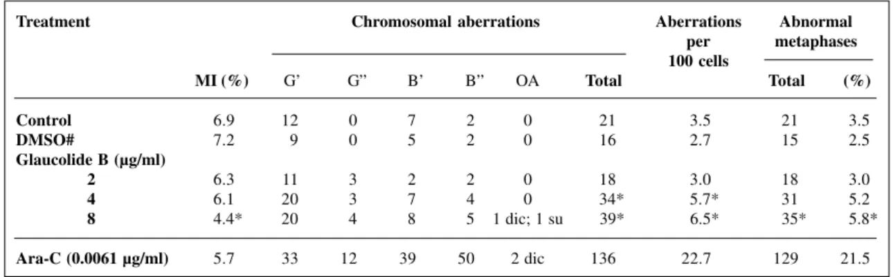
The chromosomal aberrations in human lympho-cytes treated with glaucolide B are summarized in Table I. Gaps were the most consistent structural aberrations. The total frequency of chromosomal aberrations at glaucolide B concentrations of 4 and 8 µg/ml was significantly higher than the controls (P = 0.0387). There were no significant differences among individuals. Compared with the nega-tive control, glaucolide B significantly increased number of cells with chromosome aberrations only at the highest concentration tested (P = 0.0407) (Table I). This concentra-tion also significantly decreased the mitotic index compared to the controls (P = 0.0206). Glaucolide B did not increase the SCE frequency nor did it alter the cell PI (Table II).
BALB/c mice bone marrow cells exposed to glaucolide B
Table II - Frequencies of sister chromatid exchange (SCE) and the proliferation index (PI) in human cultured lymphocytes treated with glaucolide B.
Treatment SCE No of metaphases PI
Mean ± SEM Total 1st cycle 2nd cycle 3rd cycle
Control 9.9 ± 0.3 1482 89 106 105 1.05
DMSO# 9.8 ± 0.4 1475 115 120 65 0.83
Glaucolide B (µg/ml)
2 8.9 ± 0.3 1338 123 97 80 0.86
4 10.7 ± 0.3 1611 104 154 42 0.79
8 10.0 ± 0.4 1496 138 110 52 0.71
A total of 150 and 200 cells were analyzed per treatment for SCE and PI, respectively. SEM = Stan-dard error of the mean. #Final concentration, 0.104%.
Table I - The mitotic index (MI) and the number of chromosomal aberrations and abnormal metaphases in human cultured lymphocytes treated with glaucolide B.
Treatment Chromosomal aberrations Aberrations Abnormal
per metaphases 100 cells
MI (%) G’ G” B’ B” OA Total Total (%)
Control 6.9 12 0 7 2 0 21 3.5 21 3.5
DMSO# 7.2 9 0 5 2 0 16 2.7 15 2.5
Glaucolide B (µg/ml)
2 6.3 11 3 2 2 0 18 3.0 18 3.0
4 6.1 20 3 7 4 0 34* 5.7* 31 5.2
8 4.4* 20 4 8 5 1 dic; 1 su 39* 6.5* 35* 5.8*
Ara-C (0.0061 µg/ml) 5.7 33 12 39 50 2 dic 136 22.7 129 21.5 A total of 600 cells were analyzed per treatment. G’ = Chromatid gap; G” = chromosome gap; B’ = chromatid break; B” = chromo-some break; OA = other aberrations; dic = dicentric; su = sister union; DMSO = dimethylsulfoxide; Ara-C = Arabinofuranosylcytosine. #Final concentration of DMSO, 0.104%. *Significantly different from the controls (P<0.05).
Table III - The mitotic index (MI) and the number and frequency of chromosomal aberrations and abnormal metaphases in bone marrow cells from BALB/c mice treated by gavage with glaucolide B.
Treatment Chromosomal aberrations Aberrations Abnormal per metaphases 100 cells
MI (%) G’ G” B’ B” Total Total (%)
Control (water) 1.50 16 0 6 0 22 3.6 20 3.33
Milk (320 mg/kg b.w.) 1.45 9 1 3 0 13 2.1 13 2.17
Glaucolide B (mg/kg)
160 1.25 10 0 10 0 20 3.3 20 3.33
320 1.47 14 0 7 1 22 3.7 22 3.67
640 1.40 12 0 9 0 21 3.5 21 3.50
CP (8.0 mg/kg) 2.45 13 0 70 0 83 13.8 57 9.5
DISCUSSION
Many plants and compounds derived from them have been tested for their toxicity and potential therapeu-tic action. Such studies have led to the discovery of new drugs which may be used in medicine. The SL are a group of plant substances with a variety of biological effects that are currently being tested for their possible therapeutic use. Evaluation of the cytotoxic and mutagenic potential of glaucolide B in human lymphocytes and in BALB/c mouse bone marrow cells is important because of the biological properties of this compound.
For many years, human lymphocyte cultures have been used to assess the clastogenic potential of test sub-stances in vitro (Henderson et al., 1997). However, since many drugs are (in)activated after their metabolization, tests in vivo of mutagenicity are recommended for assessing the clastogenic activity of certain compounds whose ac-tivity requires metabolic activation; such an approach also provides conditions closer to those found in humans (Le-gator and Ward Jr., 1991). Huggett et al. (1996) recom-mended that any assessment of genotoxicity should be based on studies in vitro and in vivo, and not simply on one or the other of these.
At concentrations above 15 µl/ml of culture medium glaucolide B was cytotoxic to lymphocytes, but did not change the frequency of chromosomal aberrations. Concen-trations of 4 and 8 µl/ml of culture medium dose-depen-dently increased the frequency of chromosomal aberrations. The frequency of chromosomal aberrations in the negative controls was 3.5/100 cells, which was within the range of 0-6.7% for spontaneous aberrations proposed by Kasuba et al. (1995).
Some authors do not recognize gaps as true chromo-somal damage and attribute them to problems with the stain-ing procedure (Preston et al., 1987a). However, several stud-ies on the clastogenic action of different compounds have suggested that gaps may indeed be associated with a mu-tagenic action (Goetz and Dohnalva, 1975; Tavares and Takahashi, 1996; Varanda et al., 1997). Anderson and Richardson (1981) reported that the frequency of gaps was dependent on the dose of the mutagenic agent and that such gaps were as sensitive as other types of aberrations for indi-cating chromosomal damage. This conclusion agrees with our findings described above, particularly when the fre-quency of chromosomal gaps in cultured cells treated with glaucolide B was compared with that of the negative control. SL may act directly on DNA and cause lethal cell damage (Jones et al., 1981), inducing chromosomal breaks in human lymphocytes (Vaidya et al., 1978). Parthenolide, a cytotoxic SL, inhibited DNA replication in HeLa cells, most likely by interfering with the DNA template. This lactone also caused single-strand breaks in HeLa cell DNA (Woynarowski and Konopa, 1981). This mechanism may explain the increased frequency of total chromosomal aber-rations in lymphocytes exposed to glaucolide B. A similar
clastogenic effect on human lymphocytes was found with the SL goyazensolide which significantly increased the num-ber of chromosomal anum-berrations at a concentration of 0.6 µg/ml of culture medium (Mantovani et al., 1993).
Concentrations of glaucolide B ≥≥≥≥≥ 15 µg/ml of cul-ture medium were cytotoxic and inhibited the growth of cultured cells. A concentration of 8 µg/ml significantly reduced the MI when compared with the negative control. This effect may reflect the fact that lactones react strongly with the sulfhydryl (Hanson et al., 1970) and thiol (Kupchan et al., 1970; Smith et al., 1972) groups of en-zymes, thereby inhibiting activities which have biologi-cally important functions. The inhibitory action of lactones may also be attributable to the presence of the O=C-C-CH2 group which, according to Picman (1986), is respon-sible for the cytotoxicity. The inhibition of metabolic and enzymatic activities by SL may involve Michael-type re-actions (Lee et al., 1977).
SCE are frequently considered a parameter for as-sessing genotoxicity, since their frequency is clearly in-creased by exposure to many mutagenic and carcinogenic chemicals. However, SCE can occur spontaneously in an above normal quantity, even when there is no exposure to a known genotoxin (Bender et al., 1992).
Glaucolide B did not increase the number of SCE compared to the controls. The frequency of SCE for the negative controls and DMSO were 9.9 and 9.8 SCE/cell, respectively, which is within the 5-10 SCE/cell basal fre-quency suggested by Natarajan and Obe (1986). There is considerable evidence that the mechanisms involved in the production of chromatid breaks are different from those causing SCE (Benedict and Jones, 1979).
In experiments with other lactones such as erematholide-C and 15 deoxygoyazensolide under the same conditions as in the present study, the frequency of SCE in human lymphocytes in vitro did not increase significantly (Vicentini-Dias, 1992). Similar results were observed for the pseudoguayanolide goyazensolide (Mantovani et al., 1993) and eremanthine (Dias et al., 1995).
The PI is a parameter which analyzes the kinetics of cellular division. The cytotoxicity of a compound can be assessed using PI because interference with the cell cycle delays or accelerates this index. The PI values obtained above show that there was little variation among the glaucolide B treated and control cells. Thus, glaucolide B does not influence the kinetics of cell growth.
Glaucolide B had no clastogenic effect in the as-say in mice. Similar results were reported for eremanthine in BALB/c mouse bone marrow cells (Dias et al., 1995), and for goyazensolide in Wistar rat bone marrow cells (Mantovani et al., 1993).
pharmacokinetics of glaucolide B in mice. It is also pos-sible that bone marrow cells may not be the target organ for this compound.
Although various studies demonstrate the possibil-ity of SLs having mutagenic activpossibil-ity, Woynarowski et al. (1981) suggested that SLs undergo some type of cellular metabolic transformation to be able to acquire the ability to damage DNA. However, no evidence of direct interac-tion between SL and DNA has been observed. DNA may not be a target molecule for the direct action of SLs, since most studies have demonstrated that SLs mainly act by inhibiting enzymes which play an important role in the maintenance of the integrity of cells and, consequently, of the organism.
In addition, the lactones can react with the nucleo-philic centers of intracellular macromolecules (Jones et al., 1981). Such a reaction with the thiol group of glutathione (Picman et al., 1979; Schmidt, 1997), an important intracel-lular compound which participates in the inactivation of chemical substances (Hayes and Pulford, 1995), may be sufficient to protect the cell, and macromolecules such as DNA, from the effects of glaucolide B at the concentrations tested here. Woerdenbag et al. (1989) observed that glu-tathione depletion increased the extent of DNA damage caused by the SL eupatoriopicrin in tumor cells.
The MI of BALB/c mice showed that glaucolide B did not interfere with the growth and division of bone marrow cells. At the highest dose tested (640 mg/kg b.w.), no deathes were recorded.
Because it is only soluble in DMSO, intraperito-neal administration of glaucolide B requires a much greater volume of DMSO than is recommended by guidelines (0.1 ml) (Preston et al., 1987b), so we opted for gavage, using powdered milk solution, which, besides not being toxic, was better accepted by the animals, assuring total drug ingestion. Milk was not cytotoxic or clastogenic, as ob-served by Tavares and Takahashi (1994) and Dias et al. (1995), who used milk to dilute the alkaloid boldine and the SL eremanthine, respectively.
In conclusion, glaucolide B had no significant clastogenic effect in vivo. However, since the compound did show cytotoxic and clastogenic effects in human lym-phocytes in vitro, caution should be exercised in the use of this substance as a medicine.
ACKNOWLEDGMENTS
We are grateful to Miss S.A. Neves and Mr. L.A. Costa Jr. for valuable technical assistance. This research was supported by CNPq and publication was supported by FAPESP.
RESUMO
O glaucolido B é uma lactona sesquiterpênica, γ-lactona
α,β-insaturada, isolada da Vernonia eremophila Mart. (Verno-nieae, Asteraceae); apresenta atividade esquistossomicida e anti-microbiana, além de atividade analgésica. A aceitação de uma
substância para uso medicinal também depende de dados sobre sua toxicidade, além de sua eficiência medicinal. Assim, o objetivo deste trabalho foi testar a atividade clastogênica e citotóxica do composto glaucolido B in vitro e in vivo, utilizando linfócitos em cultura temporária e células da medula óssea de camundongos BALB/c, respectivamente. Analisaram-se o índice mitótico (MI) e as aberrações cromossômicas nos sistemas in vitro e in vivo, e trocas entre cromátides irmãs (SCE) e índice proliferativo (PI) somente no ensaio in vitro. Nas culturas de linfócitos humanos as concentrações superiores a 15 µg/ml de meio de cultura inibiram totalmente o crescimento celular. Os testes realizados com as concentrações 2, 4 e 8 µg/ml de meio de cultura demonstraram que o glaucolido B induziu aumento significativo na freqüência de aberrações cromossômicas nas culturas tratadas com as duas maiores concentrações, e mostrou-se citotóxico em concentrações iguais ou superiores a 8 µg/ml de meio de cultura, mas não aumentou a freqüência basal de SCE. A análise das células de medula óssea de camundongos não revelou aumento significativo na freqüência de aberrações cromossômicas com a administração de diferentes concentrações de glaucolido B (160, 320 e 640 mg/kg de peso corpóreo), e também não interferiu na divisão celular. Assim, este composto não apresentou ação clastogênica sobre células de mamíferos in vivo, no entanto teve efeito citotóxico e clastogênico in vitro, sendo necessário cautela no seu possível uso como medicamento.
REFERENCES
Alarcon, M.C.B.V., Lopes, J.L.C. and Herz, W. (1990). Glaucolide B, a molluscicidal sesquiterpene lactone, and other constituents of Ver-nonia eremophila. Planta Med. 56: 271-273.
Ames, B.N. (1983). Dietary carcinogens and anticarcinogens. Oxygen radi-cals and degenerative diseases. Science 221: 1256-1263.
Anderson, D. and Richardson, C.R. (1981). Issues relevant to the assess-ment of chemically induced chromosome damage in vivo and their relationship to chemical mutagenesis. Mutat. Res. 90: 261-272.
Bender, M.A., Preston, R.J., Leonard, R.C., Pyatt, B.E. and Gooch, P.C.
(1992). On the distribution of spontaneous SCE in human peripheral blood lymphocytes. Mutat. Res. 281: 227-232.
Benedict, W.F. and Jones, P.A. (1979). Mutagenic, clastogenic and oncogenic effects of 1-β-D-arabinofuranosylcytosine. Mutat. Res. 65: 1-20.
Degrassi, F., De Salvia, R., Tanzarella, C. and Palitti, F. (1989). Induc-tion of chromosomal aberraInduc-tions and SCE by camptothecin, an in-hibitor of mammalian topoisomerase I. Mutat. Res. 211: 125-130.
Dias, F.L., Takahashi, C.S., Sakamoto-Hojo, E.T., Vichnewski, W. and
Sarti, S.J. (1995). Genotoxicity of the natural cercaricides “sucupira” oil and eremanthine in mammalian cells in vitro and in vivo. Environ. Mol. Mutagen. 26: 338-344.
Ford, C.E. and Hamerton, J.L. (1956). A colchicine hypotonic citrate squash sequence for mammalian chromosomes. StainTechnol. 31: 247-251.
Gilbert, B., Souza, J.P., Fascio, M., Kitagawa, M., Nascimento, S.S.C., Fortes, C.C., Seabra, A.P. and Pellegrino, J. (1970). Esquistos-somose: proteção contra infecção por terpenóides. An. Acad. Bras. Ciênc. 42 (Suppl.): 397-400.
Goetz, P. and Dohnalva, J. (1975). Relationship between experimental re-sults in mammals and man. I. Cytogenetic analysis of bone marrow injury induced by a single dose of cyclophosphamide. Mutat. Res. 31: 247-254.
Hanson, R.L., Lardy, H.A. and Kupchan, S.M. (1970). Inhibition of phos-phofructokinase by quinose methide and α-methylene lactone tumor inhibitors. Science 168: 378-380.
Henderson, L., Jones, E., Brooks, T., Chételat, A., Ciliutti, P., Freemantle, M., Howard, C.A., Mackay, J., Phillips, B., Riley, S., Roberts, C., Wotton, A.K. and Van de Waart, E.J. (1997). Industrial genotoxicology group collaborative trial to investigate cell cycle pa-rameters in human lymphocyte cytogenetics studies. Mutagenesis 12: 163-167.
Hollander, M. and Wolf, D.A. (1973). Nonparametric Statistical Methods. John Wiley & Sons, Inc., New York.
Huggett, A.C., Schilter, B., Roberfroid, M., Antignac, E. and Koeman, J.H. (1996). Comparative methods of toxicity testing. Food Chem. Toxicol. 34: 183-192.
Jones, D.H., Kim, H.L. and Donnelly, H.C. (1981). DNA damaging ef-fects of three sesquiterpene lactones in repair deficient mutants of Ba-cillus subtilis. Res. Commun. Chem. Pathol. Pharmacol. 34: 161-164.
Kasuba, V., Sentija, K., Garaj-Vrhovac, V. and Fucic, A. (1995). Chro-mosome aberrations in peripheral blood lymphocytes from control individuals. Mutat. Res. 346: 187-193.
Konstantopoulou, I., Vassilopoulou, L., Mavragani-Tsipidou, P. and
Scouras, Z.G. (1992). Insecticidal effects of essential oils. A study of the effects of essential oils extracted from eleven Greek aromatic plants on Drosophila auraria. Experientia 48: 616-619.
Korenberg, J.R. and Freedlender, E.F. (1974). Giemsa technique for the detection of sister chromatid exchanges. Chromosoma 48: 355-360.
Kupchan, S.M., Fessler, D.C., Eakin, M.A. and Giacobre, T.J. (1970). Reactions of alpha methylene lactone tumor inhibitors with model biological nucleophiles. Science 168: 376-378.
Lee, J.H., Hall, T.H., Mar, E.C., Starnes, C.O., El Gebaly, S.A., Waddel, T.G., Hadgraft, R.I., Ruffner, C.G. and Weidner, I. (1977). Ses-quiterpene antitumor agents: inhibitors of cellular metabolism. Sci-ence 196: 533-536.
Legator, M.S. and Ward Jr., J.B. (1991). Use of in vivo genetic toxicity data for risk assessment. Mutat. Res. 250: 457-465.
Mantovani, M.S., Takahashi, C.S. and Vichnewski, W. (1993). Evalua-tion of the genotoxic activity of the sesquiterpene lactone goyazensolide in mammalian systems in vitro and in vivo. Rev. Bras. Genét. 16: 967-975.
Moorhead, O.S., Nowell, P.C., Mellman, W.J., Battips, D.M. and
Hungerford, D.A. (1960). Chromosome preparations of leukocytes cultured from human peripheral blood. Exp. Cell Res. 20: 613-616.
Natarajan, A.T. and Obe, G. (1986). How do in vivo mammalian assays compare to in vitro assays in their ability to detect mutagens? Mutat. Res. 167: 189-201.
Perry, P. and Wolff, S. (1974). New Giemsa method for differential stain-ing sister chromatids. Nature 251: 156-158.
Picman, A.K. (1986). Biological activities of sesquiterpene lactones.
Biochem. Syst. Ecol. 14: 255-281.
Picman, A.K., Rodriguez, E. and Towers, G.H.N. (1979). Formation of adducts of parthenin and related sesquiterpene lactones with cys-teine and glutathione. Chem. Biol. Interact. 28: 83-89.
Preston, R.J., San Sebastian, J.R. and McFee, A.F. (1987a). The in vitro
human lymphocyte assay for assessing the clastogenicity of chemi-cal agents. Mutat. Res. 189: 175-183.
Preston. R.J., Dean, B.J., Galloway, S., Holden, H., McFee, A.F. and
Shelby, M. (1987b). Mammalian cytogenetic assays. Analysis of chromosome aberrations in bone marrow cells. Mutat. Res. 189: 157-165.
Rabello-Gay, M.N. (1991). Teste de metáfases da medula óssea. Teste com linfócitos do sangue periférico. In: Mutagênese, Teratogênese e Carcinogênese (Rabello-Gay, M.N., Regina Rodrigues, M.A. and Monteleone-Neto, R., eds.). Sociedade Brasileira de Genética, Ribeirão Preto, SP, pp. 77-106.
Rodriguez, E., Towers, G.H.N. and Mitchell, J.C. (1976). Biological ac-tivities of sesquiterpene lactones. Phytochemistry 15: 1573-1580.
Schmidt, T.J. (1997). Helenanolide-type sesquiterpene lactones - III. Rates and stereochemistry in the reaction of helenalin and related helenanolides with sulfhydryl containing biomolecules. Bioorg. Med. Chem. 5: 645-653.
Smith, C.H., Larner, J., Thomas, A.M. and Kupchan, M. (1972). Inacti-vation of glycogen synthesis by the tumor inhibitor vernolepin.
Biochim. Biophys. Acta 276: 94-104.
Tavares, D.C. and Takahashi, C.S. (1994). Evaluation of the genotoxic potential of the alkaloide boldine in mammalian cell system in vitro
and in vivo. Mutat. Res. 321: 139-145.
Tavares, D.C. and Takahashi, C.S. (1996). Effects of the amino acid glutamine on frequency of chromosomal aberrations induced by gamma radiation in Wistar rats. Mutat. Res. 370: 121-126.
Vaidya, V.G., Kulkarni, I. and Nagasampagi, B.A. (1978). In vitro and in vivo cytogenetic effects of sesquiterpene lactone parthenin derived from Parthenium hysterophorus Linn. Indian J. Exp. Biol. 16: 1117-1118.
Varanda, E.A., Raddi, M.S.G., Dias, F.L., Araújo, M.C.P., Gibran, S.C.A., Takahashi, C.S. and Vilegas, W. (1997). Mutagenic and cytotoxic activity of an isocoumarin (paepalantine) isolated from
Paepalanthus vellozioides. Teratog. Carcinog. Mutagen. 17: 85-95.
Vicentini-Dias, V.E.P. (1992). Estudo da ação citotóxica das lactonas sesquiterpênicas eremantolido C e 15-deoxigoyazensolido, sobre medula óssea de ratos e linfócitos humanos. Ph.D. thesis, Faculdade de Medicina de Ribeirão Preto, Universidade de São Paulo, Ribeirão Preto, São Paulo.
Woerdenbag, H.J., Lemstra, W., Malingre, T.M. and Konings, A.W.T.
(1989). Enhanced cytostatic activity of the sesquiterpene lactone eupatoriopicrin by glutathione depletion. Br. J. Cancer 59: 68-75.
Woynarowski, J.M. and Konopa, J. (1981). Inhibition of DNA biosynthe-sis in HeLa cells by cytotoxic and antitumor sesquiterpene lactones.
Mol. Pharmacol. 19: 97-102.
Woynarowski, J.W., Beerman, T.A. and Konopa, J. (1981). Induction of deoxyribonucleic acid damage in HeLa S3 cells by cytotoxic and
an-titumor sesquiterpene lactones. Biochem. Pharmacol. 30: 3005-3007.