Description of
Culicoides
(
Mataemyia
)
felippebauerae
sp. n.,
Forcipomyia musae
immatures, and occurrence of
F. genualis
,
breeding in banana stems in Brazilian Amazonia
(Diptera: Ceratopogonidae)
Gustavo R Spinelli/
+, María M Ronderos, Pablo I Marino, Daiane Silveira Carrasco*,
Ruth L Menezes Ferreira*
División Entomología, Museo de La Plata, Paseo del Bosque s/n, 1900 La Plata, Argentina *Instituto Nacional de Pesquisas da Amazônia, Manaus, AM, Brasil
The following three species of Ceratopogonidae were collected breeding in the rhizomatous herb Phenakospermum guyannense Endl., 1833 in the vicinity of Manaus, Brazil, a new species, Culicoides (Mataemyia) felippebauerae Spinelli,
Forcipomyia (Forcipomyia) genualis (Loew), and F. (Phytohelea) musae Clastrier & Dellécole. C. (M.) felippebauerae
is described and illustrated as adult, pupa, and fourth instar larva, the adult compared with the adult of C. barthi
Taveres and Souza and larva and pupa with those of C. dicrourus Wirth & Blanton and C. macieli Tavares & Ruiz, the only species with known immatures in the subgenus. The pupa and fourth instar larva of F. (P.) musae
are described and illustrated and compared with immatures of F. (P.) edwardsi Saunders.
Key words: Culicoides - Forcipomyia - biting midges - new species - immatures - “bananeiro” - Manaus
The Amazonian tropical rain forest houses a variety of small-sized vegetal substrates containing water. These microhabitats include treeholes, broken or damaged bam-boo, axils of bromeliads, epiphytes, rotten banana stems, inflorescences, fruit husks, etc., in which breed, among other taxa, insects belonging to different families of Diptera (Fish 1976, 1983, Winder 1977). Among them, immatures of Ceratopogonidae are one the most con-spicuous inhabitants of these environments (Fish & Soria 1978, Wirth & Soria 1981, Vitale et al. 1981, Fish 1983, Mercer et al. 2003).
Phenakospermum guyannense Endl., 1833 (Stre-litziaceae) is a rhizomatous herb that can grow to 2 m or more tall. It is widely but patchily distributed in tropical South America; the leaves are large, alternate, and distichously arranged, differentiated into sheat, petiole, and blade. Large populations of this plant function as breeding habitats for insects, and are very common bor-dering roads in Amazonia, where they are commonly re-ferred as “floresta de sororoca” (Ribeiro et al. 1999).
The purpose of this paper is to record three species of Ceratopogonidae recently collected in the axils of the “bananeiro” P. guyannense in the vicinity of Mana-us, Brazil, to describe and illustrate a new species of
Culicoides Latreille, as well as the larva of Forcipomyia
(Phytohelea) musae Clastrier & Dellecole and re-describe the pupa of this latter species.
MATERIALS AND METHODS
Immatures were collected near Manaus, Brazil, on BR-174 road, at km 31 (02º08’58.7’’S, 60º00’05.9’’W), km 45 (02º35’10.5’’S, 60º01’57.1’’W), km 95 (02º08’58.7’’S, 60º00’05.9’’W), and km 123 (01º54’20.6’’S, 60º03’41.1’’W).
The sheats of P. guyannense located in the pseu-dostem 30 cm above the ground were cut and plants were opened on a tray. Afterwards, the water with the im-matures was collected inside plastic containers and car-ried to the laboratory. Larvae were observed daily to record amd monitor pupal development.Pupae were in-dividually placed in small plastic cups, and were checked daily for emergence of adults.
Larvae were slide-mounted in Canada balsam, with their ventral side upward to facilitate examination of the epipharynx within the head capsule with a phase contrast microscope and oil immersion. Pupae and adults were also slide-mounted in Canada balsam, examined, mea-sured, and drawn using a binocular microscope with cam-era lucida. Wing and scutum photomicrographs were taken with a Pentax Optio S 40, digital camera through a Leitz Wetzlar SM-LUX, binocular microscope.
Terminology of immatures follows Díaz et al. (2005) for Culicoides and Spinelli et al. (2005) for Forci-pomyia. Terms for adult structures are those in the
Manual of Nearctic Diptera (McAlpine et al. 1981), with the modifications for wing veins and cells proposed by Szadziewski (1996).
The holotype of the new species is deposited in the collection of the Instituto Nacional de Pesquisas da Amazônia (Inpa). Paratypes and other studied specimens are deposited in the collection of the Museo de La Plata, Argentina (MLPA).
+ Corresponding author: spinelli@fcnym.unlp.edu.ar
RESULTS
Culicoides (Mataemyia) felippebauerae Spinelli, n. sp. (Figs 1-30)
Diagnosis - Only species in the subgenus Mataemyia
with one spermatheca. Male with stout, sinuate parameres with filiform tip; posteromedial projection of aedeagus tapered to slender, narrow, blunt tip, with a pair of well developed subapical points. Pupal respiratory horn with 8 apical spiracles.
Description
Male - Similar to female with the usual sexual dif-ferences. Wing (Fig. 1) length 0.87 (0.83-0.90, n = 3) mm; width 0.31 (0.30-0.32, n = 3) mm; CR 0.60 (n = 3). Wing pattern similar to female, with distal pale spot in cell r3 broadly abbuting anterior wing margin; distal pale spot in cell m1 abbuting wing margin. Genitalia (Fig. 9). Tergite 9 long, with stout, triangular apicolateral pro-cesses, posteromedial margin slightly notched; sternite 9 with shallow posteromedial excavation. Gonocoxite elongate, with ventral root foot-shaped, the posterior heel not developed, dorsal root slender; gonostylus nearly straight, tapering gradually distally, apex abruptly curved 90º, sharply pointed. Parameres (Fig. 10) separate, each with slightly knobbed base; midportion stout, sinuate; distal portion bent laterally, narrowing to slender, simple, filiform tip that are ventrally directed. Aedeagus with high, rounded basal arch extending 0.50 of total length; basal arm slender, moderately recurved; posteromedial projection tapered abruptly to slender, narrow, blunt tip, with pair of lateral, well developed subapical points.
Female
Head - Dark brown. Eyes (Fig. 5) bare, separated by distance equal to diameter of one ommatidium. Flagel-lum (Fig. 6) brown, flagellomeres 2-12 vasiform, 9-12 more elongate than 2-8, 13 elongate with bluntly pointed tip; AR 0.94 (0.88-0.97, n = 9); sensilla coeloconica on flagellomeres 1, 5-8. Palpus (Fig. 7) brown, third seg-ment moderately swollen, with broad, shallow, subapical pit bearing capitate sensilla; PR 2.09 (2.00-2.23, n = 4); P/H ratio 0.63 (0.54-0.73, n = 5). Mandible with 15-17 teeth.
Thorax - Scutum and scutellum with markings as in photo (Fig. 2), major central area with broad, long, H-shaped mark. Legs dark brown; fore and midfemora with subapical, tibiae with subbasal pale rings, hind tibia pale distally; hind tibial comb with four spines, the sec-ond from the spur longest. Wing (Figs 3-4) length 1.07 (0.96-1.20, n = 7) mm; width 0.49 (0.43-0.56, n = 7) mm; CR 0.66 (0.64-0.67, n = 12); second radial cell and distal third of first radial cell in dark spot; pale spot over crossvein r-m large, broadly abutting costal wing mar-gin; second radial cell elongate, with broad lumen; poststigmatic pale spot in cell r3 L-shaped, nearly iso-lating a small dark spot behind second radial cell; distal pale spot in cell r3 oval, nearly filling apical portion of cell, usually not abbuting anterior wing margin (Fig. 3), but broadly abutting wing margin in some specimens (Fig. 4); cell m1 with two extensive, elongate pale spots, proxi-mal one merges into transverse band of wing, distal one
separated from wing margin by a short distance (Fig. 3), or abbuting wing margin in some specimens (Fig. 4); cell m2 with pale spot behind medial fork and with two distal pale spots, distal most one broadly abutting wing mar-gin; cell cua1 with rounded pale spot broadly abutting wing margin; anal cell with two large distal pale spots. Macrotrichia scattered on distal third of wing, also a few present in cell cua1 and anal cell. Halter pale brown.
Abdomen - Brown, pleura of segments 1-7 darkish. One ovoid spermatheca (Fig. 8) with sclerotized neck, measuring 51 (44-62, n = 4) by 40 (34-44, n = 2) µ, neck 9 (8-10, n = 4) µ; a sclerotized ring present.
Culicoidesfelippebauerae - Fig. 1: male. Figs 2-4: female. Figs 1, 3-4: wing. Fig. 2: scutum and scutellum, dorsal view.
Culicoidesfelippebauerae - Figs 5-8: female. Figs 9-10: male. Fig. 5: head. Fig. 6: flagellum; Fig. 7: palpus. Fig. 8: spermatheca and ring. Fig. 9: genitalia (parameres removed). Fig. 10: parameres.
Larva - Head capsule (Figs 19-23) yellowish brown; elongated, slightly tapering to blunt apex; chaetotaxy as in Figs 20-22; HL 0.145 (0.144-0.146; n = 2) mm; HW 0.976 (0.965-0.987; n = 2) mm; HR 0.148 (0.147-0.149; n = 2); SGW 0.765 (0.754-0.777; n = 2) mm; SGR 1.27 (n = 2). Labrum (Figs 21-22, 25-26) membranous; pala-tum (Figs 21-23, 25-26) with two groups of three sensillae styloconica each, two pairs of sensillae chaetica, one immediately under the other, immediately underneath one pair of sensillae trichoidea; messors (Figs 25-26, 28) small, thin, well sclerotized; six well developed scopae (Fig. 28). Maxilla (Figs 25-27) scle-rotized, with one long, thick, stout seta on galeolacinia; maxillary palpus (Fig. 27) cylindrical, with 3 papillae of
different length (one long, two of medium length). Man-dible (Figs 19, 25-27) large, broad at base, strongly scle-rotized; length 0.040 mm; one well defined, subapical, inner tooth; base wide with prominent, rounded point of articulation; one sensory pit, one seta on the aboral sur-face, both evident under SEM. Hypostoma (Figs 22-23, 25, 28) with rounded, mesal elevation; 3-4 prominent lateral teeth. Epipharynx (Figs 19, 23) poorly massive, with two combs: dorsal comb sclerites with with 6 lan-ceolate, subequal teeth/sclerite; DCW 0.030 mm; comb 4 massive, small, with 20 very small pointed teeth; each lateral arm membranous, without curtain or fringe; LAW 0.070 mm. Hypopharynx (Figs 19, 23) short, lightly scle-rotized; posterior end of each arm blunt, hypo-pharingeal fringe with 24-26 long, fine teeth.
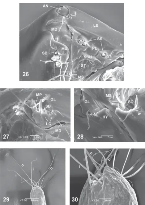
Thoracic pigmentation yellowish, diffuse. Caudal segment (Figs 29-30) wide; CSL 0.327 mm; CSW 0.178 mm. One seta “o” 0.303 mm long, the other 0.357 mm, one seta “i” 0.191 mm long, the other 0.216 mm; tance between bases of setae “o” subequal to the dis-tance between bases of setae “i”.
Distribution - Brazil (Amazonas).
Taxonomic discussion - C. felippebauerae is a mem-ber of the subgenus Mataemyia Vargas, and represents the first species in the subgenus with a single spermath-eca. The only available key for Mataemyia was provided by Wirth and Soria (1981) (as discrepans group) for only 8 of the hitherto 15 recognized species in the subgenus (Borkent & Spinelli 2000).
The adult of C. felippebauerae is very similar to C.
barthi Tavares & Souza from Rio de Janeiro, Brazil, from which it can be distinguished by the single spermatheca, longer second radial cell and tip of parameres without fringe. The wing patterns of C. azureus Wirth & Blanton and C. mojingaensis W. & B., both from Panama, are very similar to that of C. felippebauerae except for the shape of the distal pale spot in cell r3 (irregular in C.
azureus, oval in C. mojingaensis).
The larva and pupa of only two species of the subge-nus Mataemyia are presently known: C. dicrourus Wirth & Blanton and C. macieli Tavarez & Ruiz, which were described by Wirth and Soria (1981). The pupa of both species bear 25-30 spiracles on the respiratory organ, and the basal sensillum in the am tubercle lack setae. Additionaly, the pupa of C. dicrourus differs from that of C. felippebauerae in that the dl tubercle bears 3 se-tae. The descriptions of the larvae of C. dicrourus and
C. macieli are very incomplete, so no comparison with the larva of C. felippebauerae is presently possible. However, the setae of the caudal segment of both spe-cies are shorter than those of C. felippebauerae.
felippebauerae exhibits characters typical of carnivo-rous species, such as an elongate head with the mouth-parts directed anteriorly, a highly sclerotized labium, a lightly built pharynx, and broad, pointed, hooked man-dible (Mullen & Hribar 1988).
Types - Holotype male, allotype female (with pupal exuviae), Brazil, Amazonas, Manaus, km 123 BR 174, 29-VII-2005, D Carrasco. Paratypes, 3 males, 12 males, 1 larva, as follows; same data as holotype, 2 fe-males (one with pupal exuviae); same data except 21-VI-2006, 1 larva; same data except km 45, 1 male, 1 fe-male (with pupal exuviae); same data except km 45, 4-IX-2005, 4 females (3 with pupal exuviae); same data except km 95, 21-VI-2006, 2 males, 5 females.
Culicoides felippebauerae - Figs 11-18: pupa. Fig. 19: larva. Fig. 11: anterodorsal tubercle (ad). Fig. 12: dorsolateral tubercle (dl), respiratory horn (PRH), pedicel (P). Fig. 13: ventrolateral setae (vl), ventromedian setae (vm). Fig. 14: dorsal tubercles (d). Fig. 15: operculum, anteromarginal tubercle
(am), basal sensillum (bs). Fig. 16: fourthabdominal segment, abdominal tubercles: lateral anterosubmarginal tubercle (lasm), dorsal posteromarginal
tubercle (dpm), dorsal anterosubmarginal tubercle (dasm), ventral posteromarginal tubercle (lpm). Fig. 17: female caudal segment (ventral view). Fig. 18: male caudal segment (ventral view), posterolateral processes (PP). Fig. 19: head capsule, mandible (MD), epipharynx (EP), hypopharynx (HP).
Material examined by SEM - Brazil, Amazonas, Ma-naus, km 123 BR 174, 21-VI-2006, D Carrasco, 2 larvae.
Derivation of specific epithet - This species is named after our friend and colleague María Luiza Felippe-Bauer (Instituto Oswaldo Cruz, Rio de Janeiro) in recognition of her important contribution to the ceratopogonid tax-onomy in the Neotropics.
Forcipomyia (Forcipomyia) genualis (Loew).
Ceratopogon genualis Loew, 1866: 128 (male; Cuba).
Ceratopogon propinqua Williston, 1896: 279 (male; St. Vincent).
fe-male; Trinidad); Saunders, 1957: 660 (redescription; all stages; Trinidad).
Forcipomyia genualis: Johannsen, 1943: 777 (com-bination); Wirth, 1965: 125 (synonym: F. raleighi); Wirth, 1969: 575 (Galápagos islands record; distribution); Wirth & Soria, 1975: 22 (redescription; distribution); Borkent, 1991: 104 (redescription, notes; Galápagos islands).
Forcipomyia (Forcipomyia) genualis: Wirth, 1974: 5 (in catalog; synonym: F. propinqua; distribution); Spinelli & Marino, 1998: 39 (Argentina record).
Specimens examined - Brazil, Amazonas, Manaus, km 45 BR 174, 29-VII-2005, D Carrasco, 1 male; same data except km 95, 1 male.
Culicoides felippebauerae, larva - Fig. 20: head capsule (dorsal view). Fig. 21: head capsule (anterodorsal view). Fig. 22: head capsule (lateroventral view). Fig. 23: head capsule (ventral, internal view). Fig. 24: head capsule (basal ventral, internal view). Fig. 25: head capsule (frontal view). Head capsule chaetotaxy: j: collar pits; p: posterior perifrontal setae; q: postfrontal setae; s: anterior perifrontal setae; t: prefrontal setae; o: parahypostomal setae; v: posterolateral setae; y: ventral setae; w: anterolateral setae; x: paranntenal setae; antennae (AN); epipharynx (EP); labrum (LB); hypopharynx (HP) hypostoma (HY); hypopharingeal fringe (HF); mandible (MD); messors (MS); maxilla (MX), palatum (PL).
Distribution - Widely distributed, from US (Louisi-ana to Florida) to Northern Argentina.
Forcipomyia (Phytohelea) musae Clastrier & Delécolle (Figs 31-57)
Forcipomyia (Phytohelea) musae Clastrier & Delé-colle, 1994: 51 (male, female, pupa; French Guiana).
with short, stout seta, (ii) with minute, stout seta, (iii) pore; one ventromedian (vm) medium length setae, thin, one pore. Respiratory horn (Figs 31, 45) length 0.20 (0.19-0.22, n = 7) mm; amber brown, with scale-like spicules on basal half, with 13-14 apical spiracles; pedicel short, hyaline, length 0.025 (0.022-0.028, n = 7) mm; P/H 0.12 (0.11-0.14, n = 7). Operculum (Fig. 34) 0.5 as long as greatest breadth, apex broadly rounded; surface covered by rounded tubercles; anteromarginal tubercles (am) well developed, with single very short, stout seta; OL 0.17 (0.15-0.18, n = 5) mm; OW 0.30 (0.28-0.31, n = 5) mm; OW/OL 1.81 (1.70-2.12, n = 5). Cephalothorax (Fig. 46) with four dorsal cuticular
pro-cesses, two well developed (m3, m4), two rudimentary (m5, m6). Abdominal segments with scarse anterior spinules. Fourth abdominal segment (Fig. 35) with tu-bercles as follows: dorsal anterosubmarginal tubercle (dasm) with long, posteriorly directed seta, tuberculate broad base; dorsal posterosubmarginal tubercle (dpm) without seta, base elongate, triangular; lateral pos-teromarginal tubercle (lpm) with two setae, one long, one short, base broad tuberculate; four ventral pos-teromarginal tubercles (vpm): (i-ii) with medium length, strong seta, base triangular, (iii) with strong seta, base tuberculate, iv with thin seta, base poorly developed. Female caudal segment (Fig. 36) length 0.62 (0.56-0.67,
n = 4) mm, width 0.25 (0.21-0.28, n = 4) mm, approxi-mately two times longer than width; ventrolateral sur-face with band of posteriorly directed spicules; dorsal surface with posteriorly directed spicules except later-ally and on a small, closed, mesal area; posterolateral processes long, tip pointed, base with outer long setae. Male caudal segment (Figs 37, 47) length 0.58 (0.54-0.60, n = 3) mm, width 0.24 (0.21-0.26, n = 3) mm. Genital
pro-cesses ventral, stout, with distal wrinkles.
Description of larva - Exuviae pale brown. Head cap-sule brown, well developed, prognathous, HL 0.364 (0.288-0.392, n = 7) mm; HW 0.254 (0.192-0.312, n = 8) mm; HR 1.394 (1.256-1.50, n = 7); SGW 0.204
(0.16-0.24, n = 8) mm; SGR 1.24 (1.20-1.31, n = 8). Antennae rounded, reduced. Head chaetotaxy (Figs 48-51) as fol-lows: nine sensory setae, three pits; “j” pore simple; se-tae “p” short, thin; seta “q” long; seta “s” very long; seta “t” as long than “q”;seta “v” long; “r” pore simple; setae “o” thin, one long, one short; seta “u” long; setae “x” minute, thin; seta “y” very long, stout; “z” pore simple. Labrum (Figs 48, 50-51) short, not extending beyond hypostoma; palatum (Figs 51-53) with a group of three sensillae styloconica on the outer edge, one postero-medial sensilla coeloconica; immediately underneath on medial surface two groups of three sensillae trichodea, one long, two short, one posterolateral sensilla
coe-Forcipomyia (Phytohelea) musae, pupa - Fig. 31: dorsolateral tubercle (dl), respiratory horn (PRH), pedicel (P). Fig. 32: ventromedian setae (vm). Fig.
33: dorsal tubercles (d). Fig. 34: operculum, anteromarginal tubercle (am). Fig. 35: fourthabdominal segment, abdominal tubercles: dorsal anterosubmarginal
loconica, behind of these with tuft sensillae trichodea of different length. Messors (Fig. 38) stout, comma-shaped. Mandible (Figs 52-53) stout, not articulated with head capsule, with seven teeth, one apical, blunt, one subapical, truncate, posterior five elongated, slender, ML 0.089 (0.074-0.104, n = 8) mm; MW 0.032 (0.026-0.044, n = 7) mm; hypostoma (Fig. 54) membranous, serrate. Epipharynx (Fig. 39) massive, strongly sclero-tized, with median sclerite bearing numerous pointed, strong teeth; comb 3 with fringe; comb 2 with 20 small teeth; dorsal comb with approximately 40 fine teeth; lat-eral arms stout, with short apical teeth. LAW 0.154 (0.142-0.168, n = 6) mm; DCW 0.067 (0.064-0.074, n = 7) mm. Hypopharynx (Fig. 40) sclerotized, broad, lat-eral arms hyaline, thin. Maxilla bilobated (Figs 52-53, 55) with conspicuous basal fringe; galeolacinia with stout
seta; maxillary palpus rounded, flattened, with four small papillae, lateral sclerite. Prothoracic pseudopod (Fig. 56) bifid, each ramus with two rows of eight golden thorn like hooks, 12-13 pairs of anterior hairs. Chaetotaxy of second abdominal segment (Fig. 41): dorsally, one seta “a” very long, stout, base poorly developed; ventrally, eight setae, three very long, thin, as long as dorsal seta (“e”, “f”, “g”), the other five (“b”, “c” , “d”, “h”, “i”) very short, thin. Eigth abdominal segment (Figs 42, 57) bear-ing a very long dorsal setae measurbear-ing 0.42 (0.40-0.44, n = 7) mm, arising from crossbar, surpassing the anal segment; cuticle devoid of spicules. Anal segment (Fig. 43) with four apical strong, blade-like setae, lateral mar-gins serrate. Anal pseudopod with two rows of five pairs of laterally directed, sclerotized hooks, the ones of an-terior row short, stout, with anan-teriorly recurved tips;
hooks of posterior row elongated, slender, slightly paler. Anal papillae (Fig. 42) hyaline, bilobated, each lobe with two pairs of papillae; CSL 0.333 (0.312-0.360, n = 7) mm, CSW 0.264 (0.224-0.296, n = 5) mm, CSR 1.304 (1.162-1.501, n = 5).
Specimens examined - Brazil, Amazonas, Manaus, km 31 BR 174, 2-IX-2005, D Carrasco, 1 female (with larval and pupal exuviae); same data, 1 male, 1 female (with pupal exuviae); same data except 14-VI-2006, 4 larvae; same data except km 123, 29-VII-2005, 2 males (with larval and pupal exuviae); same data, 1 male, 1 fe-male (with pupal exuviae); same data except km 95, 14-VI-2006, 1 pupa, 1 larva.
Other specimens examined (preserved in alcohol 70%) - Brazil, Amazonas, Manaus, 54 males (29 with pupal exuviae, 2 with larval exuviae), 55 females (31 with pupal exuviae, 1 with larval exuviae), 11 pupae, 7 larvae from the above mentioned localities and dates.
Material examined by SEM - Brazil, Amazonas, Manaus, km 95 BR 174, 14-VI-2006, D Carrasco, 7 pupae, 7 larvae.
Distribution - Brazil (Amazonas), French Guiana.
Taxonomic discussion - This species belongs to the
F. bromelicola species group, as defined by de Meillon and Wirth (1979). It keys out to couplet 5 in de Meillon and Wirth (1979), where it may be distinguished from F.
(Phytohelea) edwardsi (Saunders) from Brazil, by the slightly unequal sized, pyriform spermathecae with scle-rotized necks (subequal sized, subspherical with uns-clerotized necks in F. edwardsi). The male genitalia of
F. musae are also very similar to those of F. edwardsi, but can be distinguished from the later species by the gonostylus shorter than gonocoxite (subequal in F.
edwardsi), and by the recurved posterior processes of parameres (spoon-like in F. edwardsi). Immatures of F.
edwardsi can be distinguished from immatures of F.
musae by the respiratory horn of the pupa expanded lat-erally and bearing 12 apical spiracles (not expanded, with 14 apical spiracles in F. musae), by the larger head ratio
(HR) of the larvae, and by the larval lateral setae with basal fin-like expansions (without these expansions in F. musae). Adults of this species also resemble F. bromelicola
(Lutz) by virtue of their similar male genitalia, but dif-fer from this species by the following characters: fe-male palpus with subapical sensory pit (at midlength in
F. bromelicola), humeral areas and scutellum pale (brown in F. bromelicola) and pyriform spermathecae with sclerotized neck (elliptical with unsclerotized neck in F. bromelicola). The description of the larva and pupa of F. bromelicola by Saunders (1925) is very incom-plete, and therefore it is very difficult to compare the immature stages of both species.
ACKNOWLEDGMENTS
To Dr William Grogan for detailed critical review of the manu-script acting as a journal referee.
REFERENCES
Borkent A 1991. The Ceratopogonidae (Diptera) of the Galápagos Islands, Ecuador with a discussion of their phylogenetic relati-onships and zoogeographic origins. Entomol Scan 22: 97-122. Borkent A, Spinelli GR 2000. Catalog of the New World biting
midges south of the United States of American (Diptera: Ceratopogonidae). Contrib Entomol Intern 4: 1-107. Clastrier J, Delécolle JC 1994. Description de Forcipomyia
(Phytohelea) musae n. sp. de la Guyane francaise (Diptera, Ceratopogonidae). Rev Franc Entomol 16: 51-56. De Meillon B, Wirth WW 1979. A taxonomic review of the
sub-genus Phytohelea of Forcipomyia (Diptera: Ceratopo-gonidae). Proc Entomol Soc Wash 81: 178-206.
Díaz F, Ronderos MM, Spinelli GR 2005. The immatures of the Neotropical species Culicoides venezuelensis Ortiz & Mirsa (Diptera: Ceratopogonidae). Trans Am Entomol Soc 131: 375-385.
Fish D 1976. Structure and Composition of the Aquatic In-vertebrate Community Inhabiting Epiphytic Bromeliads in South Florida and the Discovery of an Insectivorous Bro-meliad, Ph D Thesis, University of Florida, Gainesville, 78 pp. Fish D 1983. Phytotelmata: flora and fauna. In JH Frank, LP
Lounibos, Phytotelmata: Terrestrial Plants as Hosts of Aquatic Insect Communities, Plexus, Medford, p. 161-190.
Fish D, Soria SJ 1978. Water-holding plants (Phytotelmata) as larval habitats for ceratopogonid pollinators of cacao in Bahia, Brazil. Revta Theobroma 8: 133-146.
Johannsen OA 1943. A generic synopsis of the Ceratopogoni-dae (HeleiCeratopogoni-dae) of the Americas, a bibliography, and a list of the North American species. Ann Entomol Soc Am 36: 763-791. Loew H 1866. Diptera Americae septentrionalis indigena. Berl
Entomol Zeits 9: 127-186.
Macfie JWS 1938. Notes on Ceratopogonidae (Diptera). Proc R Entomol Soc London (B) 7: 157-166.
McAlpine JF, Peterson BV, Shewell GE, Teskey HJ, Vockeroth JR, Wood DM 1981. Manual of Nearctic Diptera, Vol. 1, Agriculture, Monograph 27, Canada, 674 pp.
Mercer DR, Spinelli GR, Watts DM, Tesh RB 2003. Biting rates and developmental substrates for biting midges (Diptera: Ceratopogonidae) in Iquitos, Peru. J Med Entomol 40: 807-812.
Mullen GR, Hribar LJ 1988. Biology and feeding behavior of Ceratopogonidae larvae (Diptera: Ceratopogonidae) in North America. Bull Soc Vector Ecol 13: 60-81.
Murphree CS, Mullen GR 1991. Comparative larvae morpholo-gy of the genus Culicoides Latreille (Diptera: Ceratopogo-nidae) in North America with a key to species. Bull Soc Vector Ecol 16: 269-399.
Ribeiro JELS, Hopkins MJG, Vicentini A, Sothers CA, Costa MAS, Brito JM, Souza MAD, Martins LHP, Lohmann LG, Assunçao PACL, Pereira EC, Silva CF, Mesquita MR, Procópio LC 1999. Flora da Reserva Ducke. Guia de Identificação das Plantas Vasculares de uma Floresta de Terra-firme na Amazônia Central, 19th ed., INPA-DFID, Manaus, 816 pp.
Ronderos MM, Spinelli GR 2000. The larvae and pupa of
Culicoides bambusicola Lutz observed with SEM, and ad-ditional notes on the adults (Diptera: Ceratopogonidae). Trans Am Entomol Soc 126: 133-144.
Saunders LG 1925. On the life history, morphology and system-atic position of Apelma Kieff. and Thyridomyia n. g. (Diptera, Nemat. Ceratopogoninae). Parasitology 17: 252-277. Saunders LG 1957. Revision of the genus Forcipomyia based
on characters of all stages (Diptera, Ceratopogonidae).
Canad J Zool 34: 657-705.
Spinelli GR, Marino PI 1998. First records for Argentina of three species of Forcipomyia (Diptera : Ceratopogonidae). Revta Soc Entomol Argent 57: 39-40.
Spinelli GR, Marino PI, Ronderos MM 2005. The fourth instar larva and pupa of the Neotropical species Forcipomyia
(Forcipomyia) rioplatensis Marino & Spinelli (Diptera: Ceratopogonidae). Proc Entomol Soc Wash 107: 108-114. Szadziewski R 1996. Biting midges from Lower Cretaceous
amber of Lebanon and Upper Cretaceous Siberian amber of Taimyr (Diptera, Ceratopogonidae). Studia Dipterol 3: 23-86. Vitale GC, Wirth WW, Aitken THG 1981. New species and records of Culicoides reared from arboreal habitats in Panama, with a synopsis of the debilipalpis group (Diptera: Ceratopogonidae). Proc Entomol Soc Wash 83: 140-159. Williston SW 1896. On the Diptera of St. Vincent (West Indies).
Trans Entomol Soc London 1896: 253-446.
Winder JA 1977. Some organic substrates which serve as insect breeding sites in bahian cocoa plantations. Revta Brasil Biol 37: 351-356.
Wirth WW 1965. Family Ceratopogonidae. In A Stone, A Cata-log of the Diptera of America North of Mexico, Agricul-ture Handbook 276, Washington, D.C., p. 121-142. Wirth WW 1969. New species and records of Galapagos
Dip-tera. Proc Calif Acad Sci 36: 571-594.
Wirth WW 1974. Family Ceratopogonidae. In A Catalogue of the Diptera of the Americas South of the United States, Fasc. 14, 89 pp.
Wirth WW, Soria SJ 1975. A new Neotropical Forcipomyia mid-ge closely related to F. (F.) genualis (Loew) (Diptera: Ceratopogonidae). Revta Theobroma 5: 19-27.