A new species of the
Eigenmannia trilineata
(Gymnotiformes:
Sternopygidae) species group from the río Orinoco basin, Venezuela
Luiz A. W. Peixoto
1and Brandon T. Waltz
2A new species of the Eigenmannia trilineata species group is described from the río Orinoco basin, Venezuela. The new
species is distinguished from congeners by a unique set of characters including an ossified basibranchial 1; 198-217 anal-fin rays; suborbital depth, 21.3-26.1% HL; length of anterodorsal process of maxilla equal to the width of the posterior nostril; premaxilla with 17 teeth distributed in three rows; hyaline pectoral and anal fins; and number of scale series above lateral line, 9-10. It raises the number of species allocated to the Eigenmannia trilineata species group to 13 and the number of species within the genus to 18.
Keywords: Biodiversity,Electric-fishes, Taxonomy, Tuvira.
Se describe una nueva especie del grupo Eigenmannia trilineata de la cuenca del río Orinoco, Venezuela. La nueva especie se distingue de sus congéneres por una combinación única de caracteres, incluyendo el basibranquial 1 osificado; número de radios de la anal, 198-217; profundidad del suborbital, 21.3-26.1% HL; longitud del proceso anterodorsal de la maxila igual al ancho de la narina posterior; 17 dientes premaxilares distribuidos en tres hileras; aletas pectoral y anal hialinas; y 9-10 hileras de escamas sobre la serie de la línea lateral. La presente contribución eleva el número de especies del grupo Eigenmannia trilineata a 13, y a 18 aquellas dentro del género.
Palabras Clave: Biodiversidad, Pez cuchillo, Peces eléctricos, Taxonomía.
1Setor de Ictiologia, Museu de Zoologia da Universidade de São Paulo, Av. Nazaré, 481, Ipiranga, 04299-970 São Paulo, SP, Brazil. [email protected] (corresponding author)
2Department of Biology, University of Louisiana at Lafayette, 410 E. St. Mary Blvd., LA, 70504-2451 Lafayette, USA. [email protected] Introduction
Commonly known as electric glass knifefishes or ituí
transparente, Eigenmannia Jordan & Evermann, 1896 is a genus of weakly electric freshwater fishes broadly distributed throughout most of the Neotropics on both sides
of the Andes, from Panama to northern Argentina (Albert,
2001, 2003). Species within the genus exhibit a wave-type electric organ discharge and can be found in diverse habitats, from small streams and tributaries to floodplain lakes and large river channels (Crampton, Albert, 2006).
Following a series of contributions (Eigenmann, 1893; Jordan, Evermann, 1896; Ellis, 1913; Mago-Leccia, 1994; Triques, 1996; Albert, 2001; Peixoto et al., 2015; Peixoto, Wosiacki, 2016; Campos-da-Paz, Queiroz, 2017), seventeen species are currently recognized as valid in
the genus Eigenmannia: E. antonioi Peixoto, Dutra & Wosiacki, 2015 from therio Anapu, rio Amazonas basin; E. besouro Peixoto & Wosiacki, 2016 from tributaries in the rio São Francisco basin; E. correntes Campos-da-Paz & Queiroz, 2017 from the rio Correntes, rio Paraguay
basin; E. desantanai Peixoto, Dutra & Wosiacki, 2015
from the rio Cuiabá, rio Paraguay basin; E. guairaca Peixoto, Dutra & Wosiacki, 2015 from the riacho Água
do Ò, upper rio Paraná basin; E. humboldtii (Steindachner,
1878) from the río Magdalena; E. limbata (Schreiner &
Miranda Ribeiro, 1903) from the Amazonas basin; E. macrops (Boulenger, 1897)from the Amazonas, Orinoco,
and Guyanas basins; E. matintapereira Peixoto, Dutra &
Wosiacki, 2015 from the rio Uneiuxi and rio Urubaxi, rio Negro basin; E. microstoma (Reinhardt, 1852) from the rio São Francisco basin; E. muirapinima Peixoto, Dutra & Wosiacki, 2015 from small tributaries of the Amazon
River; E. nigra Mago-Leccia, 1994 from the rio Negro, río Orínoco, and rio Amazonas basins; E. pavulagem Peixoto, Dutra & Wosiacki, 2015 from tributaries of the rio Capim,
rio Guamá basin; E. trilineata López & Castello, 1966 from the río de La Plata and rio Paraná; E. vicentespelaea Triques, 1996 from the São Vicente I and II caves, rio Tocantins basin; E. virescens (Valenciennes, 1836) from
vicentespelaea,and E. waiwai (thus removing E. microstoma from the E. microstoma species group and E. trilineata from the E. virescens species group). Most recently, two species were described for the group, E. besouro and E. correntes (Peixoto, Wosiacki, 2016; Campos-da-Paz, Queiroz, 2017).
All current members of the Eigenmanniatrilineata species group are found in the Amazon, Paraguay, Paraná, La Plata,
and São Francisco drainages.
Based on analysis of material collected in the río Orinoco basin, we describe a new species of Eigenmannia belonging to the E. trilineata species group.
Material and Methods
Morphometric, meristic, and osteological nomenclature follow Lundberg, Mago-Leccia (1986) and Peixoto et al. (2015). Measurements were taken point-to-point to the nearest 0.1 mm using digital calipers under a stereomicroscope, preferentially utilizing the left side of
each specimen. Abbreviations reported in this manuscript
are total length (TL), the distance from the tip of the snout to
distal margin of the caudal filament; length to end of anal fin
(LEA), from the tip of the snout to the insertion of the last
anal-fin ray; and head length (HL), from the tip of the snout to the posterior-most margin of the branchial opening. In the
description, the frequencies of each count are expressed in parentheses and holotype data are indicated by an asterisk.
In the pectoral and anal-fin ray counts, unbranched rays are represented by lower case Roman numerals and branched
rays are indicated by Arabic numerals. Specimens were cleared and stained (c&s) following the protocol of Taylor,
Van Dyke (1985). Pre-caudal vertebrae counts include the four vertebrae of the Weberian apparatus. Herein, the “lateral line stripe” refers to the dark stripe along the lateral line; the “superior midlateral stripe” refers to the concentration of
small separate chromatophores with diffuse margins located
below the lateral line; the “inferior midlateral stripe” refers to the dark stripe located over the proximal portion of anal-fin pterygiophores; and the “anal-anal-fin base stripe” refers to the dark stripe located on the base of the anal fin (Peixoto et al., 2015; Peixoto, Wosiacki, 2016). Morphological data of E. correntes were based on Campos-da-Paz, Queiroz (2017). Infraorbitals, premaxillary, dentary, and maxillary bones
urn:lsid:zoobank.org:act:F127F01B-AC21-4D1B-BC4F-EBAB9EA64854
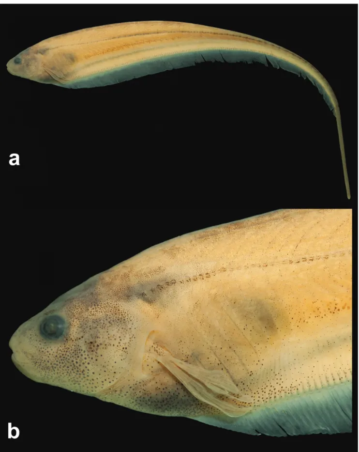
Fig. 1
Holotype. MZUSP 96497, 131.8 mm LEA, Venezuela, Bolivar, Cedeño, río Parguaza, río Orinoco basin, near the community of Puente Parhueña, 5°53’30”N 67°24’14”W, 19 Jul 2004, M. de Pinna & C. Oliveira.
Paratypes. All from Venezuela. Bolivar: FMNH 130239, 1, 95.4 mm LEA; MPEG 33926, 1, 103.7 mm LEA; MZUSP 119711, 6 + 2 c&s, 27.8-116.2 mm LEA, collected with the holotype. MZUSP 96465, 1, 116.2 mm LEA, Caicara del Orinoco, río Orinoco, Laguna de Castilleros, 7°30’51”N 66°09’20”W, 22 Jul 2004, M. de Pinna & C. Oliveira.
Non-types. All listed specimens were directly preserved in alcohol (except USNM specimens). Venezuela. Apure:
USNM 260225, 13, 47.1-68.5 mm LEA, Caño Caicara. Bolivar: LBP 2225, 1, 54.7 mm LEA, Caicara del Orinoco, río Orinoco, Laguna de Castilleros. LBP 2311, 3, 91.1-107.5 mm LEA, Cedeño, río Orinoco, río Parguaza. LBP 3083, 4, 53.6-65.1 mm LEA, Caicara del Orinoco, río Orinoco. LBP 9976, 3, 57.7-89.9 mm LEA (two specimens measured;
one specimen damaged), Caicara del Orinoco, río Orinoco, Laguna de Castilleros.
Diagnosis.Eigenmannia sayona can be distinguished from its congeners, except species of the E. trilineata species group, by the presence of a superior midlateral stripe (vs. absence). Eigenmannia sayona can be differentiated from congeners in the E. trilineata species group by the
ossification of the first basibranchial (Fig. 2) (vs. unossified). The new species can be distinguished from congeners in the E. trilineata species group, except E. correntes, by the unique
E. matintapereira, E. microstoma, E. muirapinima, E. pavulagem, and E. trilineata, by the number of anal-fin rays, 198-217 (vs. 150-181 in E. besouro; 143-164 in E. correntes; 170-196 in E. desantanai; 151-170 in E. guairaca; 169-191 in E. vicentespelaea; 167-195 in E. waiwai). The new species can be distinguished from E. besouro, E. desantanai, E. muirapinima, E. vicentespelaea, and E. waiwai by the number of tooth rows on the endopterygoid, 1 (vs. 2). The new species can be differentiated from E. besouro, E. correntes, E. desantanai, E. guairaca, E. microstoma, and E. trilineata by the number of precaudal vertebrae, 13 (vs. 14; 14; 11-12; 15; 14-15; and 14, respectively). Eigenmannia sayona can be differentiated from E. antonioi, E. correntes, E. desantanai, E. guairaca, E. muirapinima, E. pavulagem, E. vicentespelaea, and E. waiwai by the length of the anterodorsal process of maxilla being equal to the width of the posterior nostril (Fig. 5) (vs. equal to 1.5 times the width of the posterior nostril in E. waiwai; or approximately 20-50% of the width of the posterior nostril in aforementioned species - Fig. 4b of Peixoto et al., 2015). Eigenmannia sayona can be further differentiated from E. besouro, E. correntes, E. matintapereira, E. trilineata, and E. waiwai by the depth of the posterodorsal expansion on
infraorbitals 1+2, approximately equal to the total length of infraorbitals 1+2 (Fig. 6) (vs. less than 50% of the length of infraorbitals 1+2). The new species differs from E. besouro, E. correntes, E. vicentespelaea and E. waiwai by possessing a terminal mouth (vs. subterminal). The new species can be distinguished from E. microstoma and E. trilineata by
the suborbital depth, 20.6-26.8% HL (vs. 29.9-40.8%; and 32.5-46.6%, respectively). Eigenmannia sayona can be distinguished from E. microstoma and E. vicentespelaea by
the number of scales above lateral line, 9-10 (vs. 11-15; and 7-8, respectively). Eigenmannia sayona can be differentiated from E. matintapereira, E. trilineata, and E. vicentespelaea by the number of pectoral-fin rays, ii,12-13 (vs. ii,16-17; ii,14-15; and ii,15-17, respectively). Eigenmannia sayona can be differentiated from E. matintapereira, E. muirapinima, and E. waiwai,by oral width, 18.7-21.7% HL (vs.12.6-16.1%; 13.2-18.1%; and 9.5-14.6%, respectively). Eigenmannia sayona can be further distinguished from E. guairaca and E. waiwai by orbital diameter, 17.3-22.4% HL (vs.11.4-15.0%; and 22.6-28.8%, respectively). Eigenmannia sayona differs
Fig. 2. Dorsal view of anterior portion of gill arches of Eigenmannia sayona (MZUSP 119711, paratype, 90 mm LEA). BH= basihyal; BB1= basibranchial 1; BB2= basibranchial 2; HB1= hypobranchial 1; CB1=
ceratobranchial 1. Cartilage represented by gray coloration. Scale bar: 0.5 mm.
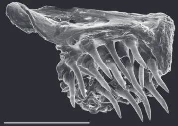
Fig. 3. Scanning electron micrograph. Ventral view of left premaxilla, showing the dentition pattern of Eigenmannia sayona (MZUSP 119711, paratype, 116.2 mm LEA). Scale bar: 0.5 mm.
Description. Species of small/medium size, maximum
length recorded 131.8 mm TL. Morphometric data is
presented in Tab. 1. Body elongate and laterally compressed.
slightly concave along anterior half of abdominal cavity; then posteriorly angled to last anal-fin ray. Ventral profile of caudal filament straight. Greatest body depth at vertical line through distal margin of pectoral fin. Head laterally
compressed, greatest width at posterior opercular region and greatest depth at posterior margin of supraoccipital.
Dorsal profile of head slightly convex or nearly straight from upper lip to vertical through branchial opening. Ventral profile of head slightly concave from anterior margin of lower lip to branchial opening. Snout rounded in profile. Mouth terminal. Upper lip slightly overlapping lower lip or lips equal in length. Premaxilla teeth 17(2) in three rows [outermost row with 4(2) teeth; median row with 6(2); innermost row with 7(2) teeth, Fig. 3]. Maxilla with
sickle-shaped anterodorsal process equal to the width of posterior
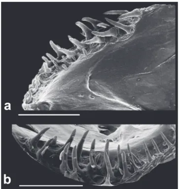
nostril. Dentary teeth 19(1) or 26(1) in two irregular rows [outermost row with 11(1) or 14(1); innermost row teeth, 8(1) or 12(1), Fig. 4]. Dentary teeth increasing abruptly in size from the sixth or seventh tooth of outermost row towards rictus. Coronomeckelian bone equal to 30% of length of Meckel’s cartilage. Endopterygoid teeth 8(1) or 9(1) in a single row. Mouth rictus at vertical line through
anterior nostril or in region between nostrils. Anterior nares
tubular, posterior margin at vertical line through posterior
margin or middle portion of rictus. Posterior nares elliptical,
non-tubular, and closer to anterior margin of eye than tip of snout. Eye approximately circular, covered by skin,
lateral on anterior half of head. Antorbital and infraorbitals
1-4 enlarged partial cylinders with slender osseous arches.
Fifth and sixth infraorbitals slender and tubular. Depth of
posterodorsal expansion on infraorbitals 1+2 equal to total length of infraorbitals 1+2 (Fig. 5). Branchial opening
moderately elongate. Branchial membrane joined with isthmus. Anus and urogenital papilla shifting anteriorly
during ontogeny, from vertical line through posterior portion of opercle in juveniles [minimum examined 47.1 mm LEA] to vertical line through posterior portion of eye in adults. Anus and urogenital papilla at vertical line through posterior
margin of eye in mature specimens.
Cycloid scales present from posterior margin of head
to distal portion of caudal filament. Lateral line complete, 122(2), 124(1), 125(2), 127(2), 128*(1), 132(1), 134(2) or 136(1) perforated scales to vertical line through end of anal fin (N=12). Longitudinal series of scales above lateral line at distal margin of pectoral fin, 9*(5), 10(7) or 11(4). Scales over anal-fin pterygiophores approximately one-half size of others.
Pectoral-fin rays, ii,12*(6) or ii,13(11); distal margin slightly rounded; tip reaching vertical through 16th to 27th
anal-fin ray. Anal-fin origin posterior to vertical line through pectoral-fin base; total anal-fin rays, 198*-217 (N=14); distal margin of anal fin slightly convex. Caudal filament cylindrical, tapering gradually distally, relatively short and approximately 40% of LEA in mature specimens.
Precaudal vertebrae 13(2). Anterior vertebrae 10(1) or 12(1), transitional vertebrae 1(1) or 3(1). Displaced hemal spines 3(2).
Fig. 4. Scanning electron micrographs. (a) lateral view and (b) dorsal view of right dentary of Eigenmannia sayona (MZUSP 119711, paratype, 116.2 mm LEA), showing the
dentition pattern. Scale bar: 1 mm.
Fig. 5. Scanning electron micrograph of left maxilla
(inverted) of Eigenmannia sayona in lateral view, anterior to left (MZUSP 119711, paratype, 116.2 mm LEA). CT= connective tissue on leading edge of maxilla. Arrow
Fig. 6. Scanning electron micrograph of right infraorbital
1+2 of Eigenmannia sayona in lateral view, anterior to right (MZUSP 119711, paratype, 116.2 mm LEA). Arrow
indicates the posterodorsal expansion. Scale bar: 1 mm.
vertical line approximately between base of 25th to 30h
anal-fin ray to posterior-most one-third of body. Anal-fin base stripe thick, two scales deep, extending from vertical between base of first anal-fin ray to last anal-fin ray. Pectoral and anal fins hyaline, scattered tiny chromatophores on
interradial membranes.
Geographic distribution. Eigenmannia sayona is known from río Orinoco basin, from río Parguaza, río Apure, and
Laguna de Castilleros, Venezuela (Fig. 7).
Fig. 7. Map of northern South America, illustrating the geographic distribution of Eigenmannia sayona. Star indicates type locality. Some points represent more than one locality.
Etymology. The specific epithet “sayona” is assigned to
the new species in reference to “La Sayona”, a spirit of philanderous vengeance in Venezuelan lore. A noun in
apposition.
Discussion
Currently, Eigenmannia lacks diagnostic characters
common to all species within the genus (Mago-Leccia, 1994; Albert, 2001), resulting in confusion regarding the
generic limits within Sternopygidae. Thus, we opt to identify E. sayona as a member of Eigenmannia by its possession of all synapomorphies common to the Eigenmanniinae (Albert, 2001), and the absence of most characters diagnostic to other genera of Sternopygidae.
Tab. 1. Morphometric data for Eigenmannia sayona, new
species. Min= minimum; Max= maximum; SD= standard deviation; N= number of specimens measured.
Holotype Min Max Mean SD N Total length (mm) 160.0 113.8 160.0 - - 13
Length to end of anal fin (mm) 131.8 65.3 131.8 - - 14 Head length (mm) 13.6 9.2 15.8 - - 14
Percentage of LEA
Head length 10.3 10.3 15.1 12.3 1.7 14 Preanal distance 13.5 12.7 18.9 15.2 1.9 14 Prepectoral distance 11.3 11.3 16.3 13.1 1.8 14
Snout to anus 5.6 5.6 8.0 6.8 0.9 14
Body depth at pectoral fin 13.3 12.9 18.8 15.6 2.1 14
Body depth at anal fin 12.1 11.7 16.0 13.9 1.5 14
Body width 4.2 4.1 6.1 4.9 0.8 14
Anal-fin length 89.6 80.0 89.6 85.4 2.1 14
Pectoral-fin length 7.7 7.3 11.2 8.6 1.5 13
Caudal-filament length 22.6 21.2 44.5 34.8 7.9 11
Caudal-filament depth 1.6 1.2 2.6 1.4 0.1 11
Caudal-filament width 1.0 0.7 1.1 0.9 0.1 11
Percentage of HL
Snout length 25.1 22.5 28.3 25.3 1.8 16
Internasal distance 9.5 7.2 10.5 9.0 1.1 16 Snout to posterior naris distance 19.8 16.3 21.0 19.0 1.6 16 Posterior naris to orbit distance 7.3 6.9 9.9 8.1 1.1 16
Internarial width 17.3 14.6 18.4 16.8 1.1 16 Orbital diameter 20.6 17.3 22.4 20.1 1.2 16 Postorbital distance 52.3 50.1 55.5 52.4 1.9 16 Opercular opening 25.9 23.9 32.1 28.2 1.9 16 Suborbital depth 21.3 20.6 26.8 23.2 2.1 16
Interorbital distance 34.0 27.9 37.2 33.2 1.9 16
Head width at opercle 63.3 58.3 66.1 61.4 2.6 16 Head width at orbits 50.9 39.5 50.9 46.3 2.9 16
Head depth at supraoccipital 77.9 74.2 82.3 79.1 2.3 16 Head depth at orbits 58.4 53.9 64.2 58.5 3.1 16 Maxilla length 23.2 15.9 23.2 19.4 2.5 16
Correa et al. (2006) proposed six synapomorphies for Rhabdolichops, none of which are present in E. sayona. Another eigenmanniin genus, Distocyclus, was recently distinguished from other genera in the subfamily by four autapomorphies (see Dutra et al., 2014), of which E. sayona shares none. Vari et al. (2012) proposed four synapomorphies for Archolaemus, all of which are absent in E. sayona, except
for the ventral surface of the upper lip porous, possibly representing a case of convergence (Vari et al., 2012
noted another possible convergence of upper lip surface
morphology between Archolaemus and an undescribed species in the E. trilineata group). In the same study, Vari et al. (2012) proposed six synapomorphies for Japigny, all of which are absent in E. sayona. Therefore, E. sayona can be excluded from all other diagnosed genera in Eigenmaniinae
and is assigned provisionally to the genus Eigenmannia. Approximately 50 species of gymnotiforms inhabit the
río Orinoco basin (Mago-Leccia, 1994; Campos-da-Paz, 1995; Albert, 2003; Campos-da-Paz, 2003; de Santana, Crampton, 2007, 2011; de Santana, Vari, 2009, 2010a, 2010b, 2012, 2014; Lundberg et al., 2013; Ivanyisky, Albert, 2014). Five species of Eigenmannia have been reported, among them: E. limbata, E. humboldtii, E. macrops, E.nigra,and E. virescens (e.g. Leccia, 1978; Mago-Leccia, 1994; Albert, 2001, 2003; Lasso et al., 2004).
In the most complete taxonomic study of sternopygids from Venezuela, Mago-Leccia (1978) recorded the presence
of E. virescens and E. macrops (aside from E. humboldtii, which may refer to E. limbata, and E. nigra), in the río
Orinoco basin. However, the specific delimitations of E. macrops and E. virescens are historically controversial due to taxonomic confusion resulting from insufficient
descriptions and inadequate diagnostic characters. The lack
of type material and type-locality information regarding E. virescens has led us to restrict the application of this name only to populations without stripes from the La Plata and Paraná basin (see additional comments in Peixoto et al., 2015). Furthermore, Mago-Leccia (1978) mentions two or three longitudinal stripes associated with and
near the lateral line in specimens identified by him as E. virescens; however, this information does not correspond to Valenciennes (1836; plate xiii), which includes a drawing
of a specimen without longitudinal stripes on body surface. Thus, the identity of the specimens listed as E. virescens in
Mago-Leccia (1978) remains uncertain.
Similarly, analyses performed on the holotype and topotypes of E. macrops raise concern regarding the identity of specimens listed as E. macrops from the data presented
in Mago-Leccia (1978). Mago-Leccia (1978) described the
presence of two or three stripes on the body surface of E. macrops; however, this character contradicts the original description by Boulenger (1897), which described the body
surface of E. macrops as uniform pale brownish. Because
of these conflicting descriptions, we opt to temporarily consider the material listed in Mago-Leccia (1978) to be
two unnamed species of Eigenmannia. Unfortunately, the
listed specimens could not be analyzed during this study due
to difficulty in accessing the material. A study is currently in development to clarify the taxonomic uncertainty
surrounding the identity of E. macrops and E. virescens.
Comparative material examined. Material examined in addition to that listed in Peixoto et al. (2015). Eigenmannia macrops: Guyana: BMNH 1897.8.6.1, holotype of Sternopygus macrops,
128.6 mm LEA. USNM 405266, 6 of 16, 65.7-163.9 mm LEA.
Eigenmannia guairaca: Brazil: LBP 9911, 1 of 3, 104.9 mm LEA.
Eigenmannia nigra: Brazil: MPEG 2430, 7+1c&s, 154.0-262.5
mm. Eigenmannia limbata: Brazil: MZUSP 75569, 1 of 2, 231.2
mm LEA.
Acknowledgments
The authors ackowledge James Maclaine (BMNH); Leo Smith, Mary Rogers, Susan Mochel, and Caleb McMahan (FMNH); Claudia Uribe (IAvH); Lúcia Py-Daniel and Renildo de Oliveira (INPA); Gustavo Chiaramonte and Ricardo Ferriz (MACN); Zilda Lucena and Carlos Lucena (MCP); Marcelo Britto (MNRJ); José Figueredo, Naércio Menezes, Mário de Pinna, Osvaldo Oyakawa, Aléssio Datovo and Michel Gianeti (MZUSP); Carla Pavanelli (NUP); Richard Vari, Lynne Parenti, Jeff Clayton, and Sandra Raredon (USNM); Cláudio Oliveira (LBP); Wolmar Wosiacki and Izaura Guimarães (MPEG) for the loan of specimens and assistance during visits to their institutions.
The authors also acknowledge Fernando Dagosta for
assistance in preparation of the holotype figure and Gustavo Ballen for providing the “Resumen”. The authors thank the Laboratório de Microscopia Eletrônica do Museu de Zoologia and Lara Guimarães (MZUSP) for the technical
support during the preparation of scanning electron
micrographs images. This manuscript has benefitted by
helpful comments and suggestions from James Albert and
Maxwell Bernt. BTW acknowledges his wife, Kassandra
Waltz, for her patience and helpful comments throughout the
development of the study. LAWP is supported by FAPESP (processes# 2013/09926-3 and BEPE 2015/24709-4).
References
Albert JS. Species diversity and phylogenetic systematics of American knifefishes (Gymnotiformes, Teleostei). Ann Arbor: University of Michigan; 2001. (Museum of Zoology, University of Michigan. Miscellaneous Publications; No. 190). Albert JS. Family Sternopygidae. In: Reis RE, Kullander SO, Ferraris
CJ, Jr., organizers. Check list of the freshwater fishes of South and Central America. Porto Alegre: Edipucrs; 2003. p.487-491. Boulenger GA. Description of a new gymnotine fish of the genus
Sternopygus. Ann Mag Nat Hist. 1897; 6(20):305.
Campos-da-Paz R. Revision of the South American freshwater fish
genus Sternarchorhamphus Eigenmann, 1905 (Ostariophysi: Gymnotiformes: Apteronotidae), with notes on its relationships.
Kapoor BG, editors. Communication in fishes. Enfield: Science Publishers; 2006. p.647-731.
Dutra GM, de Santana CD, Vari RP, Wosiacki WB. The South american electric glass knifefish genus Distocyclus
(Gymnotiformes: Sternopygidae): redefinition and revision. Copeia. 2014; 2014(2):345-54.
Eigenmann CH. V. – Notes on some South American fishes. Ann N Y Acad Sci. 1893; 7:625-37.
Ellis MM. The gymnotid eels of Tropical America. Mem. Carnegie Mus. 1913; 6(3): 109-95.
Ivanyisky III SJ, Albert JS. Systematics and biogeography
of Sternarchellini (Gymnotiformes: Apteronotidae):
diversification of electric fishes in large Amazonian rivers. Neotrop Ichthyol. 2014; 12(3):565-84.
Jordan DS, Evermann BW. The fishes of North and Middle America: a descriptive catalogue of the species of fish-like vertebrates found in the waters of North America, north of the isthmus of Panama. Bull U S Natl Mus. 1896; 47(1):1-1240. Lasso CA, Mojica JI, Usma JS, Maldonado-O JA, DoNascimento
C, Taphorn DC et al. Peces de la cuenca del río Orinoco.
Parte I: lista de especies y distribución por subcuencas. Biota Colombiana. 2004; 5(1):95-158.
Lundberg JG, Mago-Leccia F. A review of Rhabdolichops
(Gymnotiformes, Sternopygidae), a genus of South American
freshwater fishes, with descriptions of four new species. Proc Acad Nat Sci Philadelphia. 1986; 138(1):53-85.
Lundberg JG, Cox-Fernandes C, Campos-da-Paz R, Sullivan
JP. Sternarchella calhamazon n. sp., the Amazon’s most
abundant species of apteronotid electric fish, with a note on the
taxonomic status of Sternarchella capanemae Steindachner,
1868 (Gymnotiformes, Apteronotidae). Proc Acad Nat Sci Philadelphia. 2013; 162:157-73.
Mago-Leccia F. Los peces de la familia Sternopygidae de Venezuela. Act Cient Venez. 1978; 29(1):1-91.
Mago-Leccia F. Electric fishes of the continental waters of America: classification and catalogue of the electric fishes of the order
Gymnotiformes (Teleostei: Ostariophysi), with descriptions of
new genera and species. Caracas: FUDECI; 1994. (Biblioteca de la Academia de Ciencias Físicas, Matemáticas y Naturales; vol 29). Peixoto LAW, Dutra GM, Wosiacki WB. The electric glass knifefishes
of the Eigenmannia trilineata species-group (Gymnotiformes:
Sternopygidae): monophyly and description of seven new species. Zool J Linnean Soc. 2015; 175(2):384-414.
de Santana CD, Crampton WG. Phylogenetic interrelationships,
taxonomy, and reductive evolution in the Neotropical electric fish genus Hypopygus (Teleostei, Ostariophysi,
Gymnotiformes). Zool J Linnean Soc. 2011; 163(4):1096-156. de Santana CD, Vari RP. The South American electric fish genus
Platyurosternarchus (Gymnotiformes: Apteronotidae). Copeia.
2009; 2009(2):233-44.
de Santana CD, Vari RP. New rheophilic species of electric knifefish from the rapids and waterfalls of the lower rio Xingu, Brazil (Gymnotiformes, Apteronotidae). Copeia. 2010a; 2010(1):160-64.
de Santana CD, Vari RP. Eletric fishes of the genus
Sternarchorhynchus (Teleostei, Ostariophysi, Gymnotiformes):
phylogenetic and revisionary studies. Zool J Linnean Soc. 2010b; 159(1):223-371.
de Santana CD, Vari RP. New species of Adontosternarchus
(Gymnotiformes, Apteronotidae) from the rio Purus basin,
Brazil. Copeia. 2012; 2012(3):535-40.
de Santana CD, Vari RP. Brown ghost electric fishes of the
Apteronotus leptorhynchus species-group (Ostariophysi,
Gymnotiformes): monophyly, major clades, and revision. Zool J Linnean Soc. 2013; 168(3):564-96.
Taylor WR, Van Dyke GC. Revised procedures for staining and clearing small fishes and other vertebrates for bone and cartilage study. Cybium. 1985;9(2):107-19.
Triques ML. Eigenmannia vicentespelaea, a new species of
cave dwelling electrogenic neotropical fish (Ostariophysi: Gymnotiformes: Sternopygidae). Revue Fr Aquariol. 1996; 23(1-2):1-4.
Vari RP, de Santana CD, Wosiacki WB. South American electric knifefishes of the genus Archolaemus (Ostariophysi,
Gymnotiformes): undetected diversity in a clade of reophiles. Zool J Linnean Soc. 2012; 165(3):670-99.
Valenciennes A. Poissons. In: d’Orbigny A, editor. Voyage dans l’Amérique Méridionale (le Brésil, la République Orientale de l’Uruguay, la République Argentine, la Patagonie, la République du Chili, la République de Bolivia, la République du Pérou), exécuté pendant les années 1826, 1827, 1828, 1829, 1830, 1832 et 1833. Paris: Bertrand et Levrault; 1836. p.1-11.