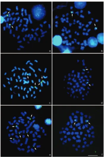
Fluorescent in situ hybridization (FISH) was per- formed with a biotinyled 18S rDNA probe from the fish Prochilodus argenteus (Hatanaka and Galetti Jr, 2004) and
Texto
Imagem

Documentos relacionados
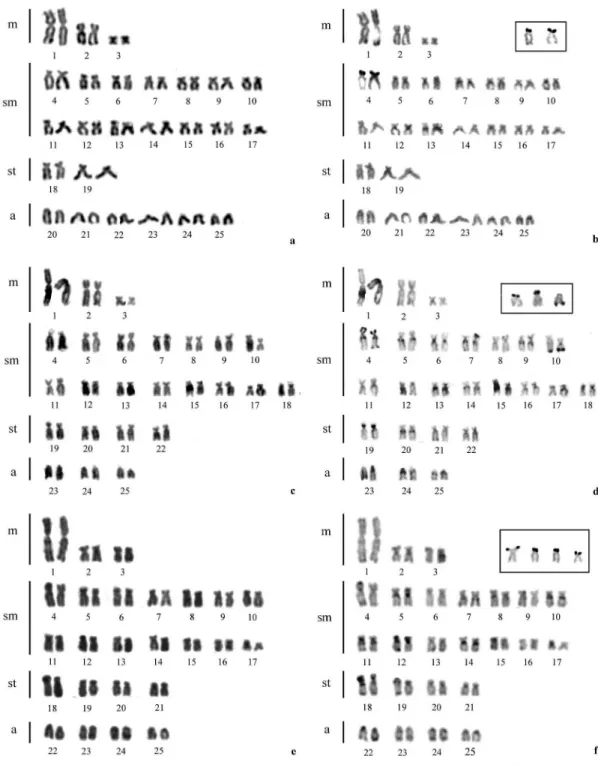
The nucleolar organizing regions (NORs) were studied by silver nitrate staining and rDNA fluorescence in situ hybridiza- tion (FISH) and were found to be located in the telomeric
In situ hybridization with the 45S rDNA probe re- vealed four signals which co-localized with the CMA + bands of the short arms of the acrocentrics, while the 5S rDNA probe
specimens from the Mogi Guaçu river (São Paulo State, Brazil) Jesus and Moreira-Filho (2003) detected not only a large metacentric pair but also a variable number of ribo- somal
The aim of this study was to describe mitotic and meiotic chromosomes of Cycloneda sanguinea using C-banding, fluorescent in situ hybridization (FISH) rDNA probes, and
and fluorescence in situ hybridization with a 45S rDNA probe were used to determine the chromosome number and the number and physical position of GC-rich heterochromatin and 45S
Species with a single pair of chromosomes bearing the nucleolar or- ganizing regions (NORs) were identified, as well as species with multiple NORs, up to a maximum of seven 18S
In situ hybridization by fluorescence (FISH) with the 18S rDNA probe showed only one pair of stained chromosomes, confirming the heteromorphism observed with
regions (CSRs) and fluorescent in situ hybridization (FISH) with rDNA probes. We found that the karyotypes of all four species was 2n = 2x = 16, with, except for the eighth
