Molecular analysis of holoprosencephaly in South America
Clarice Pagani Savastano
1,2, Kênia Balbi El-Jaick
3, Marcelo Aguiar Costa-Lima
4,
Cristina Maria Batista Abath
5, Sebastiano Bianca
6, Denise Pontes Cavalcanti
7, Têmis Maria Félix
8,
Gioacchino Scarano
9, Juan Clinton Llerena Jr.
10, Fernando Regla Vargas
3,11, Miguel Ângelo Martins
Moreira
12, Hector N. Seuánez
12, Eduardo Enrique Castilla
2,13and Iêda Maria Orioli
1,21
Estudo Colaborativo Latino Americano de Malformações Congênitas, Departamento de Genética,
Universidade Federal do Rio de Janeiro, Rio de Janeiro, RJ, Brazil.
2
Instituto Nacional de Genética Médica Populacional, Rio de Janeiro, RJ, Brazil.
3
Departamento de Genética e Biologia Molecular, Universidade Federal do Estado do Rio de Janeiro,
Rio de Janeiro, RJ, Brazil.
4
Departamento de Genética, Universidade do Estado do Rio de Janeiro, Rio de Janeiro, RJ, Brazil.
5Maternidade Cândida Vargas, Instituto Cândida Vargas, João Pessoa, PB, Brazil.
6
Centro di Consulenza Genetica e di Teratologia della Riproduzione, Dipartimento Materno Infantile,
ARNAS Garibaldi Nesima, Catania, CT, Italy.
7
Departamento de Genética Médica, Universidade Estadual de Campinas, Campinas, SP, Brazil.
8Serviço de Genética Médica, Hospital das Clínicas de Porto Alegre, Porto Alegre, RS, Brazil.
9
Registro Campano Difetti Congeniti, Azienda Ospedaliera “Gaetano Rummo”, Benevento, BN, Italy.
10Centro de Genética Médica, Instituto Fernandes Figueira, Fundação Oswaldo Cruz, Rio de Janeiro, RJ,
Brazil.
11
Estudo Colaborativo Latino Americano de Malformações Congênitas, Laboratório de Epidemiologia de
Defeitos Congênitos, Instituto Oswaldo Cruz, Fundação Oswaldo Cruz, Rio de Janeiro, RJ, Brazil.
12
Programa de Genética, Instituto Nacional de Câncer, Rio de Janeiro, RJ, Brazil.
13
Estudio Colaborativo Latino Americano de Malformaciones Congenitas,
Centro de Educación Médica e Investigación Clínica, Buenos Aires, Argentina.
Abstract
Holoprosencephaly (HPE) is a spectrum of brain and facial malformations primarily reflecting genetic factors, such as chromosomal abnormalities and gene mutations. Here, we present a clinical and molecular analysis of 195 probands with HPE or microforms; approximately 72% of the patients were derived from the Latin American Collabo-rative Study of Congenital Malformations (ECLAMC), and 82% of the patients were newborns. Alobar HPE was the predominant brain defect in almost all facial defect categories, except for patients without oral cleft and median or lat-eral oral clefts. Ethmocephaly, cebocephaly, and premaxillary agenesis were primarily observed among female pa-tients. Premaxillary agenesis occurred in six of the nine diabetic mothers. Recurrence of HPE or microform was approximately 19%. The frequency of microdeletions, detected using Multiplex Ligation-dependant Probe Amplifica-tion (MLPA) was 17% in patients with a normal karyotype. Cytogenetics or QF-PCR analyses revealed chromosomal anomalies in 27% of the probands. Mutational analyses in genesSHH, ZIC2, SIX3 and TGIF were performed in 119 patients, revealing eight mutations inSHH, two mutations in SIX3 and two mutations in ZIC2. Thus, a detailed clinical description of new HPE cases with identified genetic anomalies might establish genotypic and phenotypic correla-tions and contribute to the development of additional strategies for the analysis of new cases.
Keywords: holoprosencephaly, ECLAMC,SHH,ZIC2,SIX3. www.sbg.org.br
Send correspondence to Iêda Maria Orioli. Instituto de Biologia, Universidade Federal do Rio de Janeiro, Caixa Postal 68.011, 21941-617 Rio de Janeiro, RJ, Brazil. E-mail: orioli@centroin.com.br.
Introduction
Holoprosencephaly (HPE, MIM 236100) is a com-plex brain malformation affecting both the forebrain and the face. This condition can be described according to cere-bral malformation severity: lobar, semi-lobar, alobar and middle interhemispheric variant (MIH-HPE) (Demyer and Zeman, 1963; Simonet al., 2002). Facial anomalies are variable, ranging from cyclopia, ethmocephaly or ceboce-phaly to mild forms, such as ocular hypotelorism or single median maxillary central incisor (SMMCI) (Cohen Jr, 1989; Richieri-Costa and Ribeiro, 2006; El-Jaick et al., 2007a). In HPE families, mild facial anomalies can occur without cerebral malformation, and these anomalies are considered risk factors for HPE in subsequent offspring (Berryet al., 1984; El-Jaicket al., 2005). Cerebral malfor-mations without facial evidence occur in 10-20% of HPE patients (Cohen Jr, 1989).
The etiology of HPE is complex, including both ge-netic and environmental factors (Barr Jret al., 1983; Bel-loniet al., 1996; Cohen Jr and Shiota, 2002; Dubourget al., 2007). Chromosomal anomalies represent approximately 45% of HPE cases. Multiple malformation syndromes with normal karyotypes, such as Smith-Lemli-Optiz, Pallister Hall and velo-cardio-facial Syndrome, correspond to 25% of HPE cases (reviewed by Dubourget al., 2007). Numer-ous mutations in the genes involved in the development of the forebrain and midline face during embryogenesis have been described. The four genes typically associated with HPE cases areSHH[HPE3, MIM 142945] (Roessleret al., 1996),ZIC2[HPE5, MIM 609637] (Brown et al., 1998), SIX3[HPE2, MIM 157170] (Walliset al., 1999), andTGIF [HPE4, MIM 142946] (Grippet al., 2000). Mutations in one of these genes correspond to 10-20% of non-syndromic cases (Orioli et al., 2001; Dubourg et al., 2004). Other genes associated with HPE, although to a lesser degree, in-clude PTCH [HPE7, MIM 610828] (Ming et al., 2002), TDGF1[MIM 187395] (De la Cruz et al., 2002), GLI2 [HPE9, MIM 610829] (Roessler et al., 2003), DHCR7 [SLOS, MIM 270400] (Shimet al., 2004),FAST1/FOXH1 [MIM 603621] (Roessler et al., 2008), DISP1 [MIM 607502] (Roessler et al., 2009a), FGF8 [HH6, MIM 612702] (Arauzet al., 2010), andCDON[HPE11, MIM 614226] (Baeet al., 2011).
The phenotypic variability and incomplete penetran-ce make it difficult to conduct genetic counseling in HPE. Families typically present autosomal dominant, but also autosomal recessive and possibly X-linked inheritance (Ming and Muenke, 1998; Wallis and Muenke, 1999; Mue-nke and Beachy, 2001). Autosomal dominant inheritance has an estimated penetrance of 80% (Odentet al., 1998), and the same mutation segregating within a family can present asymptomatic, mild and severe forms of the disease (Roessleret al., 1996; El-Jaicket al., 2005). We recently analyzed HPE cases from the ECLAMC (Estudio Latino-americano de Malformaciones
Congénitas-Latin-American Collaborative Study of Congenital Malforma-tions) since 2000 (Orioliet al., 2001; El-Jaicket al., 2005, El-Jaicket al., 2007a), and in the present study, we evaluate the contribution of chromosomal anomalies and mutations in the four main HPE genes (SHH,SIX3,TGIF, andZIC2).
Materials and Methods
ECLAMC
Most of the families studied (140/195; 72%) were de-rived from the ECLAMC (Estudio Latinoamericano de Malformaciones Congénitas-Latin American Collabora-tive Study of Congenital Malformations). ECLAMC is a hospital-based registry, which examines births in South America (Castilla and Orioli, 2004). In 1998, the ECLAMC initiated a molecular epidemiological program, called MOLECLAMC, and biological samples from newborns presenting congenital malformations were collected for molecular analysis.
Samples from MOLECLAMC primarily comprise blood spots on filter paper (Schleicher and Schuell no. 903 or IsoCode), and a pediatrician interviewed the mothers of the malformed infants using a form with 50 questions con-cerning risk factors, including environmental exposure, history of other familial congenital defects, and parental consanguinity (Castilla and Orioli, 2004). The DNA repos-itory of MOLECLAMC also receives samples from pedia-tricians and geneticists associated with the ECLAMC. These cases occasionally involve older patients, represent-ing 28% (55/195) of the families presented herein.
Patients
The patients were referred based on typical HPE fa-cial dysmorphisms, with or without confirmation through brain imaging, on SMMCI or a family history of HPE. Aneuploidy analyses were performed on some HPE pa-tients with no informed karyotype. Chromosomal micro-rearrangements were evaluated in some HPE and SMMCI patients without mutations in the four main HPE genes and no known chromosomal anomalies.
The patients referred to the Birth Defects Laboratory at the Federal University of Rio de Janeiro were analyzed in the last 14 years. Initially, we only analyzed mutations in the four main HPE genes. In 2008, the laboratory imple-mented the MLPA (Multiplex Ligation-Dependent Probe Amplification) technique, and in 2010, we initiated studies to screen for trisomies using QF-PCR. Not all patients could be studied using all techniques, reflecting the lack of adequate DNA quantity and quality, particularly for older samples.
The Ethics Committee of the ECLAMC (CEMIC: Centro de Educación Médica e Investigación Clínica, Bue-nos Aires, Argentina, IORG-0001315) approved the pro-ject, and written informed consent was obtained from adult patients and the parents of infant patients.
DNA extraction
The DNA from blood spotted onto Schleicher and Schuell no. 903 filter paper (Schleicher & Schuell Inc., Keene, NH, USA) was extracted using QIAamp DNA Mini Kit (Qiagen, Valencia, CA, USA) or DNeasy 96 Tissue Kit (Qiagen, Valencia, CA, USA), and DNA from blood spot-ted onto IsoCode filter papers (Schleicher & Schuell Inc., Keene, NH, USA) was extracted through boiling in water. The DNA from peripheral blood samples on EDTA was ex-tracted through salting out (Milleret al., 1988).
PCR and direct sequencing
The coding region and exon-intron boundaries of SHH,SIX3, TGIF, andZIC2were amplified as previously described (El-Jaick 2007a,b; Savastanoet al., 2014). Auto-mated sequencing was performed using MegaBACE 1000 (Amersham Pharmacia Biotech In, London, UK) or ABI Prism 377 (Applied Biosystems, Foster City, CA, USA) DNA sequencing systems. The electropherograms were an-alyzed using Sequencher software (Gene Codes Corpora-tion, Ann Arbor, MI, USA). SIFT and PolyPhen2 online software programs were used to predict the effects of non-synonimous mutations. The gene mutation nomenclature used in this study was consistent with the recommendations of den Dunnen and Antonarakis (2000), and updates are available on the website for the Human Genome Variation Society (HGVS).
Micro-rearrangements analysis
Submicroscopic deletions or gains were screened through Multiplex Ligation-Dependent Probe Amplifica-tion (MLPA) (Schoutenet al., 2002), using the MLPA Kit P187 or the MLPA Kit P187 B1 (MRC-Holland, Amster-dam, Netherlands). These kits contain probes against the HPE genesSHH, SIX3, TGIF, ZIC2, PTCH, GLI2,and can-didate genesTRAPPC10andFBXW11.Normalization and analysis were performed using Coffalyser (Coffa et al., 2008) or GeneMarker (Softgenetics, State College, PA, USA) software programs.
QF-PCR analysis of trisomies
HPE patients without confirmed karyotypes were fur-ther tested for aneuploidies on chromosomes 13, 18, 21 and X through QF-PCR, using the Aneufast Multiplex QF-PCR Kit (Genomed AG, Wollerau, Switzerland). Patients with alterations in more than one marker for the same chromo-some were subjected to further tests using chromochromo-some specific back-up markers. The analyses were performed us-ing GeneMarker (Softgenetics, State College, PA, USA).
Results
The series
From 1998 to 2012 a total of 195 probands were re-ferred for genetic analysis of the HPE genes. Most of the
samples (160/195; 82.1%) were obtained from newborns, livebirths (145/195; 74.4%) or stillbirths (15/195; 7.7%), but the sample also included children of different ages (26/195; 13.3%), adults (3/195; 1.5%), four fetuses (4/195; 2.1%), and two (1.0%) of unknown ages. Females were predominant, with 112 females (58.6%) vs. 79 males (41.4%), and this predominance was significant when con-sidering the sex ratio expected at birth (c2= 7.10; P = 0.008;
GL = 1). One patient had no information, and three patients were intersexes. There were 110 patients with isolated HPE or microform (“isolated cases”), and 85 patients (43.6%) with other malformations unrelated to the HPE spectrum (“associated cases”). The occurrence of HPE was verified through pre or postnatal brain imaging or autopsy in 155 probands (79.5%). Forty-one cases with specified HPE types presented with alobar HPE type (41/79; 51.9%), 29 cases presented with semilobar HPE type (29/79; 36.7%), and nine cases presented with lobar HPE type (9/79; 11.4%). In 97 probands HPE was not specified, although there was a suspicion of interhemispheric variant of HPE in three cases. Nineteen patients (19/195; 9.7%) had no HPE in cerebral imaging: one patient presented premaxillary agenesis and anophtalmia, two patients were obligated car-rier mothers, and 16 patients had SMMCI. Five of the 16 SMMCI patients exhibited a mild phenotype without men-tal deficiency and additional defects, such as microcephaly, choanal atresia, lacrimal conduct atresia, or ocular hypo-telorism.
The facial descriptions of the 195 patients, including the two obligated carrier mothers, were classified as cyclo-pia (19 cases), ethmocephaly (6 cases), cebocephaly (17 cases), and premaxillary agenesis (41 cases). The facial types in 172 patients were distributed among seven addi-tional categories: median cleft lip (8 cases), bilateral or uni-lateral cleft lip with or without cleft palate (8 cases), cleft palate (5 cases), no oral cleft (60 cases), no oral cleft with SMMCI (16 cases), atypical facial cleft (4 cases), and no specified facial type (11 cases). The numbers of patients in these eleven facial type categories are presented in Table 1.
years (95% CI: 25.8-27.8), and did not vary among the fa-cial groups. There were nine cases of maternal diabetes among the 139 probands (6.5%, 95% CI: 3.2-12.3), and six of these cases exhibited premaxillary agenesis. The propor-tion of chromosomal anomalies or gene mutapropor-tions did not differ among the facial categories (Table 1).
Micro-rearrangement analysis and chromosomal anomalies
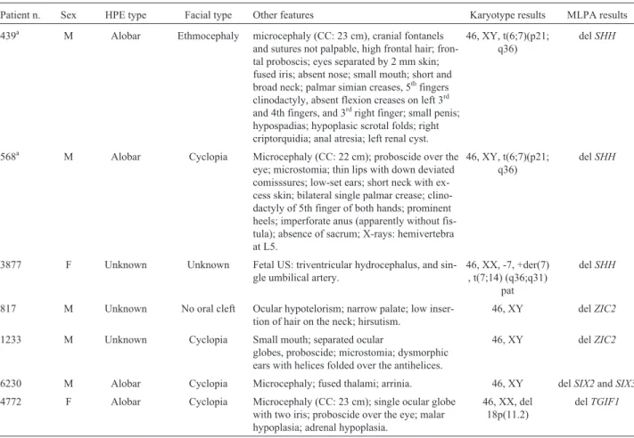
MLPA screening was performed in 58 patients: ten patients with common trisomies (chromosomes 13 and 18), five patients with structural chromosomal anomalies, 23 patients with normal karyotypes, and 20 patients without karyotype information, showing failure in 15 cases and in-conclusive results in eight cases. Among the 35 cases with MLPA results, 28 patients had HPE and seven patients had microforms. We also observed normal MLPA results in 27 cases, in which 14 patients had normal karyotypes, one pa-tient exhibited 46,XX, inv(5) (p14.3; q23.1), and 12 cases showed no karyotype information. Altered MLPA results were observed in three of the 17 cases with normal karyo-types (17.6%; 95% CI: 4.7-44.2) or in three of the 29 cases with or without informed karyotypes (10.3%; 95% CI: 2.7-28.5). We detected twoZIC2deletions and one deletion encompassing theSIX3andSIX2genes. The other altered MLPA results were observed in two cases of trisomy 13, and one case of 18p-, t(6;7) and t(7;14). An affected sibling of the patient with t(6;7) was also examined to verify the occurrence of theSHHdeletion. Table 2 shows the pheno-type and MLPA alterations observed in individuals with gene deletions.
Informed karyotypes were observed in 65 of the probands, and 20 patients had chromosome anomalies. We successfully performed QF-PCR analysis on 26 probands without informed karyotypes and detected chromosomal aneuploidies in five patients: one case of trisomy 21, one case of trisomy 18, and three cases of trisomy 13. Con-sidering informed karyotype and the QF-PCR analysis, we obtained karyotype information for 91 patients (46.7%). Chromosomal anomalies, including aneuploidies or struc-tural anomalies, were observed in 25/91 patients (27.5%; 95% CI: 18.9-38.0). The increased frequency of chromo-somal abnormalities was observed in patients with associ-ated HPE (22/51; 43.1%). Only three patients with isolassoci-ated HPE or microform (3/40; 7.5%) showed chromosomal ab-errations: 18p-, inv(5) and an unspecified chromo-somopathy. The inv(5) observed in a SMMCI patient, was also observed in her father and brother, both exhibiting a normal phenotype.
Mutation screening
Mutation analyses were successfully performed in 120 patients forSHHandTGIF, 125 patients forSIX3, and 151 patients forZIC2. All four genes were successfully se-quenced in 119 patients: 45 patients had HPE, or
micro-Savastanoet al. 253
form, and also other malformations not associated with the HPE spectrum, and 74 patients had isolated HPE or micro-form. Most of the studied patients (86/119; 72.3%) were newborns (81 livebirths, and five stillbirths), but the sample also included 25 children of different ages (25/119; 21.0%) ranging from 4 months to 17 years, three adults (3/119; 2.5%), and four fetuses (4/119; 3.4%). We could not re-trieve information for the age of one patient. Among the 119 studied patients, 36 patients had normal karyotypes (18 males and 18 females), 19 patients had normal QF-PCR re-sults, and seven patients had chromosomal anomalies. The remaining 57 patients had unknown karyotypes (18 associ-ated, and 39 isolated HPE or microform), representing 47.9% of the studied cohort.
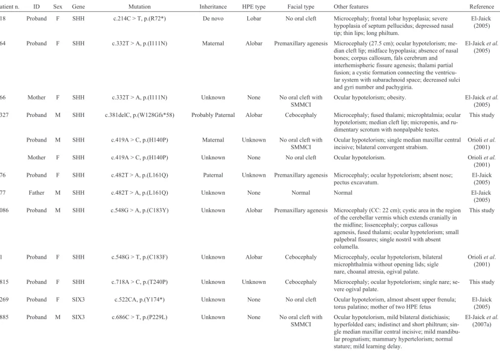
In total, 47 variants were detected, 35 variants were classified as benign or of unknown significance based on several features, including the presence of the variant in population SNP databases, synonymous benign variants predicted using PolyPhen and SIFT software, or UTR and intronic variants with unknown significance. We detected 12 damaging mutations in 119 probands (10.1%; 95% CI: 5.6-17.3) (Table 3). The pathogenetic mutations included eightSHHgene mutations (66%), twoSIX3gene mutations
(17%), and twoZIC2gene mutations (17%). All 12 patients presented isolated HPE or microform.
The mutation frequency is similar (10.9%) among the probands with normal karyotype or QF-PCR results (six mutations out of 55 cases). The pedigrees of the six families with mutation recurrences are presented in Figure 1. The parents from six probands were available for study to deter-mine the mutation inheritance. The clinical characteriza-tion and mutacharacteriza-tions identified in the probands and parents are summarized in Table 3. Figure 2 shows clinical photos and eletropherograms from the three most recently studied mutated patients. The other patients have been previously described (Orioliet al., 2001; El-Jaicket al., 2005, 2007a).
SHHmutations
We detected eight mutations in theSHHgene. Three patients have been previously described. El-Jaick et al. (2005) described a newborn girl presenting the mutation c. 332T > A, p.(I111N), with alobar HPE and premaxillary agenesis, inherited from the mother affected with SMMCI and hypotelorism. Orioliet al.(2001) described the other two patients: 1) a 8-year-old male presenting the mutation c. 419A > C, p.(H140P) without specified HPE type, ocular Table 2- Chromosome deletions in patients with holoprosencephaly (HPE).
Patient n. Sex HPE type Facial type Other features Karyotype results MLPA results
439a M Alobar Ethmocephaly microcephaly (CC: 23 cm), cranial fontanels
and sutures not palpable, high frontal hair; fron-tal proboscis; eyes separated by 2 mm skin; fused iris; absent nose; small mouth; short and broad neck; palmar simian creases, 5thfingers
clinodactyly, absent flexion creases on left 3rd
and 4th fingers, and 3rdright finger; small penis;
hypospadias; hypoplasic scrotal folds; right criptorquidia; anal atresia; left renal cyst.
46, XY, t(6;7)(p21; q36)
delSHH
568a M Alobar Cyclopia Microcephaly (CC: 22 cm); proboscide over the
eye; microstomia; thin lips with down deviated comisssures; low-set ears; short neck with ex-cess skin; bilateral single palmar crease; clino-dactyly of 5th finger of both hands; prominent heels; imperforate anus (apparently without fis-tula); absence of sacrum; X-rays: hemivertebra at L5.
46, XY, t(6;7)(p21; q36)
delSHH
3877 F Unknown Unknown Fetal US: triventricular hydrocephalus, and sin-gle umbilical artery.
46, XX, -7, +der(7) , t(7;14) (q36;q31)
pat
delSHH
817 M Unknown No oral cleft Ocular hypotelorism; narrow palate; low inser-tion of hair on the neck; hirsutism.
46, XY delZIC2
1233 M Unknown Cyclopia Small mouth; separated ocular
globes, proboscide; microstomia; dysmorphic ears with helices folded over the antihelices.
46, XY delZIC2
6230 M Alobar Cyclopia Microcephaly; fused thalami; arrinia. 46, XY delSIX2andSIX3
4772 F Alobar Cyclopia Microcephaly (CC: 23 cm); single ocular globe with two iris; proboscide over the eye; malar hypoplasia; adrenal hypoplasia.
46, XX, del 18p(11.2)
delTGIF1
aPatient n. 568 is brother of patient n. 439. Chomosomes were reanalyzed after the birth of the second affected son in this family.
Savastano
et
al.
255
Table 3- Clinical characterization and mutations in HPE patients.
Patient n. ID Sex Gene Mutation Inheritance HPE type Facial type Other features Reference
818 Proband F SHH c.214C > T, p.(R72*) De novo Lobar No oral cleft Microcephaly; frontal lobar hypoplasia; severe hypoplasia of septum pellucidus; depressed nasal tip; thin lips; long philtum.
El-Jaick (2005)
564 Proband F SHH c.332T > A, p.(I111N) Maternal Alobar Premaxillary agenesis Microcephaly (27.5 cm); ocular hypotelorism; me-dian cleft lip; midface hypoplasia; absence of nasal bones; corpus callosum, fals cerebrum and interhemispheric fissure agenesis; thalami partial fusion; a cystic formation connecting the ventricu-lar system with subarachnoid space; decreased sulci and gyri number and pachygiria.
El-Jaicket al.
(2005)
566 Mother F SHH c.332T > A, p.(I111N) Unknown None No oral cleft with
SMMCI
Ocular hypotelorism; obesity. El-Jaicket al.
(2005) 5327 Proband M SHH c.381delC, p.(W128Gfs*58) Probably Paternal Alobar Cebocephaly Microcephaly; fused thalami; microphtalmia; ocular
hypotelorism; median cleft lip; micropenis, and ru-dimentary scrotum with nonpalpable testes.
This study
2 Proband M SHH c.419A > C, p.(H140P) Maternal Unknown No oral cleft with
SMMCI
Ocular hypotelorism; single median maxillar central incisive; bilateral convergent strabism.
Orioliet al.
(2001)
6 Mother F SHH c.419A > C, p.(H140P) Unknown None No oral cleft Ocular hypotelorism. Orioliet al.
(2001) 576 Proband F SHH c.482T > A, p.(L161Q) Paternal Unknown Premaxillary agenesis Microcephaly; ocular hypotelorism; absent nose;
pectus excavatum.
El-Jaick (2005)
577 Father M SHH c.482T > A, p.(L161Q) Unknown None Normal Normal El-Jaick
(2005) 2086 Proband M SHH c.548G > A, p.(C183Y) Unknown Alobar Premaxillary agenesis Microcephaly (CC: 22 cm); cystic area in the region
of the cerebellar vermis which extends cranially in the midline; lissencephaly; corpus callosus agenesis, fused thalami; ocular hypotelorism; small palpebral fissures; single nostril with absent columella.
This study
81 Proband F SHH c.548G > T, p.(C183F) Unknown Alobar Cebocephaly Microcephaly, ocular hypotelorism, bilateral microphthalmia without opening lids; sigle nare, choanal atresia, ogival palate.
Orioliet al. (2001)
2815 Proband F SHH c.718A > C, p.(T240P) Unknown Unknown Cebocephaly Microcephaly; ocular hypotelorism; single nare; se-vere ogival palate.
This study
1269 Proband F SIX3 c.522CA, p.(Y174*) Unknown None No oral cleft Ocular hypotelorism, almost absent upper frenula; torus palatino; mother of two HPE fetus
El-Jaick (2005)
2885 Proband M SIX3 c.686C > T, p.(P229L) Unknown None No oral cleft with
SMMCI
Ocular hypotelorism, mild bilateral distichiasis; hyperfolded ears; indistinct and short philtrum; sin-gle median maxillar central incisive; mild mandibu-lar prognatism; mammary hypertelorism; normal stature; mild learning delay.
El-Jaicket al.
hypotelorism, SMMCI, and strabism, inherited from the mother affected with mild ocular hypotelorism; and 2) a newborn male presenting the mutation c. 548G > T, p.(C183F) with alobar HPE, microcephaly, ocular hypo-telorism, bilateral microphthalmia without lid opening, a single nostril with choanal atresia, and an ogival palate.
The mutation c. 214C > T, p.(R72*) likely represents a de novo mutation in a female infant with lobar HPE, microcephaly, and ocular hypotelorism (El-Jaick, 2005). The mutation c. 381delC, p.(W128Gfs*58) was observed in a male newborn prenatally diagnosed with HPE, present-ing a typical face, with microcephaly, ocular hypotelorism, cleft lip, micropenis, and rudimentary scrotum with non-palpable testes (Figure 2 e, f, and g). An analysis of the fam-ily history revealed a paternal half-brother with HPE and a father with ocular hypotelorism. Unfortunately, DNA from the father and half-brother was not available. The mutation c. 482T > A, p.(L161Q) was observed in a female proband with HPE, microcephaly, ocular hypotelorism, premaxil-lary agenesis, flat nose, and pectus excavatum, presenting the karyotype 46,XX, 16qh+. The family history revealed a sister with HPE. The mutation was inherited from the fa-ther, who was not affected (El-Jaick, 2005). The mutation c. 548G > A, p.(C183Y) was detected in a male proband with alobar HPE, showing the absence of the corpus callo-sum and fusion of the thalami, a cystic area in the region of the cerebellar vermis, extending cranially through the midline, lissencephaly, microcephaly, ocular hypotelorism, small palpebral fissures, single nostril, and premaxillary agenesis (Figure 2, a and b). The patient presented CC = 22 cm at birth (< 3rdcentile) and 29.8 cm at 13 months of age (< 3rdcentile), with a normal karyotype (46,XY). The pa-tient evolved with significant psychomotor retardation, feeding difficulties and difficult to control seizures, dying at age five. The parents were not available for this study. The mutation c. 718A > C, p.(T240P) was present in a
fe-Patient
n.
ID
Sex
Gene
Mutation
Inheritance
HPE
type
Facial
type
Other
features
Reference
469
Proband
F
ZIC2
c.857_858delAC,
p.(H286Rfs*80)
De
Novo
Semilobar
No
oral
cleft
Microcephaly;
at
six
months
old:
trigonocephaly;
upslanting
palpebral
fissures;
ligth
iris;
bilateral
epicantic
folds;
high
palate.
Orioli
et
al.
(2001)
6721
Proband
M
ZIC2
c.1411_1415delGTGTC,
p.(V471Rfs*
57)
Paternal
Alobar
Atypical
facial
cleft
Microcephaly;
fused
thalami;
corpus
callosus
agenesis;
bilateral
and
symmetrical
increase
of
the
echogenicity
of
the
periventricular
white
matter;
oc-ular
hypertelorism;
bulky
nose
with
left
alar
cleft
(Tessier
n.
1)
and
a
small
protuberance
at
the
upper
right
lateral
wall;
well
delineated
philtrum;
large
mouth.
Savastano
et
al.
(2014)
6724
Father
M
ZIC2
c.(=/1411_1415delGTGTC)
p.(V471Rfs*57)
Unknown
None
No
oral
cleft
Normal.
Savastano
et
al.
(2014)
Table
3
(cont.)
male infant with microcephaly, ocular hypotelorism, single nostril, and pronounced ogival palate (Figure 2, c and d). The family history revealed a nephew of the maternal grandmother with hydrocephalus, and a paternal uncle with a clubfoot. No relatives were available for further analysis in this study.
SIX3mutations
SIX3mutations were identified in two probands. The mutation c. 522CA, p.(Y174*) was detected in a woman with ocular hypotelorism and normal intelligence (El-Jaick, 2005). The patient was pregnant with two fetuses af-fected with HPE. The mutation c. 686C > T, p.(P229L) was observed in an eight-year-old male patient (El-Jaicket al., 2007a) with SMMCI, ocular hypotelorism, mild bilateral distichiasis, mammary hypertelorism, hyperfolded helices, an indistinct and short philtrum, mild mandibular prognatism, normal stature, and a mild learning delay but normal cerebral CT.
ZIC2mutations
Mutations in the ZIC2 gene were detected in two probands. Orioli et al. (2001) previously described a 6-month-old female, presenting the mutation c. 857_858delAC, p.(H286Rfs*80), with semilobar HPE, microcephaly, trigonocephaly and a relatively non-dys-morphic face.
The mutation c. 1411_1415delGTGTC,
p.(V471Rfs*57) was detected in a male infant with a
prena-tal diagnosis of semilobar HPE, presenting microcephaly, ocular hypertelorism, and a rare cleft nose. The family his-tory revealed a paternal half-sister with HPE. The father presented the same mutation, likely in mosaicism, with mild ocular hypotelorism (Savastanoet al., 2014).
Discussion
More than 80% (160/195) of the patients in this study were newborn, and two-thirds of these patients were ob-tained from the ECLAMC, except for the older patients and those with SMMCI. The controlled epidemiological vari-ables, such as sex, maternal age, maternal diabetes, parental consanguinity, chromosomal or gene defects, and the recur-rence of HPE or microform in the family, showed that this sample did not differ from that previously studied at ECLAMC (Orioli and Castilla, 2007).
The phenotypic variables, such as type of HPE, facial types, presence of other cerebral defects or the presence of other defects not associated with the HPE spectrum, dif-fered from those previously observed (Orioli and Castilla, 2007) when newborn patients may not present features, such as SMMCI and/or mental retardation. The expansion of the classical facial classification in HPE (DeMyerset al., 1964) resulted in 11 facial types. We examined some epide-miological and phenotypic variables according to these 11 facial types, to detect useful differences among them, as a facial description is more easily obtained than cerebral im-aging or karyotype. The semilobar HPE type was less
fre-Savastanoet al. 257
quently observed among the four classical facial types, but the sample is too small to access the significance of this finding. Because the semilobar HPE type was more fre-quently observed in trisomy 13 cases (four among five with HPE type specified), we attempted to characterize the other chromosome anomalies with specified HPE types and ob-served only alobar HPE in four cases (18 trisomy, 21 trisomy, t(6;7), and del 18p).
The slight female predominance of 1.4:1 (fe-male:male) observed in the present study was consistent with that previously observed in other studies (Croenet al., 1996; Forrester and Merz, 2000; Mercieret al., 2011). This female predominance was restricted to three facial types: ethmocephaly, cebocephaly, and premaxillary agenesis. Patients with cyclopia showed a normal male proportion. This sexual difference might reflect causal differences of HPE associated with facial types. The increased frequency to be confirmed of maternal diabetes in patients with pre-maxillary agenesis, andZIC2mutations typically associ-ated with normal or mildly affected faces (Brownet al., 2001), might be examples of association between cause and facial type.
Chromosomal anomalies are described in 32% to 41% of HPE cases, and trisomy 13 is the most frequently observed anomaly (reviewed by Solomonet al., 2010a). In the present study, karyotyping or QF-PCR analyses re-vealed chromosomal anomalies in 27.5% (25/91) of the in-formative patients. This frequency must be underestimated as 104 patients (53.3%) were not studied or had inconclu-sive results through QF-PCR. Furthermore, structural ano-malies were not detected using this method. As expected, an increased frequency of chromosomal anomalies was ob-served in patients with HPE and associated malformations.
In addition to the usual cytogenetic study, tracing microdeletions involving genes associated with HPE through techniques, such as MLPA, contributed signifi-cantly to the diagnosis. The rate of chromosomal micro-anomaly detection through MLPA in the present study, ranging from 10.3% (3/29) to 17.6% (3/17 with normal karyotype), is not significantly different from the 5% fre-quency of microdeletions involving the main four HPE genes in newborns (Bendavidet al., 2006a) or the 9.8% fre-quency observed in patients without point mutations (Ben-david et al., 2006b). The frequency of micro-rearrangements and mutations in the four main HPE genes varied between cohorts of fetuses and live-born children. While mutations are more frequent in livebirths, submicro-scopic deletions occur more frequently in fetuses (Ben-david et al., 2006a, 2006b, 2009). In the present study, which primarily included livebirths, the frequency of microdeletions, 10.3% (2.7-28.5), and mutations, 12/119 (10.1%; 5.6-17.3), are similar in both groups.
Array-CGH studies have reported frequencies of 17% to 22% losses or gains in the genome of HPE patients, con-sidering known and new HPE loci (Bendavidet al., 2009;
Mercier et al., 2011). However, the meaning of these candidate loci in the etiology of HPE was uncertain. In a ge-notype-first approach study, Rosenfeldet al.(2010) identi-fied 136 individuals with deletions in any of the 35 selected HPE loci identified in a cohort of 26,922 patients studied through array CGH. The authors observed HPE in 13 indi-viduals, 11 patients with deletions involving the four pri-mary HPE genes, one patient with a deletion in the HPE8 locus (14q13), and one patient with a deletion inFGF8. The other six loci involving HPE genes or candidates were only associated with HPE microforms, but most of the deletions identified in HPE candidate genes showed no HPE, nor mi-croforms of HPE.
The phenotype of patients with microdeletions in one of the four main HPE genes is highly heterogeneous. Simi-lar to intragenic mutations, these patients present pheno-types ranging from alobar HPE to microforms (Bendavidet al., 2007; Rosenfeldet al., 2010). No microdeletions were observed in the seven patients with HPE-microform or the patient with 46,XX, inv(5) (p14.3; q23.1), whose brother and father were carriers with the same inversion, showing normal results using the P-187 HPE Kit for MLPA, where probes against the candidate HPE gene,FBXW11, were lo-cated in 5q35.1. The patient presented with SMMCI, age-nesis of corpus callosum, iris colobome, ocular hypo-telorism, and a mild mental deficiency. Additional studies in this family will be important to determine whether the gene in this locus is associated with HPE.
The mutation frequency observed in the present study was 12/119 (10.1%; 5.6-17.3). Dubourget al.(2004) re-ported a similar rate of 17% in fetuses and living children with normal karyotypes, while Nanniet al.(2000) observed mutation in only one case studied, representing 4.3% of the sample of newborn cases in a population from California. Lazaroet al.(2004) observed a similar frequency of 16% when considering syndromic and nonsyndromic HPE and patients with midline facial and/or cerebral anomalies with-out neuroradiological HPE. Although we did not conduct a functional analysis of the identified mutations, these anom-alies affected conserved protein regions and therefore can be considered as contributors to the HPE phenotype.
The mutation ratio is higher in liveborn children (20% to 23%) than in fetuses (12.5% to 14%), who usually present more severe phenotypes due to chromosomal ano-malies (Dubourget al., 2004; Bendavidet al., 2009). How-ever, even if most of the sample comprised newborns, the mutational frequency is low compared with the cited litera-ture, likely reflecting the high frequency of cases without karyotype observed in the present study (53.3%).
Paulussenet al.(2010) observed a lower frequency of 3.5% in a Dutch cohort of non-syndromic HPE, including fe-tuses, neonatal deceased children, children and adults. Roessleret al.(2012) included the data from Dubourget al. (2004) and Paulussenet al.(2010) in their analyses and ob-served an aggregated frequency of 5.9% in 475 studied in-dividuals. With the exception of the cohort from Paulussen et al. (2010),SHH is the most commonly mutated gene among HPE individuals.
Consistent with the study from Solomonet al.(2012), we observed the prevalence of missense mutations (6/8, 75%) and a similar prevalence of nonsense (1) and frameshift (1) mutations in theSHHgene. A nonsense mu-tation in codon 72 was detected in the patient 818 (Table 2). A quarter of the mutations described inSHHwere nonsense or frameshift mutations (Roessleret al., 2009b; Solomonet al., 2012). Except for the mutation p.M457Rfx*18, these anomalies eliminated the autocatalytic processing site from the protein and are considered as null alleles (Roessleret al., 2009b). The mutation observed in the patient 5327 (Ta-ble 2) was a deletion of the nucleotide at position c.381 in SHH, resulting in a change in the reading frame and gener-ating a stop codon at 58 residues after the alteration. This frameshift mutation resulted in a truncated protein termi-nating 14 residues before the autocatalytic cleavage site, likely generating a non-functional allele. The variant c. 482T > A, p.(L161Q), detected in the proband 576 (Table 2), was inherited from her father. This mutation is consid-ered damaging (score 1.00). It is located in domain N-SHH, a conserved region that occurs among species, causes the substitution of a hydrophobic amino acid (leucine) to a hy-drophilic amino acid (glutamine) and was not observed in the Exome Variant Server. Solomonet al.(2012) described the same mutation in a patient with unknown HPE and no other information about the phenotype. Mutation c. 548G > A, p.(C183Y) was identified in patient number 2086 (Ta-ble 2) and was also identified in a female HPE patient de-scribed by Roessleret al.(2009b), with no detailed clinical information, who inherited the mutation from her father (Solomonet al., 2012). This mutation occurred in the same position of the mutation observed in patient number 81 (Ta-ble 2) previously described by Orioliet al.(2001). Indeed, codon 183 is a hot spot for mutations, as this codon was al-tered in at least four unrelated families: mutation p.(C183R), detected in a family described by Roessleret al. (2009b) and Solomonet al.(2012); mutation p.(C183F), detected in a patient previously described by Orioliet al. (2001); and the two unrelated probands with mutation p.(C183Y) described in the present study, Roessleret al. (2009b), and Solomonet al.(2012). The altered region was localized in domain N-SHH and is highly conserved among species. The PolyPhen2 analysis revealed the mutation p.(C183R) as likely damaging (score of 1.00). The variant c. 548G > A was not described in the Exome Variant Server among more than 6,000 sequenced individuals. The
alter-ation c. 718A > C, p.(T240P), detected in patient 2815 (Ta-ble 2), affects the C-terminal domain of the SHH protein. Roessleret al.(2009b) reported two mutations affecting the adjacent amino acid. This region is conserved among dif-ferent phyla, and this change leads to the substitution of a threonine, an amino acid with hydrophilic characteristics, with a hydrophobic proline. This mutation was not de-scribed in the Exome Variant Server, and the PolyPhen2 analysis considered this mutation as likely damaging (score of 0.773).
In families where theSHHgene mutation is segregat-ing, widely variable expressivity among affected members is observed (Solomonet al., 2012). Indeed, in the present study, at least half of the probands with mutations in the SHHgene inherited the mutation from one parent who was mildly affected. There was no disproportion of sex in the probands with theSHHmutation (proportion was 1:1), con-sistent with the results of previous studies (Mercieret al., 2011; Solomonet al., 2012). The small sample size pre-vented comparisons of the distribution of HPE types or fa-cial features.
The SIX3 gene presented a mutation frequency of 2/119 (1.7%; 0.3-6.5), which is not different from the 4% frequency described by Dubourget al.(2004) in 200 indi-viduals with normal karyotypes, including fetuses and liv-ing children and is also not different from the 5.1% frequency reported by Mercieret al.(2011). Paulussen et al. (2010) observed a higher frequency of 10.5%. Muta-tions in theSIX3gene have been predominantly observed in exon 1 (Lacbawanet al., 2009).
TheZIC2gene has been previously described as the second most frequently mutated gene in HPE cases (Dubourget al., 2007). In the present study, the frequency was only 2/119 (1.7%; 0.3-6.5), a similar frequency of mu-tations as observed in theSIX3gene. Paulussenet al.(2010) also observed a 10.5% mutation frequency inZIC2and in SIX3genes, although these frequencies were higher than the frequencies observed in the present study. Other studies have reported mutation frequency between 3% and 8.4% (Brownet al., 2001; Dubourget al., 2004; Solomonet al., 2010b; Mercieret al., 2011; Roessleret al., 2012). Patients presenting mutations in theZIC2gene typically have mild facial defects or no facial anomalies (Brownet al., 2001). It is likely that there was a bias in the present study towards more typical faces, which would lower mutation frequency in geneZIC2and increase chromosomal defect rate. The two mutations inZIC2detected in the present study were observed in patients with semilobar HPE and microce-phaly, one patient with a relatively non-dysmorphic face, and another patient presenting a nose cleft (Table 2) (Orioli et al., 2001; Savastanoet al., 2014). Interestingly, four pa-tients presented with atypical facial clefts. However, the other patient with a nose cleft showed normal results in the mutational analysis. Recently, there have been attempts to establish genotype-phenotype relations in HPE patients
through studies including hundreds of mutated individuals for SHH (Solomon et al., 2012), ZIC2 (Solomon et al., 2010b) andSIX3genes (Lacbawanet al., 2009). Although the number of cases in the present study identified with a chromosomal or gene defect is small (40/195; 20.5%), cer-tain features that were previously identified in the geno-type-first approach study of Rosenfeldet al.(2010) as the possible common cause between Dandy-Walker anomaly and HPE were observed in the present study. A total of 47 patients presented other cerebral defects besides or instead of HPE, and eight patients showed Dandy-Walker anoma-lies: two patients with trisomy 13, one patient with trisomy 18, one patient with a normal karyotype, and the remaining four patients did not have informed karyotypes.
The absence of mutations in theTGIF gene in our sample likely reflects sample size constraints, as the fre-quencies of mutations in this gene have been reported as ap-proximately 1% (Dubourget al., 2004).
The frequency of chromosome anomalies and muta-tions observed in the present study suggests that patients di-agnosed with HPE must first be karyotyped, as previously recommended by other authors (Mercieret al., 2010; Pine-da-Alvarez et al., 2010). Molecular methods, such as QF-PCR, are efficient for the diagnosis of associated HPE cases when premature death of newborn precludes cyto-genetics studies. Isolated HPE cases might present struc-tural alterations not detected using QF-PCR. In these cases, the MLPA technique is an alternative for the detection of gains or losses in key genes associated with HPE. Current recommendations for the diagnosis of HPE patients also in-clude array-CGH analyses, as a sensitive and efficient tech-nique for the identification of gains and losses in known or new HPE loci, which could clarify the etiology of many ad-ditional cases (Mercieret al., 2010). A mutation in one of the four main HPE genes explains only 10% to 20% of cases, and therefore mutation screening is recommended after the exclusion of chromosomal anomalies.
Acknowledgments
This research was partially supported through grants
from CNPq (476978/2008-4, 573993/2008-4,
554755/2009-2, 402045/2010-6, 481069/2012-7, 306396/2013-0) and FAPERJ (E-26/102.797/2012, E26/110.140/2013), Brazil. The authors would like to thank Maura S. da Silva and Kelli C. M. Mendes from UFRJ, and Leila S. Monnerat, Kelly R. L. de Souza, and Carolina Furtado from INCa, for technical support.
References
Arauz R, Solomon BD, Pineda-Alvarez DE, Gropman AL, Par-sons JA, Roessler E and Muenke M (2010) A hypomorphic allele in the FGF8 gene contributes to holoprosencephaly and is allelic to Gonadotropin-Releasing Hormone Defi-ciency in humans. Mol Syndromol 1:59-66.
Bae GU, Domené S, Roessler E, Schachter K, Kang JS, Muenke M and Krauss RS (2011) Mutations in CDON, encoding a hedgehog receptor, result in holoprosencephaly and defec-tive interactions with other hedgehog receptors. Am J Hum Genet 89:231-240.
Barr Jr M, Hanson JW, Currey K, Sharp S, Toriello H, Schmickel RD and Wilson GN (1983) Holoprosencephaly in infants of diabetic mothers. J Pediatr 102:565-568.
Belloni E, Muenke M, Roessler E, Traverso G, Siegel-Bartelt J, Frumkin A, Mitchell HF, Donis-Keller H, Helms C, Hing AV,et al.(1996) Identification of Sonic hedgehog as a can-didate gene responsible for holoprosencephaly. Nat Genet 14:353-356.
Bendavid C, Haddad BR, Griffin A, Huizing M, Dubourg C, Gicquel I, Cavalli LR, Pasquier L, Shanske AL, Long R,et al.(2006a) Multicolour FISH and quantitative PCR can de-tect submicroscopic deletions in holoprosencephaly patients with a normal karyotype. J Med Genet 43:496-500. Bendavid C, Dubourg C, Gicquel I, Pasquier L, Saugier-Veber P,
Durou MR, Jaillard S, Frébourg T, Haddad BR, Henry C,et al. (2006b) Molecular evaluation of foetuses with holo-prosencephaly shows high incidence of microdeletions in the HPE genes. Hum Genet 119:1-8.
Bendavid C, Dubourg C, Pasquier L, Gicquel I, Le Gallou S, Mottier S, Durou MR, Henry C, Odent S and David V (2007) MLPA screening reveals novel subtelomeric rear-rangements in holoprosencephaly. Hum Mutat 28:1189-1197.
Bendavid C, Rochard L, Dubourg C, Seguin J, Gicquel I, Pasquier L, Vigneron J, Laquerrie A, Marcorelles P, Jeanne-Pasquier C,et al.(2009) Array-CGH analysis indicates a high preva-lence of genomic rearrangements in holoprosencephaly: An updated map of candidate loci. Hum Mutat 30:1175-1182. Berry SA, Pierpont ME and Gorlin RJ (1984) Single central
inci-sor in familial holoprosencephaly. J Pediatr 104:877-880. Bertolacini CD, Richieri-Costa A and Ribeiro-Bicudo LA (2009)
Sonic hedgehog (SHH) mutation in patients within the spec-trum of holoprosencephaly. Brain Dev 32:217-222. Brown LY, Odent S, David V, Blayau M, Dubourg C, Apacik C,
Delgado MA, Hall BD, Reynolds JF, Sommer A, et al.
(2001) Holoprosencephaly due to mutations in ZIC2: Ala-nine tract expansion mutations may be caused by parental somatic recombination. Hum Mol Genet 10:791-796. Brown SA, Warburton D, Brown LY, Yu CY, Roeder ER,
Stengel-Rutkowski S, Hennekam RC and Muenke M (1998) Holoprosencephaly due to mutations in ZIC2, a homologue of Drosophila odd-paired. Nat Genet 20:180-183.
Castilla EE and Orioli IM (2004) ECLAMC: The Latin-American Collaborative Study of Congenital Malformations. Comm Genet 7:76-94.
Coffa J, van de Wiel MA, Diosdado B, Carvalho B, Schouten J and Meijer GA (2008) MLPAnalyzer: Data analysis tool for reliable automated normalization of MLPA fragment data. Cell Oncol 30:323-335.
Cohen Jr MM (1989) Perspectives on holoprosencephaly: Part III. Spectra, distinctions, continuities, and discontinuities. Am J Med Genet 34:271-288.
Croen LA, Shaw GM and Lammer EJ (1996) Holoprosencephaly: Epidemiologic and clinical characteristics of a California population. Am J Med Genet 64:465-472.
De la Cruz JM, Bamford RN, Burdine RD, Roessler E, Barkovich AJ, Donnai D, Schier AF and Muenke M (2002) A loss-of-function mutation in the CFC domain of TDGF1 is asso-ciated with human forebrain defects. Hum Genet 110:422-428.
Demyer W and Zeman W (1963) Alobar holoprosencephaly (arhinencephaly) with median cleft lip and palate: Clinical, electroencephalographic and nosologic considerations. Confin Neurol 23:1-36.
den Dunnen JT and Antonarakis SE (2000) Mutation nomencla-ture extensions and suggestions to describe complex muta-tions: A discussion. Hum Mutat 15:7-12.
Dubourg C, Lazaro L, Pasquier L, Bendavid C, Blayau M, Le Duff F, Durou MR, Odent S and David V (2004) Molecular screening of SHH, ZIC2, SIX3, and TGIF genes in patients with features of holoprosencephaly spectrum: Mutation re-view and genotype-phenotype correlations. Hum Mutat 24:43-51.
Dubourg C, Bendavid C, Pasquier L, Henry C, Odent S and David V (2007) Holoprosencephaly. Orphanet J Rare Dis 2:8. El-Jaick KB (2005) Análise mutacional em pacientes com
holo-prosencefalia. Doctoral thesis (Doctoral Program in Biolog-ical Sciences-Genetics). Instituto de Biologia, Universidade Federal do Rio de Janeiro, 157 pp.
El-Jaick KB, Brunoni D, Castilla EE, Moreira MA and Orioli IM (2005) SHH Ile111Asp in alobar holoprosencephaly in a proposita, whose mother had only a solitary median maxil-lary incisor. Am J Med Genet A 136A:345.
El-Jaick KB, Fonseca RF, Moreira MA, Ribeiro MG, Bolognese AM, Dias SO, Pereira ET, Castilla EE and Orioli IM (2007a) Single median maxillary central incisor: New data and mu-tation review. Birth Defects Res A Clin Mol Teratol 79:573-580.
El-Jaick KB, Powers SE, Bartholin L, Myers KR, Hahn J, Orioli IM, Ouspenskaia M, Lacbawan F, Roessler E, Wotton D,et al.(2007b) Functional analysis of mutations in TGIF
associ-ated with holoprosencephaly. Mol Genet Metab 90:97-111. Forrester MB and Merz RD (2000) Epidemiology of holopro-sencephaly in Hawaii, 1986-97. Paediatr Perinat Epidemiol 14:61-63.
Gripp KW, Wotton D, Edwards MC, Roessler E, Ades L, Mei-necke P, Richieri-Costa A, Zackai EH, Massague J, Muenke M, et al. (2000) Mutations in TGIF cause holoprosen-cephaly and link NODAL signalling to human neural axis determination. Nat Genet 25:205-208.
Lacbawan F, Solomon BD, Roessler E, El-Jaick K, Domene S, Velez JI, Zhou N, Hadley D, Balog JZ, Long R,et al.(2009) Clinical spectrum of SIX3-associated mutations in holo-prosencephaly: Correlation between genotype, phenotype and function. J Med Genet 46:389-398
Lazaro L, Dubourg C, Pasquier L, Le Duff F, Blayau M, Durou MR, de la Pintiere AT, Aguilella C, David V and Odent S (2004) Phenotypic and molecular variability of the holopro-sencephalic spectrum. Am J Med Genet A 129A:21-24. Mercier S, Dubourg C, Belleguic M, Pasquier L, Loget P, Lucas J,
Bendavid C and Odent S (2010) Genetic counseling and “molecular” prenatal diagnosis of holoprosencephaly
(HPE). Am J Med Genet C Semin Med Genet 154C:191-196.
Mercier S, Dubourg C, Garcelon N, Campillo-Gimenez B, Gic-quel I, Belleguic M, Ratié L, Pasquier L, Loget P, Bendavid C,et al.(2011) New findings for phenotype-genotype corre-lations in a large European series of holoprosencephaly cases. J Med Genet 48:752-760.
Miller SA, Dykes DD and Polesky HF (1988) A simple salting out procedure for extracting DNA from human nucleated cells. Nucleic Acids Res 16:1215.
Ming JE and Muenke M (1998) Holoprosencephaly: From Homer to Hedgehog. Clin Genet 53:155-163.
Ming J, Kaupas M, Roessler E, Brunner H, Golabi M, Tekin M, Stratton RF, Sujansky E, Bale SJ and Muenke M (2002) Mu-tations in PATCHED-1, the receptor for SONIC HEDGEHOG, are associated with holoprosencephaly. Hum Genet 110:297-301.
Muenke M and Beachy PA (2001) Holoprosencephaly. In: Scriver CR, Beaudet AL, Sly WS, Valle D, Childs B, Kinzler KW, Vogelstein B (eds) The Metabolic and Molecular Basis of Inherited Disease. McGraw-Hill Companies, Inc., New York, pp 6203-6262.
Nanni L, Ming JE, Bocian M, Steinhaus K, Bianchi DW, Die-Smulders C, Giannotti A, Imaizumi K, Jones KL, Campo MD, et al. (1999) The mutational spectrum of the sonic hedgehog gene in holoprosencephaly: SHH mutations cause a significant proportion of autosomal dominant holopro-sencephaly. Hum Mol Genet 8:2479-2488.
Nanni L, Croen LA, Lammer EJ and Muenke M (2000) Holopro-sencephaly: Molecular study of a California population. Am J Med Genet 90:315-319.
Odent S, Le Marec B, Munnich A, Le Merrer M and Bonaiti-Pellie C (1998) Segregation analysis in nonsyndromic holo-prosencephaly. Am J Med Genet 77:139-143.
Orioli IM, Castilla EE, Ming JE, Nazer J, Burle de Aguiar MJ, Llerena JC and Muenke M (2001) Identification of novel mutations in SHH and ZIC2 in a South American (ECLAMC) population with holoprosencephaly. Hum Genet 109:1-6.
Orioli IM and Castilla EE (2007) Clinical epidemiologic study of holoprosencephaly in South America. Am J Med Genet Part A 143A:3088-3099.
Paulussen AD, Schrander-Stumpel CT, Tserpelis DC, Spee MK, Stegmann AP, Mancini GM, Brooks AS, Collée M, Maat-Kievit A, Simon ME,et al.(2010) The unfolding clinical
spectrum of holoprosencephaly due to mutations in SHH, ZIC2, SIX3 and TGIF genes. Eur J Hum Genet 18:999-1005.
Pineda-Alvarez DE, Dubourg C, David V, Roessler E and Muenke M (2010) Current recommendations for the molec-ular evaluation of newly diagnosed holoprosencephaly pa-tients. Am J Med Genet C Semin Med Genet 154C:93-101. Richieri-Costa A and Ribeiro LA (2006) Holoprosencephaly-like phenotype: Clinical and genetic perspectives. Am J Med Genet A 140:2587-2593.
Roessler E, Belloni E, Gaudenz K, Jay P, Berta P, Scherer SW, Tsui LC and Muenke M (1996) Mutations in the human Sonic Hedgehog gene cause holoprosencephaly. Nat Genet 14:357-360.
Roessler E, Du YZ, Mullor JL, Casas E, Allen WP, Gillessen-Kaesbach G, Roeder ER, Ming JE, Ruiz i Altaba A and
Muenke M (2003) Loss-of-function mutations in the human GLI2 gene are associated with pituitary anomalies and holo-prosencephaly-like features. Proc Natl Acad Sci USA 100:13424-13429.
Roessler E, Ouspenskaia MV, Karkera JD, Velez JI, Kantipong A, Lacbawan F, Bowers P, Belmont JW, Towbin JA, Gold-muntz E,et al.(2008) Reduced NODAL signaling strength
via mutation of several pathway members including FOXH1 is linked to human heart defects and holoprosencephaly. Am J Hum Genet 83:18-29.
Roessler E, Ma Y, Ouspenskaia MV, Lacbawan F, Bendavid C, Dubourg C, Beachy PA and Muenke M (2009a) Truncating loss-of-function mutations of DISP1 contribute to holopro-sencephaly-like microform features in humans. Hum Genet 125:393-400.
Roessler E, El-Jaick KB, Dubourg C, Velez JI, Solomon BD, Pineda-Alvarez DE, Lacbawan F, Zhou N, Ouspenskaia M, Paulussen A, et al. (2009b) The mutational spectrum of
holoprosencephaly-associated changes within the SHH gene in humans predicts loss-of-function through either key struc-tural alterations of the ligand or its altered synthesis. Hum Mutat 30:E920-930.
Roessler E, Vélez JI, Zhou N and Muenke M (2012) Utilizing pro-spective sequence analysis of SHH, ZIC2, SIX3 and TGIF in holoprosencephaly probands to describe the parameters lim-iting the observed frequency of mutant genegene interac-tions. Mol Genet Metab 105:658-664.
Rosenfeld JA, Ballif BC, Martin DM, Aylsworth AS, Bejjani BA, Torchia BS and Shaffer LG (2010) Clinical characterization of individuals with deletions of genes in holoprosencephaly pathways by aCGH refines the phenotypic spectrum of HPE. Hum Genet 127:421-440.
Savastano CP, Bernardi P, Seuanez HN, Moreira MAM and Orioli IM (2014) Rare nasal cleft in patient with holopro-sencephaly due to mutation in ZIC2 gene. Birth Defects Res A [in press].
Schouten JP, McElgunn CJ, Waaijer R, Zwijnenburg D, Diepvens F and Pals G (2002) Relative quantification of 40 nucleic acid sequences by Multiplex Ligation-Dependent Prove Amplification. Nucleic Acids Res 30:e57.
Shim YH, Bae SH, Kim JH, Kim KR, Kim CJ and Paik YK (2004) A novel mutation of the human 7-dehydrocholesterol reduc-tase gene reduces enzyme activity in patients with holo-prosencephaly. Biochem Biophys Res Commun 315:219-223.
Simon EM, Hevner RF, Pinter JD, Clegg NJ, Delgado M, Kins-man SL, Hahn JS and Barkovich AJ (2002) The middle interhemispheric variant of holoprosencephaly. AJNR Am J Neuroradiol 23:151-156.
Solomon BD, Rosenbaum KN, Meck JM and Muenke M (2010a) Holoprosencephaly due to numeric chromosome abnormali-ties. Am J Med Genet C Semin Med Genet 154C:146-148. Solomon BD, Lacbawan F, Mercier S, Clegg NJ, Delgado MR,
Rosenbaum K, Dubourg C, David V, Olney AH, Wehner LE, et al.(2010b) Mutations in ZIC2 in human
holopro-sencephaly: Description of a novel ZIC2 specific phenotype and comprehensive analysis of 157 individuals. J Med Genet 47:513-524.
Solomon BD, Bear KA, Wyllie A, Keaton AA, Dubourg C, David V, Mercier S, Odent S, Hehr U, Paulussen A,et al.(2012)
Genotypic and phenotypic analysis of 396 individuals with mutations in Sonic Hedgehog. J Med Genet 49:473-479. Wallis DE and Muenke M (1999) Molecular mechanisms of
holoprosencephaly. Mol Genet Metab 68:126-138. Wallis DE, Roessler E, Hehr U, Nanni L, Wiltshire T,
Richieri-Costa A, Gillessen-Kaesbach G, Zackai EH, Rommens J and Muenke M (1999) Mutations in the homeodomain of the hu-man SIX3 gene cause holoprosencephaly. Nat Genet 22:196-198.
Internet Resources
Exome Variant Server, NHLBI GO Exome Sequencing Project (ESP), http://evs.gs.washington.edu/EVS/ (December 10, 2013).
Guidelines for Mutation Nomenclature, Human Genome Varia-tion Society (HGVS), http://www.hgvs.org/mutnomen/ (December 10, 2013).
Online Mendelian Inheritance in Man (OMIM),
http://www.ncbi.nlm.nih.gov/OMIM (November 24, 2013). Polymorphism Phenotyping v2 (PolyPhen-2),
http://genet-ics.bwh.harvard.edu/pph2/index.shtml (December 10,
2013).
Sorting Intolerant From Tolerant Program (SIFT),
http://sift.jcvi.org/ (December 10, 2013).
Universidade Federal do Rio de Janeiro-Electronic theses and
dis-sertations library,
http://fenix3.ufrj.br/50/teses/d/CCS_D_KeniaBalbiElJaick. pdf (January 11, 2014).