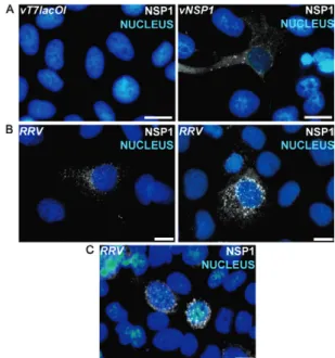
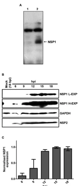
The shift from low to high non-structural protein 1 expression in rotavirus-infected MA-104 cells
Texto
Imagem


Documentos relacionados
Thus, the high expression of IFN- γ in the samples with high-grade lesions, carcinoma and with high viral load observed in our study, may have induced the recruitment of Treg
Recently, it has been demonstrated that the low levels of viral gene expression induced by a candidate HDACi may be insufficient to cause the death of infected cells by viral
No difference in serotonin secretion was observed from RRV-infected EC tumor cells silenced for VP4, VP6 and VP7 expression in comparison to infected cells transfected with siRNA
In MVM-infected cells we observed redistribution of MRN complex proteins from their diffuse nuclear localization seen in mock-infected cells into APAR bodies, the sites of ongoing
In the absence of CD8+ T cell killing, SIV-infected CD4+ T cells are long-lived, so that viral load does not decay. In vitro In vitro killing of peptide-pulsed/ vaccinia-infected
In vivo HIV-1 infection results in a distinctive mRNA transcriptome profile in CD4 + T cells that involves 260 genes in an analysis that differentiates individuals with high and
The T cell VS was initially characterized as an actin-dependent polarization of viral proteins Gag and Env to the site of cell–cell contact on infected donor cells and the
Accordingly, we observed similar levels of GFP protein in the transduced cells coupled with high expression of p27 WT , p27 1–170 , p27 T198E and p27 T198V proteins while, as
