Chronic excitotoxic lesion of the dorsal
raphe nucleus induces sodium appetite
1Departamento de Ciências Fisiológicas, Instituto de Biologia,
Universidade Federal Rural do Rio de Janeiro, Seropédica, RJ, Brasil
2Departamento de Fisiologia, Faculdade de Medicina de Ribeirão Preto,
Universidade de São Paulo, Ribeirão Preto, SP, Brasil H.R. Cavalcante-Lima1,
D. Badauê-Passos Jr.2,
W. de-Lucca Jr.2,
H.R.C. Lima1,
R.H. Costa-e-Sousa1,
E.L. Olivares1,
P.L. Cedraz-Mercez1,
R.O. Reis1, M.A. Medeiros1,
W.S. Côrtes1 and L.C. Reis1
Abstract
We determined if the dorsal raphe nucleus (DRN) exerts tonic control of basal and stimulated sodium and water intake. Male Wistar rats weighing 300-350 g were microinjected with phosphate buffer (PB-DRN, N = 11) or 1 µg/0.2 µl, in a single dose, ibotenic acid (IBO-(PB-DRN, N = 9 to 10) through a guide cannula into the DRN and were observed for 21 days in order to measure basal sodium appetite and water intake and in the following situations: furosemide-induced sodium depletion (20 mg/kg, sc, 24 h before the experiment) and a low dose of dietary captopril (1 mg/g chow). From the 6th day after ibotenic acid injection IBO-DRN rats showed an increase in sodium appetite (12.0 ± 2.3 to 22.3 ± 4.6 ml 0.3 M NaCl intake) whereas PB-DRN did not exceed 2 ml (P < 0.001). Water intake was comparable in both groups. In addition to a higher dipsogenic response, sodium-depleted IBO-DRN animals displayed an increase of 0.3 M NaCl intake compared to PB-DRN (37.4 ± 3.8 vs 21.6 ± 3.9 ml 300 min after fluid offer, P < 0.001). Captopril added to chow caused an increase of 0.3 M NaCl intake during the first 2 days (IBO-DRN, 33.8 ± 4.3 and 32.5 ± 3.4 ml on day 1 and day 2, respectively, vs 20.2 ± 2.8 ml on day 0, P < 0.001). These data support the view that DRN, probably via ascending serotonergic system, tonically modulates sodium appetite under basal and sodium depletion conditions and/or after an increase in peripheral or brain angiotensin II.
Correspondence
L.C. Reis
Departamento de Ciências Fisiológicas Instituto de Biologia, UFRuralRJ BR465, km 7
23890-000 Seropédica, RJ Brasil
Fax: +55-21-26821763 E-mail: lcreis@ufrrj.br
Research partially supported by FAPERJ (No. E-26/171.333/2002). H.R. Cavalcante-Lima was the recipient of a FAPERJ fellowship.
Publication supported by FAPESP.
Received April 22, 2004 Accepted June 1, 2005
Key words
•Sodium appetite •Water intake •Dorsal raphe nucleus •Serotonergic system •Ibotenic acid
Introduction
The organum vasculosum laminae termi-nalis, median preoptic nucleus and subforni-cal organ (SFO), structures participating in hydroelectrolytic and cardiovascular homeo-stasis, receive input from serotonergic neu-rons of the dorsal raphe nucleus (DRN)
hypovolemia induced by hemorrhage evokes changes in the electrical activity of DRN serotonergic neurons connected with the SFO (10). In agreement with this, Vivas’ group (11) reported c-Fos expression in DRN sero-tonergic neurons and laminae terminalis structures in response to salt intake under basal conditions or after sodium depletion induced by peritoneal dialysis. Furthermore, the intracerebroventricular (icv) administra-tion of serotonin (5-HT) and of the 5-HT2A/ C receptor agonists induces an increase in renal sodium excretion (12). In addition, electrolytic lesions of the DRN drastically reduce the plasma levels of atrial natriuretic peptide (ANP), both in basal and ECF vol-ume expansion situations (6,12,13). In this context, it is important to note the very im-portant role of ANP in sodium excretion and salt appetite (5,6,13-19).
Renal afferent information about tubular sodium load and plasma volume is transmit-ted toward forebrain areas relatransmit-ted to angio-tensin conversion (ANG I to ANG II) and subsequent angiotensin receptor activation (20). DRN afferent fibers arise from these areas (especially the SFO), probably involved in a feedback loop regulating sodium satiety and contributing to ECF volume and cardio-circulatory homeostasis (7,9,21).
Taken together, this evidence supports our view that central control of sodium sati-ety and excretion is regulated by the seroto-nergic system that originates from the mid-brain, but only one study acknowledged that the suppression of ascending serotonergic pathways through DRN electrolytic lesion evokes an increase in sodium appetite (21). Corroborating these findings, other investi-gators showed that basal c-Fos expression in serotonergic neurons of the DRN decreased after sodium depletion induced by perito-neal dialysis and increased after spontane-ous and induced sodium intake, suggesting that there is a tonic inhibition of sodium appetite by serotonergic cells of this nucleus (11). Although we hypothesized the same,
when working with DRN electrolytic lesion it is necessary to limit the lesion to the neuronal cell body located in the ventrome-dial portion of the DRN and to perform longer observations (21).
Thus, anatomic and physiological evi-dence clearly shows the role of the DRN in integrating viscerosensory input and fore-brain signals concerning cardiovascular and hydroelectrolyte homeostasis. In this con-text, the objective of the present study was to investigate the influence of excitotoxic DRN lesion by local ibotenic acid microinjection on sodium appetite both in the basal condi-tion and in the presence of natriorexigenic paradigms, such as furosemide-induced so-dium depletion and low-dose dietary supple-mentation with captopril.
Material and Methods
Animals
Male Wistar rats weighing 300-350 g were maintained in a room with lights on from 7:00 to 19:00 h, controlled temperature at 25ºC and free access to Purina chow and water. Before brain stereotaxic surgery the rats were housed in metabolic cages for at least 5 days. All the experimental protocols and animal procedures were carried out in accordance with current Brazilian legisla-tion.
Ibotenic lesions
so-dium phosphate buffer, pH 7.4) were per-formed during a period of 1 min. Sham-lesioned rats were microinjected with 0.2 µl phosphate buffer. A prophylactic dose of penicillin was administered to operated rats (30,000 IU, im; Fort Dodge Saúde Animal Ltda., Campinas, SP, Brazil). Data from rats with lesions outside the dorsoventromedial region of the DRN were not included in the statistical analysis.
Experimental procedures
DRN-excitotoxic-lesioned rats microin-jected with ibotenic acid (IBO-DRN) or sham-lesioned rats microinjected with so-dium phosphate buffer (PB-DRN) were used in three experimental protocols:
Group 1. IBO-DRN (N = 10) and PB-DRN (N = 11) rats were maintained in meta-bolic cages under basal conditions to meas-ure water and 0.3 M NaCl consumption for 21 days following surgery.
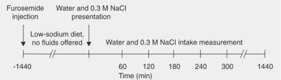
Group 2. On day 21 after microinjection, IBO-DRN (N = 9) and PB-DRN (N = 11) rats were submitted to sodium and water depletion induced by 20 mg/kg furosemide, sc (Lasix, Aventis Pharma, Suzano, SP, Brazil). During the 24 h following furosemide administration a low-sodium diet was offered to the animals (23). Distilled water and 0.3 M NaCl were then supplied and the consumption of these fluids was recorded hourly for 5 h and at the end of 24 h (Figure 1).
Group 3. A low dose (1 mg/g of chow) of dietary captopril (Bristol-Myers Squibb, São Paulo, SP, Brazil) was added every day to the chow from the 6th to the 11th days after surgery (PB-DRN, N = 11 and IBO-DRN, N = 10). This model promotes a sys-temic rise in plasma ANG I levels, which results in an elevated ANG II synthesis in circumventricular forebrain structures un-derlying the subsequent natriorexigenic re-sponse (24-29). The consumption of 0.3 M NaCl and water was determined before (3-day pretreatment period), during (captopril
treatment) and after treatment (9-day recov-ery period).
Histological analysis
Rats were anesthetized and transcardial-ly perfused with 4% paraformaldehyde to identify the lesion site. Brains were removed and serial coronal sections were examined after staining with crystal violet by the Holzer method.
Statistical analysis
Differences between the IBO-DRN and PB-DRN groups were analyzed by repeated measures two-way ANOVA followed by the Newman-Keuls test, with the level of signif-icance set at 5%.
Results
The experimental protocol is presented schematically in Figure 1. Midbrain histo-logical analyses of IBO-DRN rats revealed typical lesions in the ventromedial portion of the DRN. Fibrillar astrocytes were evi-dent along the anteroposterior axis of this nucleus (Figure 2).
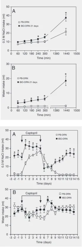
Sodium appetite was increased in IBO-DRN rats compared to PB-IBO-DRN rats through-out the experimental period, especially after 6 days post-lesion (P < 0.01). From day 11 on, 0.3 M NaCl intake reached a level al-ways higher than 18 ml (Figure 3A), what persisted up to the 35th day post-lesion (data
for 24 h (9.8 ± 1.0, 10.2 ± 1.0, and 21.6 ± 3.9
vs 13.1 ± 0.98, 15.3 ± 1.1, and 37.4 ± 3.8 ml, at 240, 300, and 1440 min in PB-DRN and IBO-DRN, respectively, P < 0.04; Figure 4A). Renal sodium and water loss-induced volume depletion evoked an increased water intake in IBO-DRN animals during the ex-perimental period up to 24 h after fluid pres-entation (P < 0.002; Figure 4B).
Low-dose captopril treatment promoted a still higher increase in 0.3 M NaCl intake in IBO-DRN animals and was significantly dif-ferent compared to the PB-DRN group on the first 2 days of treatment (13.6 ± 1.7 and 20.8 ± 2.2 vs 33.8 ± 4.3 and 32.5 ± 3.4 ml on the 1st and 2nd days in PB-DRN and IBO-DRN, respectively, P < 0.002; Figure 5A). On the first day after the end of captopril treatment there was an abrupt fall of 0.3 M NaCl intake in the IBO-DRN group from 30.1 ± 3.3 to 7.9 ± 3.3 ml. In contrast, PB-DRN rats showed a slight reduction com-pared to IBO-DRN rats (from 27.5 ± 3.0 to 19.4 ± 4.1 ml), with higher levels of 0.3 M NaCl intake than IBO-DRN at this time point. The 0.3 M NaCl intake was gradually recov-ered after day 12 (6 days after the end of captopril treatment) in both groups, return-ing to levels similar to those observed before captopril treatment. There was no difference in water intake between the groups during 14 days, except on the first two days after captopril treatment interruption when an in-crease in this parameter was observed in IBO-DRN rats (Figure 5B).
Discussion
Our results show that DRN lesions dis-rupt the functional integrity of central neural pathways that normally exert a negative drive leading to salt intake inhibition. It has been postulated that the DRN serotonergic neu-rons receive viscerosensory inputs concern-ing renal sodium load and/or ECF volume variations to trigger homeostatic regulation of sodium appetite (6,21,30). This view has Figure 2. Reactive gliosis in rats
with an excitotoxic lesion of the dorsal raphe nucleus induced by microinjection of ibotenic acid. Coronal section at the midbrain level shows fibrillar astrocytes in its ventromedial portion (ar-rowheads). Aq = aqueduct. Holzer technique. Magnification, 100X.
Figure 3. Effect of excitotoxic le-sion of the dorsal raphe nucleus produced by ibotenic acid micro-injection (IBO-DRN, N = 10) on chronic ingestion of 0.3 M NaCl (A) and water (B) by IBO-DRN rats and by sham-lesioned rats microinjected with sodium phos-phate buffer (PB-DRN, N = 11). Data are reported as mean ± SEM. *P < 0.05 compared to PB-DRN rats (two-way ANOVA fol-lowed by Newman-Keuls test).
not shown). In addition, most of the IBO-DRN rats showed a drive to eat salt, which was evident when a salt lump was offered through the metabolic cage door. This phe-nomenon was frequently observed in rats which had been previously offered the salt lump. Water intake did not differ between groups during the experimental period (Fig-ure 3B).
been shared by other investigators who have shown increased c-Fos expression in DRN serotonergic neurons after salt intake under both basal and sodium depletion conditions (11).
Low-dose captopril treatment resulted in a higher saline intake by ibotenic DRN-lesioned rats, suggesting that there is either an enhanced capacity of ANG I conversion to ANG II in forebrain structures, or an increased sensitivity to ANG II-evoked so-dium appetite (24,25,27-29). On this basis and regarding reciprocal relationships be-tween SFO and DRN, we postulated that the presumptive angiotensinergic sensitivity of the SFO is enhanced following the failure of inhibitory signals arising from the midbrain (7,10,28).
Although we did not investigate the fobrain angiotensinergic sensitivity, we re-cently demonstrated that experiments involv-ing an increase in plasma ANG II levels exarcebate the sodium appetite of ibotenic-lesioned rats (31). Hence, the view that sup-pression of neural pathways originating in the DRN reduces the threshold of the so-dium appetite response through an increase in angiotensinergic sensitivity of SFO neu-rons should be considered. That is, lesioned rats putatively respond to the natriorexigenic stimuli (induced by high plasma levels of ANG I or ANG II) which would be sub-threshold for sham-lesioned rats.
Also, we did not rule out the possibility that the deficit in the ascending regulation of sodium appetite is due, at least in part, to disruption of interconnections between the DRN and lateral parabrachial nucleus (LPBN) (32,33). This presumptive interac-tion was suggested by Lind’s work (7), which presupposed that viscerosensory informa-tion relayed from the pontine nucleus to-wards the raphe would be transmitted to the SFO through ascending serotonergic path-way from the DRN. The LPBN is an impor-tant pontine circuit that receives homeo-static viscerosensory signals from the right
Figure 4. Effect of furosemide treatment (20 mg/kg, sc, 24 h before the beginning of the ex-periment) on the cumulative in-gestion of 0.3 M NaCl (A) and water (B) by rats with excitotoxic lesion of the dorsal raphe nu-cleus produced by ibotenic acid microinjection (IBO-DRN, N = 9) and sham-lesioned rats microin-jected with phosphate buffer (PB-DRN, N = 11). Data are re-ported as mean ± SEM. *P < 0.05 compared to PB-DRN rats (two-way ANOVA followed by Newman-Keuls test).
Figure 5. Effect of dietary capto-pril treatment (1 mg/g of food) on chronic ingestion of 0.3 M NaCl (A) and water (B) by rats with excitotoxic lesion of the dorsal raphe nucleus produced by ibotenic acid microinjection (IBO-DRN, N = 10) and by sham-lesioned rats microin-jected with phosphate buffer mi-croinjection (PB-DRN, N = 11). Data are reported as means ± SEM. *P < 0.05 compared to PB-DRN rats (two-way ANOVA fol-lowed by Newman-Keuls test). atrium and vena cava stretch receptors and
The blockade of 5-HT2A/C receptors in the LPBN exacerbates the ingestion of hy-pertonic saline induced by icv administra-tion of ANG II or by a combinaadministra-tion of furo-semide + low dose of captopril (23). Micro-injection of the AT1 antagonist losartan into the SFO prevented the additional salt intake induced by methysergide administration in the LPBN of rats submitted to furosemide + captopril treatment (35). Later, Menani and colleagues (36,37) showed a widespread in-volvement of the inhibitory serotonergic mechanism in the LPBN. Thus, bilateral in-jections of serotonergic receptor antagonist methysergide amplified hypertonic saline intake after furosemiinduced sodium de-pletion, water deprivation or central cholin-ergic stimulation.
Corroborating data obtained by electro-lytic DRN lesion (21), our results reveal that excitotoxic damage of DRN neurons induces a rise in basal NaCl intake. In that study (21), Olivares and co-workers made short-term observations (3 days only) and their findings correlated with reduced ANP level in elec-trolytic DRN-lesioned rats under basal and volume expansion conditions (13). Although the reduced ANP levels could explain the increase in sodium appetite, it has been dem-onstrated that renal sodium excretion nor-malizes 7 days after electrolytic lesion of the DRN, possibly concomitant with a restora-tion of ANP secrerestora-tion (6,13).
Therefore, on the basis of the present data, the above-mentioned observation
con-flicts with the hypothesis that DRN lesion-evoked sodium appetite occurs as a result of reduced ANP secretion. We suggested that the suppression of ascending serotonergic pathways exacerbates sodium appetite through a disinhibitory mechanism in the forebrain, possibly within the SFO, whose target neurons would be ANG II-sensitive. However, the precise mechanism that un-derlies forebrain mediation of serotonergic sodium satiety is unknown.
Our data suggest that an ascending cir-cuit, possibly serotonergic, originating in the DRN, integrates viscerosensory and fore-brain signals for sodium appetite homeosta-sis. The results obtained under basal condi-tions allow us to postulate that the DRN neural circuit constitutes an integrative sys-tem that tonically triggers modulation of the outputs to the forebrain to induce salt satiety. Furthermore, disruption of the reciprocal connection between DRN and LPBN could contribute to the deficit in forebrain signal-ing for sodium appetite modulation.
Acknowledgments
The authors are grateful to Mr. Ipojucan Pereira de Souza for animal care. We are indebted to Dr. Celso Rodrigues Franci of the Department of Physiology (Faculty of Medicine of Ribeirão Preto, University of São Paulo) for his support in the Holzer technique.
References
1. Azmitia EC & Segal M (1978). An autoradiographic analysis of the differential ascending projections of the dorsal and median raphe nuclei in the rat. Journal of Comparative Neurology, 179: 641-668. 2. Steinbusch HWM (1981). Distribution of serotonin-immunoreactivity
in the central nervous system of the rat cell bodies and terminals.
Neuroscience,6: 557-618.
3. Bosler O & Descarries L (1988). Monoamine innervation of the organum vasculosum laminae terminalis (OVLT): a high resolution radioautographic study in the rat. Journal of Comparative Neurol-ogy, 272: 545-561.
4. Marcinkiewicz M, Morcos R & Chretien M (1989). CNS connections
with the raphe nucleus: retrograde tracing with WGA-apoHRP-Gold complex in the rat. Journal of Comparative Neurology, 289: 11-35. 5. McCann SM, Gutkowska J & Antunes-Rodrigues J (2003).
Neu-roendocrine control of body fluid homeostasis. Brazilian Journal of Medical and Biological Research, 36: 165-181.
6. Antunes-Rodrigues J, Castro M, Elias LLK et al. (2004). Neuroendo-crine control of body fluid metabolism. Physiological Reviews, 84: 169-208.
7. Lind RW (1986). Bi-directional, chemically specified neural connec-tions between the subfornical organ and the midbrain raphe system.
8. Scrogin KE, Johnson AK & Herbert AS (1998). Multiple receptor subtypes mediate the effects of serotonin on rat subfornical organ neurons. American Journal of Physiology, 275: R2035-R2042. 9. Tanaka J, Ushigome A, Hori K et al. (1998). Responses of raphe
nucleus projecting subfornical organ neurons to angiotensin II in rats. Brain Research Bulletin, 45: 315-318.
10. Tanaka J, Okumura T, Sakamaki K et al. (2001). Activation of serotonergic pathways from the midbrain raphe system to the sub-fornical organ by hemorrhage in the rat. Experimental Neurology, 169: 156-162.
11. Franchini LH, Johnson AK & Vivas L (2002). Sodium appetite and Fos activation in serotonergic neurons. American Journal of Physi-ology, 282: R235-R243.
12. Reis LC, Ramalho MJ & Antunes-Rodrigues J (1991). Effect of central administration of serotoninergic agonists on electrolyte ex-cretion control. Brazilian Journal of Medical and Biological Research, 24: 633-641.
13. Reis LC, Ramalho MJ, Favaretto ALV et al. (1994). Participation of the ascending serotonergic system in the stimulation of atrial natri-uretic peptide release. Proceedings of the National Academy of Sciences, USA, 91: 12022-12026.
14. DeBold AJ, Borenstein HB, Veress AT et al. (1981). Rapid and potent natriuretic response to intravenous injection of atrial myocar-dial extract in rats. Life Sciences, 28: 89-94.
15. DeBold AJ, DeBold K, Boer PH et al. (1991). A decade of atrial natriuretic factor research. Canadian Journal of Physiology and Pharmacology, 69: 1480-1485.
16. Antunes-Rodrigues J, McCann SM & Samson WK (1985). Atrial natriuretic factor inhibits dehydration- and angiotensin II-induced water intake in the conscious, unrestrained rat. Proceedings of the National Academy of Sciences, USA, 82: 8720-8723.
17. Antunes-Rodrigues J, McCann SM, Rogers LC et al. (1986). Central administration of atrial natriuretic factor inhibits saline preference in the rat. Endocrinology, 118: 1726-1728.
18. Antunes-Rodrigues J, Ramalho MJ, Reis LC et al. (1991). Lesions of the hypothalamus and pituitary inhibit volume-expansion-induced release of atrial natriuretic peptide. Proceedings of the National Academy of Sciences, USA, 88: 2956-2960.
19. Antunes-Rodrigues J, Machado BH, Andrade HA et al. (1992). Carotid-aortic and renal baroreceptors mediate the atrial natriuretic peptide release induced by blood volume expansion. Proceedings of the National Academy of Sciences, USA, 89: 6828-6831. 20. Fitch GK & Weiss ML (2000). Activation of renal afferent pathways
following furosemide treatment. II. Effect of angiotensin blockade.
Brain Research, 861: 377-389.
21. Olivares EL, Costa-e-Sousa HR, Cavalcante-Lima HR et al. (2003). Effects of electrolytic lesion of dorsal raphe nucleus on water intake and sodium appetite. Brazilian Journal of Medical and Biological Research, 36: 1709-1716.
22. Paxinos G & Watson C (1986). The Rat Brain in Stereotaxic
Coordi-nates. 2nd edn. Academic Press, New York.
23. Menani JV, Thunhorst RL & Johnson AK (1996). Lateral parabra-chial nucleus and serotonergic mechanisms in the control of salt appetite in rats. American Journal of Physiology, 270: R162-R168. 24. Fregly MJ (1980). Effects of the angiotensin converting enzyme
inhibitor, captopril on NaCl appetite of rats. Journal of Pharmacology and Experimental Therapeutics, 215: 407-412.
25. Elfon RM, Epstein NA & Fitzsimons JT (1984). Involvement of the renin-angiotensin system in captopril-induced sodium appetite in the rat. Journal of Physiology, 354: 11-27.
26. Thunhorst RL, Fitts DA & Simpson JB (1989). Angiotensin-convert-ing enzyme in subfornical organ mediates captopril-induced drink-ing. Behavioral Neuroscience, 103: 1302-1310.
27. Thunhorst RL, Beltz TG & Johnson AK (1999). Effects of subfornical organ on acutely induced thirst and salt appetite. American Journal of Physiology, 277: R56-R65.
28. Fitzsimons JT (1998). Angiotensin, thirst, and sodium appetite.
Physiological Reviews, 78: 583-686.
29. Ventura RR, Olivares EL, Badauê-Passos Jr D et al. (2001). Effect of chronic oral administration of a low dose of captopril on sodium appetite of hypothyroid rats. Influence of aldosterone treatment.
Brazilian Journal of Medical and Biological Research, 34: 407-411. 30. Badauê-Passos Jr D, Ventura RR, Silva LFS et al. (2003). Effect of brain serotoninergic stimulation on sodium appetite of euthyroid and hypothyroid rats. Experimental Physiology, 88: 252-260.
31. Cavalcante-Lima HR, Lima HRC, Costa-e-Sousa RH et al. (2005). Dipsogenic stimulation in ibotenic DRN-lesioned rats induces con-comitant sodium appetite. Neurocience Letters, 374: 5-10. 32. Petrov T, Krukoff TL & Jhmandas JH (1992). The hypothalamic
paraventricular and lateral parabrachial nuclei receive collaterals from raphe nucleus neurons: a combined double retrograde and immunocytochemical study. Journal of Comparative Neurology, 318: 18-26.
33. Petrov T, Jhmandas JH & Krukoff TL (1992). Characterization of peptidergic efferents from the lateral parabrachial nucleus to identi-fied neurons in the dorsal raphe nucleus. Journal of Chemical Neu-roanatomy, 5: 367-373.
34. Johnson AK & Thunhorst RL (1997). The neuroendocrinology of thirst and salt appetite: Visceral sensory signals and mechanisms of central integration. Frontiers in Neuroendocrinology, 18: 292-353. 35. Menani JV, Colombari DS, Beltz TG et al. (1998). Salt appetite:
interaction of forebrain angiotensinergic and hindbrain serotonergic mechanisms. Brain Research, 801: 29-35.
36. Menani JV, De Luca Jr LA & Johnson AK (1998). Lateral parabra-chial nucleus mechanisms and salt appetite induced by sodium depletion. American Journal of Physiology, 274: R555-R560. 37. Menani JV, De Luca Jr LA, De Gobbi JI et al. (2002). Serotonergic