UNIVERSIDADE DE LISBOA
Faculdade de Medicina Veterinária
USE OF ANTIMICROBIALS AND CEPHAMICIN RESISTANCE IN COMPANION ANIMALS
DOLORES CRISTINA CONCEIÇÃO SAIAL
ORIENTADORA
CO-ORIENTADORA
2013 LISBOA
Doutora Maria Inês Sanches Falcão da Fonseca
Doutora Maria Constança Matias Ferreira Pomba
Doutora Ana Luísa Mateus CONSTITUIÇÃO DO JÚRI
Doutora Berta Maria Fernandes Ferreira São Braz Doutora Maria Constança Matias Ferreira Pomba Doutor José Ricardo Dias Bexiga
UNIVERSIDADE DE LISBOA
Faculdade de Medicina Veterinária
USE OF ANTIMICROBIALS AND CEPHAMICIN RESISTANCE IN COMPANION ANIMALS
DOLORES CRISTINA CONCEIÇÃO SAIAL
DISSERTAÇÃO DE MESTRADO INTEGRADO EM MEDICINA VETERINARIA
ORIENTADORA
CO-ORIENTADORA
2013 LISBOA
Doutora Maria Inês Sanches Falcão da Fonseca
Doutora Maria Constança Matias Ferreira Pomba
Doutora Ana Luísa Mateus CONSTITUIÇÃO DO JÚRI
Doutora Ana Luísa Mateus
Professora Doutora Berta Ferreira São Brás Professora Doutora Maria Constança Matias Ferreira Pomba
Professor Doutor Ricardo Bexiga CONSTITUIÇÃO DO JÚRI
Doutora Berta Maria Fernandes Ferreira São Braz
Doutora Maria Constança Matias Ferreira Pomba
Doutor José Ricardo Dias Bexiga CONSTITUIÇÃO DO JÚRI
Doutora Berta Maria Fernandes Ferreira São Braz Doutora Maria Constança Matias Ferreira Pomba Doutor José Ricardo Dias Bexiga
i
iii
Acknowledgments
First I would like to thank to Dr. Constança Pomba, my supervisor. I am very grateful for all knowledge transmitted, advices and guidance and for being an example of professionalism and leadership. It was an honour to work with you, and I hope we can continue to work together.
Secondly, to Dr. Ana Luísa Mateus, my co-supervisor, for all the help and support provided to accomplish this project.
Third, I would like to thank Prof. Luca Guardabassi for accepting me at the laboratory of Microbiology, Faculty of Life Sciences, Copenhagen, and allow me to use the infrastructures at the laboratory to develop this study. It was and honour to work with you.
To Dr. Valeria Bortolaia, an example of professionalism, leadership and friendship. I am very grateful for all the guidance and knowledge transmitted. You made everything so much easier. It was an honour to work with you.
To Adriana Belas, Madalena Centeno and Natacha Couto, for all the knowledge and support in the laboratory.
To Francesca, Dereje, Ludmilla, for all the help and advices in the laboratory work. Copenhagen it is an amazing city not only for work, but also to live. I would like to thanks to all and each one of you in the department of Life Sciences University that made my project a reality and for welcoming me so well that made feel at home.
I would like to thanks to Doctor Laurentina Pedroso, chairman of the Veterinary board (OMV) for all the help provided in the survey divulgation.
I would like to thank Dr. Patrícia Cavaco Silva for providing human isolates for characterization. And, to Prof. Jacoby for providing positive controls which were used in this study.
Again to João, to whom this work I dedicate, for believing in me and allowing me to follow my will.
v
Abstract
USE OF ANTIMICROBIALS AND CEPHAMICIN RESISTANCE IN COMPANION ANIMALS
Objectives: This work includes two separate studies. In study 1 the aim was to investigate the use of antimicrobials in companion animals in Portugal while in study 2 the objective was to evaluate and characterize the prevalence of blaCMY-2 gene in Enterobacteriaceae and the
phylogenetic relatedness among plasmids from companion animals and humans.
Materials and Methods: In study 1 in order to understand the patterns of antimicrobial prescription a national survey was submitted to veterinarians. In study 2 plasmids harboring
blaCMY-2 were transferred into GeneHog® E. coli by electroporation and typed by S1
endonuclease pulsed-field gel electrophoresis, PCR-based replicon typing, and plasmid multilocus sequence typing (pMLST).
Results: In study 1, the use of amoxicillin-clavulanate (28%) and enrofloxacin (18%) were the most common antimicrobials used in dogs and cats, whereas clindamycin (3%) cefovecin (2%) and pradofloxacin (2%) were the less prescribed. In study 2, twenty three blaCMY-2
genes were plasmid encoded. Replicon typing demonstrated that from animal isolates, thirteen isolates were IncFII plasmids, five isolates were IncI1 plasmid, one isolate carried an A/C plasmid and the remaining isolate was non-typeable by PBRT. Regarding human isolates, one isolate was IncFII, one was IncI1 and the third isolate was also non-typeable. IncI1 blaCMY-2 plasmids showed that three were sequence type (ST2), three were
non-typeable and fourteen IncFII plasmids were F2;FIA-;FIB- by pMLST.
Conclusions and Clinical Importance: This work showed that in order to understand how antimicrobials are prescribed, further studies and implementation of a surveillance system for antimicrobial usage in these species would be recommended. Plasmid encoded resistant genes are an important factor for selection and dissemination of genes such as blaCMY-2. The
transmission of resistant genes in humans and animals is due to plasmid encoding which is of great concern, and further research is still necessary to understand about the mechanisms which have led to the rapid spread of resistant bacteria worldwide.
vii
Resumo
USO DE ANTIMICROBIANOS E RESISTÊNCIA ÀS CEFAMICINAS EM ANIMAIS DE COMPANHIA
Objetivos: Este trabalho inclui 2 estudos. O objetivo do primeiro estudo consistiu em investigar o uso de antimicrobianos em animais de companhia em Portugal enquanto no segundo estudo, o objetivo consistiu na análise e caraterização da prevalência do gene
blaCMY-2 em Enterobacteriaceas, ao mesmo tempo que pretendeu determinar a semelhança
filogenética dos respetivos plasmídeos, em animais e humanos.
Materiais e Métodos: No estudo 1, para compreender os hábitos de prescrição de antimicrobianos em Portugal foi realizado um inquérito nacional aos Veterinários. No estudo 2, os plasmídeos com o gene blaCMY-2 foram transferidos para uma célula electrocompetente
GeneHog® E. coli por electroporação, e caraterizados por S1 endonuclease pulsed-field gel electrophoresis, PCR-based replicon typing e plasmid multilocus sequence typing (pMLST). Resultados: No estudo 1, os antimicrobianos mais utilizados em cães e gatos foram a amoxicilina/acido clavulânico (28%) e enrofloxacina (18%). Clindamicina (3%), cefovecina (2%) e pradofloxacina (2%) foram os menos utilizados em ambas as espécies. No estudo 2, vinte e três genes blaCMY-2 estavam codificados em plasmídeos. De acordo com o método
replicon typing, os isolados de origem animal, treze pertenciam ao plasmídeo IncFII, cinco estavam codificados no plasmídeo IncI1, um estava presente no plasmídeo A/C e um isolado foi considerado “non-typeable”. Dos 3 isolados humanos, 1 estava incorporado num plasmídeo IncI1, 1 estava inserido no plasmídeo IncFII e o terceiro foi considerado “non-typeable”. Pelo método pMLST, os plasmídeos IncI1 foram caraterizados como ST2, e três foram considerados “non-typeable”. Catorze plasmídeos IncFII foram caraterizados como sendo F2;FIA-;FIB-.
Conclusões e Importância Clínica: Para compreender os hábitos de prescrição de antimicrobianos, seriam recomendáveis estudos complementares e a implementação de um sistema de monitorização para o consumo de antimicrobianos nestas espécies. A presença de genes de resistência em plasmídeos é um fator importante para a seleção e disseminação de genes como o gene blaCMY-2. A transmissão destes genes em humanos e
animais é mediada por plasmídeos, o que é preocupante. Investigação contínua é pois necessária para entender quais os principais mecanismos que conduziram à disseminação de bactérias com genes de resistência no mundo.
viii
Table of contents
Acknowledgments ... iii
Resumo ... vii
Table of contents ... viii
List of Figures ...x
List of Tables ... xi
List of abbreviations and symbols ... xii
1. Introduction ... 1
1.1. Training period ... 2
Part 1 ... 3
2. Literature Review ... 4
2.1. The family Enterobacteriaceae ... 4
2.1.1 Typical characteristics ... 4
2.1.2. Natural habitat ... 4
2.1.3. Cell wall structure ... 5
2.2. Principals genera from Enterobacteriaceae family ... 5
2.2.1. Escherichia coli ... 5
2.2.2. Proteus spp. ... 6
2.2.3. Klebsiella spp. ... 6
2.2.4. Enterobacter spp. ... 6
2.2.5. Other Genus ... 6
2.3. Clinical significance of Enterobacteriaceae ... 7
2.4. Antimicrobials – The era of antimicrobials... 8
2.4.1. Classification of antibacterial agents ... 10
2.5. β-lactam antibiotics ... 12
2.5.1. General structure and functions ... 12
2.5.2. Mechanism of action ... 12 2.5.3. Penicillin ... 13 2.5.4 Cephalosporins ... 13 2.5.5 Monobactams ... 13 2.5.6. Carbapenems ... 14 2.5.7. β-lactamase inhibitors ... 14 2.6. Antimicrobial resistance ... 17
2.6.1. Use of antimicrobials in food-producing animals ... 17
2.6.2. Use of antimicrobials in companion animals ... 18
2.6.3. Use of antimicrobial in humans ... 20
2.7. Mechanisms of antimicrobial resistance: Vertical and Horizontal gene transfer ... 24
2.8. Resistance to β-lactams ... 26
2.8.1. Genetics of β-lactamase: inducible or constitutive ... 28
2.8.2. Transferable resistance ... 29
2.9. Most important β-lactamases ... 29
2.9.1. TEM ESBLs type ... 30
2.9.2. SHV – Type ESBLs ... 31
2.9.3. CTX - Type ESBLs ... 31
2.9.4. Metallo β-lactamases ... 32
2.9.5. AmpC β-lactamases ... 32
Part 2 ... 37
3. Use of antimicrobials in companion animals ... 38
3.1. Aim of this study ... 38
3.2. Material and methods ... 39
ix
3.2.2. Data collection ... 39
3.2.3. Statistical analysis ... 39
3.3. Results ... 40
3.3.1. Response rate... 40
3.3.2. Questionnaire based analysis ... 40
3.3.3. Appropriateness of antimicrobial use ... 46
Part 2 ... 58
4. Characterization of cephamycin resistant in Enterobacteriaceae ... 59
4.1. Aim of this study ... 59
4.2. Material and Methods ... 60
4.2.1. Bacterial strains/isolates ... 60
4.2.2. Antimicrobial susceptibility testing ... 61
4.2.2.1. The disc diffusion method ... 61
4.2.2.2. Broth micro dilution method ... 61
Table 7 - VetMIC® interpretative criteria for Enterobacteriaceae. ... 62
4.2.3. Detection and sequencing of β-lactamase genes ... 62
4.2.3.1. Total DNA isolation ... 62
4.2.3.2. pAmpC gene detection ... 63
4.2.3.3 Purification and sequencing of DNA ... 63
4.2.3.4. Data analysis of DNA sequences ... 63
4.2.3.5. Detection of Extended spectrum β-lactamases using PCR multiplex ... 64
4.2.3.6. Plasmid analysis ... 64
4.2.3.7. Transfer of resistance ... 65
4.2.3.8. Plasmid replicon typing ... 65
4.2.3.9. S1 nuclease PFGE ... 66
4.3. Results ... 68
4.3.2. Molecular Identification of β-lactamases genes ... 68
4.3.3. Transference of resistant genes ... 68
4.3.4. Plasmid Characterization ... 71
4.3.5. S1 nuclease PFGE ... 72
4.4. Discussion ... 74
5. Conclusion and further studies ... 79
6. References ... 80
Annexes ... 99
Annex 1 – Critically important antimicrobial classes in veterinary and Human Medicine .. 99
Annex 2 – Characteristics of CMY and CTX-M β-lactamases and their encoding genes increasingly reported in animal-derived bacteria. ... 100
Annex 3 – Survey - Antimicrobial use in companion animals. ... 101
Annex 4 – Tables with percentage values for all disorders for dogs and cats. ... 111
Annex 5 - Abstract of the oral communication at the OMV Conference 2012 ... 113
Annex 6 - Poster presented at the International Meeting on Emerging Diseases and Surveillance - IMED 2013. ... 115
Annex 7 – Abstract approved for Oral Communication at the ESCMID – Conference on E. coli... 117
x
List of Figures
Figure 1 - General structure of Enterobacteriaceae ... 5
Figure 2 - Discovery of antimicrobials. ... 9
Figure 3 – Principal antimicrobial action mechanisms . ... 10
Figure 4 - Antibacterial concentration vs. time graph illustrating pharmacokinetic and pharmacodynamics parameters. ... 11
Figure 5 - Different structure of b lactam antibiotics:. ... 14
Figure 6 - Scheme representing the acquisition of plasmids resistance genes. ... 26
Figure 7 - Resistance to β-lactam antibiotics. ... 27
Figure 8 - Current Ambler classification scheme ... 30
Figure 9 - Sex of veterinarians who answered the questionnaire in Portugal. ... 40
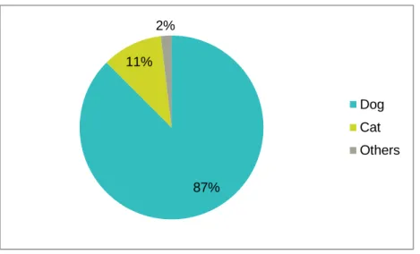
Figure 10 - Distribution of animal species in veterinary practices. ... 41
Figure 11 - Frequency of antimicrobial prescription in veterinary practices. ... 41
Figure 12 - Preference antimicrobial administration route for dogs. ... 42
Figure 13 - Preference antimicrobial administration route for cats. ... 42
Figure 14 - Frequency of antimicrobial prescription without a diagnosis confirmed. ... 42
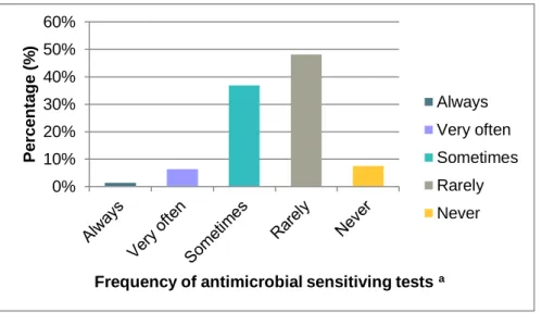
Figure 15 - Frequency of susceptibility testing performed before prescribing antimicrobials. ... 43
Figure 16 - Frequency of weighing animals for which antimicrobials are prescribed.. ... 43
Figure 17 - Main factors considered when selecting an antimicrobial for treatment.. ... 44
Figure 18 – Most common antimicrobial recommended for dogs. ... 45
Figure 19 – Most common antimicrobial recommended for cats. ... 45
Figure 20 - Skin disorders in dogs and the antimicrobials chosen by veterinarians. ... 46
Figure 21 - Skin disorders in cats and the antimicrobials chosen by veterinarians. ... 47
Figure 22 - Urinary tract infections disorders in dogs and the antimicrobials chosen by veterinarians . ... 47
Figure 23 - Urinary tract infections disorders in cats and the antimicrobials chosen by veterinarians . ... 48
Figure 24 - Gastrointestinal disorders in dogs and the antimicrobials chosen by veterinarians . ... 48
xi
Figure 25 - Gastrointestinal disorders in cats and the antimicrobials chosen by veterinarians.
... 49
Figure 26 - Respiratory tract conditions in dogs disorders and the antimicrobials chosen by veterinarians . ... 49
Figure 27 - Respiratory tract infections in cats and the antimicrobials chosen by veterinarians ... 50
Figure 28 - Orthopedic diseases – prophylaxis in dogs and the antimicrobials chosen by veterinarians. ... 50
Figure 29 - Orthopedic diseases – prophylaxis in cats and the antimicrobials chosen by veterinarians. ... 51
Figure 30 - Multiplex PCR for ampC detection. ... 68
Figure 31 - Growth of transformants on selective plates. ... 69
Figure 32 - Simplex PCR performed to confirm the presence of blaCMY-2 gene in transformants. ... 69
Figure 33 - Plasmid based replicon typing performed in all transformants. ... 71
Figure 34 - S1 nuclease PFGE performed in transformants. ... 72
List of Tables
Table 1 - Important genera and species of the family Enterobacteriaceae and most common type of infections. ... 4Table 2 - Spectrum of β-lactam antibiotics within their use in Veterinary and Human Medicine. ... 16
Table 3 - Enterobacteriaceae producing inducible AmpC β-lactamases ... 28
Table 4 - β-lactamase classification scheme ... 36
Table 5 - Origin from the isolates included in this study. ... 60
Table 6 - Diffusion disc method.. ... 61
Table 7 - VetMIC interpretative criteria for Enterobacteriaceae. ... 62
Table 8 - PCR and sequencing primers used in this study. ... 64
Table 9 - Results from resistance profile by disc diffusion, MIC and VetMIC... 70
Table 10 - PCR based-replicon typing results. ... 71
xii
List of abbreviations and symbols
APIFARMA Associação Portuguesa da Indústria Farmacêutica AVMA American Veterinary Medical Association
Bp Base pairs
BHIB Brain Heart Infusion Broth
BSAVA British Small Animal Veterinary Association CLSI Clinical Laboratories Standards Institute SvHKS Danish Small Animal Veterinary Association
DNA Deoxyribonucleic Acid
DGAV Direcção Geral de Alimentação e Veterinária dNTPs Deoxyribonucleotide Triphosphates
ECDC European Centre for Disease Prevention and Control
EFSA European Food Safety Authority
EMA European Medicines Agency
e.g. Exempli gratia
ESAC European Surveillance Antimicrobial Consume
ESBL Extended Spectrum β-Lactamases
ESVAC European Surveillance Veterinary Antimicrobial Consume EHEC Entero haemorrhagic Escherichia coli
ETEC Enterotoxigenic Escherichia coli
EU European Union
EUCAST European Committee on Antimicrobial Susceptibility testing FLUTD Feline Lower Urinary Tract Disorders
IS Inserted Sequences
MARAN Monitoring of Antimicrobial Resistance and Antibiotic Usage in Animals in the Netherlands
MBL Metallo β-lactamases
MIC Minimum Inhibitory Concentration
OMV Ordem dos Médicos Veterinários
OIE World Organization for Animal Health
pAmpC Plasmid AmpC β-lactamases
pMLST Plasmid Multilocus Sequence Typing
PBRT PCR-based Replicon Typing
PCR Polymerase Chain Reaction
PFGE Pulsed-Field Gel Electrophoresis
R Resistant
S Susceptible
SVA Swedish Veterinary Association
SWEDRES-SVARM Swedish Veterinary Antimicrobial Resistance Monitoring
ST Sequence Type
TBE Tris-borate-EDTA
UTI Urinary Tract Infection
VMD Veterinary Medicines Directorate
WHO World Health Organization
1
1. Introduction
During the World War II the production and use of penicillin was implemented for the first time as a response to treat the war causalities. In the last stage of this war the lyophilized preparations of penicillin were made available for veterinarians who reconstituted the antibiotic with saline for intramammary infusions to treat bovine mastitis. This fact represented a significant advance because penicillin proved to be more effective than treatments previously available for dairy animals. The variety of antibiotics, the routes of administration and the reasons for their use expanded during the period between 1950 and 1960 (Gustafson & Bowen, 1997).
Antimicrobials are medicines that kill or inactivate microbes. They include antibiotics, which are used against bacteria. After being exposed to an antimicrobial repeatedly, microbes can undergo changes that stop them being killed or inactivated by the treatments. This is known as antimicrobial resistance and is a growing problem in human and veterinary medicine. During the last decade a number of events have concerned the scientific community and increased the awareness of public health issues related to antimicrobial resistance. For example, the emergence of extended spectrum β-lactamases (ESBLs) it is a concern to human health due to the possibility of transfer of resistance genes across bacteria populations. The risks for humans related to antimicrobial resistance in animals are not circumscribed to foodborne risks only (European Medicine Agency [EMA], 2009a). When the microorganisms become resistant to most antimicrobials they are often referred to as “superbugs”. One the issue is that bacteria become resistant to critically important antimicrobials (World health Organization [WHO], 2011). To address the need for universal terms for the various degrees of antimicrobial resistance, the following definitions were suggested. A pandrug resistant (PDR) isolate is designated as resistant to all available classes of antimicrobials, while the definition of extensive drug resistance (XDR) refers to a pathogen which is resistant to all but one or two classes. A strain is considered a multidrug resistant (MDR), if an isolate is resistant to three or more classes of antibiotics (β-lactams, aminoglycosides, fluoroquinolones, sulfonamides, and tetracyclines) (Falagas & Karageorgopoulos, 2008).
The impact of antimicrobial use and possible misuse in animals as a public health issue is focused on food-producing animals mainly. However there are not many studies about the use of antibiotics in companion animals. The direct contact with pets makes it possible for the transference of resistant genes between companion animals and humans to occur. It is necessary to control and monitor the antimicrobial use in animals in order to avoid the increase of antimicrobial resistance. The European commission established an “Action plan against the rising threats from antimicrobial resistance” released in November 2011 to
2
promote antimicrobial surveillance in the different sectors involved actions regarding research and developing of novel antimicrobials (European Commission, 2011). The European Surveillance of Veterinary Antimicrobial Consumption (ESVAC) project was started in 2010 by the European Medicines Agency (EMA). The aim of the project is to collect information every year on the use of antimicrobials agents from Europeans state members. This information is considered critical in order to monitor and identify risk factors associated with antimicrobial usage in animals.
The awareness of this problem, together with the increase of antimicrobial resistance among companion animals and the interest by the author in this area led to this research project. The author aims to determine and characterize the resistance in Enterobacteriaceae to cephamycin in order to contribute to a better understanding of the mechanisms of development of antibiotic resistance. By understanding these mechanisms it can be possible to undertake new strategies to fight antibiotic resistance. The author also evaluates the use of antimicrobial agents in companion animals in Portugal through a national survey which was online during the time of the training period. The first part of this document will include a literature review of the main topics. The second part refers to the studies developed including its main goals, materials and methods, results, discussion and concluding remarks of this study. The terms antimicrobial and antibiotic, Rep type and Inc group to describe plasmids are used interchangeably in this thesis.
1.1. Training period
To accomplish the project, the author took a training period of approximately six months in two different locations. The first place was at the Laboratory of Antimicrobial Resistance and Biocides, Faculty of Veterinary Medicine, University of Lisbon, Portugal (FVM-UL) from September 20th to December 15th 2012, the remaining period was undertaken at LLP/Erasmus Program in the Microbiology Laboratory of the Faculty of Life Sciences, University of Copenhagen (KU-SUND) from January 10th to April 30th 2013, under the supervision of Professor Luca Guardabassi and Dr. Valeria Bortolaia.
3
Part 1
Literature Review
4
2. Literature Review
2.1. The family Enterobacteriaceae
2.1.1 Typical characteristics
The family Enterobacteriaceae is considered to be the most important family which can cause a variety of community and nosocomial infections (meaning that are acquired in hospital environment) in humans such as septicaemia, urinary tract infections (UTI), pneumonia, cholecystitis, cholangitis, peritonitis, wound infections, meningitis, and gastroenteritis. Enterobacteriaceae falls within the domain Bacteria, phylum Proteobacteria, class Gammaproteobacteria, and order Enterobacteriales. In Table 1 are listed the most common genera and species belonging to this family. The members of this family are gram-negative, rod-shaped, non-spore-forming facultative anaerobes that ferment glucose and other sugars, reduce nitrate to nitrite, and produce catalase but do not produce oxidase (Donnenberg, 2010).
2.1.2. Natural habitat
Most Enterobacteriaceae are designated as enteric because the main habitat of most of them is the lower gastrointestinal tract of animals and humans, but the environment can also be a reservoir (Farmer, Boatwright & Janda, 2008). In large intestine there is a complex and dynamic interaction with high densities of living bacteria, which achieve concentrations of up to 1011 or 1012 cells/g in the luminal contents (Guarner & Malagelada, 2003).
Table 1 - Important genera and species of the family Enterobacteriaceae and most common type of infections (adapted from Abbott, 2008; Farmer et al., 2008; Nataro, Bopp, Fields, Kaper & Stockbine, 2008; Donnenberg, 2010).
Genus Species Common type of infections
Citrobacter Freundii UTIs, pneumonia, meningitis, septicaemia, wound infections
Enterobacter cloacae, aerogenes UTIs, pneumonia, septicaemia, wound infections
Escherichia Coli UTIs, diarrhoea, septicaemia, meningitis
Klebsiella pneumonia, oxytoca UTIs, pneumonia, septicaemia
Morganella Morganii UTIs, septicaemia
Proteus mirabilis, vulgaris UTIs, pneumonia, septicaemia, meningitis, wound infections
Salmonella Enterica diarrhoea, typhoid fever, septicaemia, UTIs, osteomyelitis
Serratia Marsecens UTIs, pneumonia, wound infections, septicaemia
Shigella Dysenterii diarrhoea, dysentery
5 2.1.3. Cell wall structure
The structure from gram-negative bacteria is different from gram-positive; gram-negative bacteria cell wall is composed of a thin peptidoglycan layer, also named as murein (Beveridge & Graham, 1991; Donnenberg, 2010). The murein consists of alternating N-acetylglucosamine and N-acetylmuramic acid amino sugars joined by β-1,4 linkages, with a short peptide composed of L-alanine, glutamic acid, L-meso-diaminopalmelic acid, and D-alanine attached to the carboxyl group of the muramic acid (Donnenberg, 2010).
The periplasmatic space ranges from 20%- 40% of total cell volume in a gram-negative bacteria, and is considered an integral compartment of the gram-negative cell wall (Stock, Rauch & Roseman, 1977; Beveridge, 1999). Together the plasma membrane and the cell wall (outer membrane, peptidoglycan layer, and periplasm) constitute the gram-negative envelope (Beveridge & Graham, 1991). The envelope is responsible for maintaining the shape and osmotic stability of the cell; however in the replication and elongation process of the bacteria is always being remodelled (Figure 1) (Beveridge & Graham, 1991).
2.2. Principals genera from Enterobacteriaceae family
2.2.1. Escherichia coli
Escherichia coli are the most important bacteria in the normal microbiota of humans and
animals. It colonizes the gastro intestinal tract within hours after birth and there is a mutual relationship (symbiotic) between host and bacteria. However if the immune system or if the gastrointestinal barriers are compromised some strains can cause disease in gastro intestinal tract or urinary system for example (Nataro & Kaper, 1998). E. coli and Klebsiella
pneumonia are among the most important causes of serious hospital-acquired and
Outer membrane
Periplasm
Cytoplasmic membrane
Figure 1 - General structure of Enterobacteriaceae (reproduced with permission from Livermore & Woodford, 2006)
6
community-onset bacterial infections in humans (Paterson, 2006). The regular presence of E.
coli in the human intestine and faeces has led to tracking the bacterium in nature as an
indicator of faecal pollution and water contamination (Goyanes et al., 2007; Todar, 2008).
2.2.2. Proteus spp.
The two species most representative of this genus are: P. mirabilis and P. vulgaris which were both described in 1885 by Hauser. He noted the swarming colonies nature and classified them according to their ability to liquefy gelatine (O’Hara, Brenner & Miller, 2000). This swarming ability is related to the flagella which translocate very fast along the plates, and this characteristic allows then to differentiate from other Enterobacteriaceae. The genus Proteus spp. are lactose negative and motile (Donnenberg, 2010).
Bacteria of the genus Proteus are a part of the commensal flora of the intestinal tract from humans and animals (O’Hara et al., 2000; Giammanco, Pignato, Grimont, Grimont & Giammanco, 2011) . P. mirabilis and P. vulgaris account for the majority of clinical isolates from this genus, acting also as opportunistic pathogens causing primary and secondary infections (Donnenberg, 2010; Giammanco et al., 2011).
2.2.3. Klebsiella spp.
The genus Klebsiella was named by Trevisian in 1885 to honour microbiologist Edwin Klebs (Brisse, Grimont & Grimont, 2006). Klebsiella spp. is ubiquitous in nature, can be found in the environment (water, soil, and plants) but also in the mucosa from humans and animals.
Klebsiella pneumoniae is the most important species of the genus and is responsible
together with Klebsiella oxytoca for 8% nosocomial bacterial infections in Europe (Podschun & Ullmann, 1998).
2.2.4. Enterobacter spp.
The genus Enterobacter includes 14 species, Enterobacter aerogenes and Enterobacter
cloacae are by far the most important and relevant pathogens in humans among the genus Enterobacter. Enterobacter spp. is considered an opportunistic pathogen, and recently has
become an important cause of nosocomial infections (Sanders & Sanders, 1997).
2.2.5. Other Genus
Other important Enterobacteriaceae species are: Salmonella spp., Citrobacter spp., Shigella spp. and Serratia spp. Salmonella is a facultative intracellular pathogen acquired by consumption of water and/or contaminated food. In contrast to other pathogens Salmonella spp. requires a large number of genes to become virulent (Groisman & Ochman, 1997). The genus Salmonella includes 10 species but only two are clinically important: S. enterica, which
7
is subdivided into over 2,000 serovars, and Salmonella bongori. The most important serovars of S. enterica, are: S. typhi, which is responsible for systemic infections and typhoid fever infecting only humans; and S. typhimurium, which is a leading cause of gastroenteritis in human and other mammalian species increasing the incidence of non-typhoid Salmonella infections worldwide (McClelland et al., 2001).
The genus Citrobacter spp. includes twelve species and is an important cause for opportunistic infections. Citrobacter species are commonly found in water, soil, food, and the intestinal tracts of animals and humans (Shih, Chen, Chang, Luh, & Hsieh, 1996; Donnenberg, 2010).
In 1940, four species of the new genus Shigella spp. were recognized by Boydii as the cause for dysenteric disease in humans. Shigella and E. coli have always been considered to be very closely related, but E. coli strains became a different genus due to differences in medical significance (Lan & Reeves, 2002).
Ten species are known to belong to Serratia genus and the most important species are: S.
marsecens which can be differentiated from other bacteria by producing a red pigment
named Prodigiosin; and S. liquefascens. These species can be found in water, mammals and also in hospitalized patients (Grimont & Grimont, 2006).
2.3. Clinical significance of Enterobacteriaceae
The family Enterobacteriaceae as referred previously is indeed the most important family which colonize the gastrointestinal tract since early age and coexist with animals and humans with mutual benefits for decades. In many situations the infections caused by these species are nosocomial. As it is shown in table 1, Enterobacterial strains are responsible for a variety of clinical situations such as urinary tract infection, pneumonia, and sepsis (Kaper, Nataro & Mobley, 2004).
The species Escherichia coli causes diverse intestinal and extraintestinal diseases by means of virulence factors, named serotypes. The main causes of diarrhoea in animals and humans is triggered by enterotoxigenic E. coli (ETEC) and entero haemorrhagic E. coli (EHEC) (Kaper et al., 2004). E. coli is also considered an important pathogen in canine UTIs (Feria, Ferreira, Correia, Gonçalves & Caniça, 2002 ).
Klebsiella spp. is an opportunistic ubiquitous pathogen and is a major cause of nosocomial
infections. The targets are usually immune-compromised individuals that are hospitalized with chronic diseases. Klebsiella pneumoniae is responsible for most of the outbreaks in health care facilities. This species is also involved in urinary tract infection, pneumonia, septicaemia and soft tissue infections (Podschun & Ullmann, 1998; Struve & Krogfelt, 2004). In animals it can act as an opportunistic pathogen it has been implicated in cases of mastitis in cattle, metritis in mares, bacteraemia in calves, pneumonia and urinary tract infections in
8
dogs (Roberts, McClain, Hansen, Currin & Howerth, 2000). The importance of Klebsiella is based on the increasing reports of clinical situations caused by multiresistant strains capable of producing extended spectrum β- lactamases (Podschun & Ullmann, 1998).
Of the Yersinia genus three species Y. pestis, Y. enterocolitica and Y. pseudotuberculosis are well known animal and human pathogens. Pathogenic strains of Y. enterocolitica and Y.
pseudotuberculosis cause yersiniosis, an acute enteric disease, in humans and animals
(Stamm, Hailer, Depner, Kopp & Rau, 2013). Y. enterocolitica is regarded as a significant food-borne pathogen. Besides food producing animals, companion animals are also considered a reservoir for human Yersinia infections (Stamm et al., 2013). Y. pestis is
responsible for the plague, a zoonotic disease transmitted to human through a vector (fleas) (Linde, Neubauer, Meyer, Aleksic & Lehn, 1999). In contrast to other Enterobacterea,
Yersinia pestis circulates in the bloodstream, lymphatic vessels and organs like spleen and
liver, and may contact with Enterobactereaceas when reaching the bloodstream. To date there have been 3 major plagues and a high number of casualties (Galimand, 1997).
The family Enterobacteriaceae is the most important and relevant in clinical therapy because it is a common source of infections in animals, community and hospitals spreading easily between humans (Pitout, 2008a). These bacteria also have the ability to acquire new genetic material through horizontal gene transfer (HGT), mediated by plasmids or transposons. This combination represents an important issue regarding the emergence of multidrug resistance in Enterobacteriaceae and lights up the possibility of interspecies transfer of resistance determinants (Wise et al., 1998; Nordmann, Dortet & Poirel, 2012).
2.4. Antimicrobials – The era of antimicrobials
“One sometimes finds what one is not looking for” - Alexander Fleming
The general term “antibiotic era” is usually associated with the names of Paul Ehrlich and Alexander Fleming. Ehrlich’s wanted to find a drug against syphilis which targeted only the source of infection caused by the spirochete Treponema palladium. Syphilis was considered an endemic disease and almost incurable at the time. In 1909 after several trials with rabbits an effective compound was discovered, marketed under the name Salvarsan, and for many years was the most prescribed drug until its replacement by penicillin (Aminov, 2010).
The introduction of antibiotics in the 1940s was considered as one of the greatest advances/achievements in therapeutic medicine for treating and preventing infectious diseases in human medicine. An antibiotic can be defined as a natural compound which is produced from fungi, bacteria and other microorganisms that can kill or inhibit the growth of bacteria, while the term antimicrobial refers to a group of substances, natural or synthetic which includes also the antibiotics, antifungals, antiprotozoals and antivirals (WHO, 2011). The antimicrobials can be classified as natural when they are produced by microorganisms,
9
or synthetic when the chemical scaffolds were used later to create new generations of clinically useful antibiotics by chemical modification (Peláez, 2006). The first antibiotic named penicillin was discovered by Alexander Fleming in 1928 when he observed that a common mould (Penicillium notatum) produced a substance that inhibited the growth of colonies of
Staphylococcus spp. (Ligon, 2004).
Howard Florey and Ernst Chain, both part of an Oxford team, were the scientists who in 1940 were able to purify penicillin for clinical testing. The period from the 1950s throughout the 1970s is defined as the golden era of discovering new classes of antimicrobial agents (Aminov, 2010). Since year 1993 there has not been many new classes of antimicrobials developed by industry and that we might face a shortage of therapeutic options against multi-resistant bacteria. That is well supported by Figure 2.
Nowadays there is growing concern about the increase of antibiotic resistance increase worldwide. The use of antibiotics is considered to be the main factor for pathogenic bacteria growth which has led to the dissemination of resistant bacteria and resistant genes. This situation applies to both human and veterinary practice, since antimicrobials are used for similar purposes (e.g. prophylaxis therapy of infectious diseases) in humans and animals. This can lead to the increase of levels of resistance traits in pathogenic and commensal bacteria. Resistant bacteria can infect humans by direct contact or via food products of animal origin (Van den Bogaard & Stobberingh, 2000). So far many studies have emphasised mainly in food producing animals. To decrease the risk of transmission of antimicrobial residues and pathogens through the food chain some countries have developed and are focusing on monitoring antimicrobial usage in food-producing animals (Aarestrup & Wegener, 1999).
Sulfonamides discovered (1932)
Penicilin discovered (1928) Penicilin introduced (1942) Streptomycin discovered (1943) Bacitracin disc. (1943) Cephalosporin disc. (1945) Cloramphenicol disc.(1947) Neomycin disc. (1949) Methicilin introduced (1942) Ampicilin int. (1943) Gentamicin discovered(1943) Cephalosporin int.(1945) Vancomycin int.l (1964) Doxycicline int. (1966) Clindamycin reported (1697) Oxitetracycline discovered (1950) Erytromicin disc. (1952) Vancomycin disc.. (1956) Amoxicilin-clavulanate introduced (1984) Imipenem int. (1987) Ciprofloxacin int. (1987) Linezolid introduced (2000) Tigecycline int. (2005)
Figure 2 - Discovery of antimicrobials (adapted from http://amrls.cvm.msu.edu/pharmacology/historical-perspectives/the-golden-age-of-antibacterials). Rifampicin introduced (1971) Cephamicins discovered (1972) Amikacin introduced (1976) Azytromicin introduced (1993) Quinupristindalfopristin intr. (1999)
10 2.4.1. Classification of antibacterial agents
Antibacterial agents can be classified according to their mechanism of action (how antimicrobial growth is supressed) and by spectrum of activity. The five main target sites of antimicrobial agents are: (i) cell wall synthesis, (ii) protein synthesis, (iii) nucleic acid synthesis, (iv) metabolic pathways, and (v) cell membrane functions (Figure 3) (Tenover, 2006; Maddison, 2009). Antibacterial drugs that act by inactivating the wall synthesis include the penicillins, cephalosporins and vancomycin. Aminoglycosides and tetracycline inhibit protein synthesis by binding to the ribosomal subunit 30S, while macrolides and chloramphenicol bind to the 50S ribosomal subunit to inhibit protein synthesis. Fluoroquinolones antimicrobial agents disrupt the DNA synthesis whereas sulphonamides and trimethoprims block the pathway for folic acid synthesis (Tenover, 2006).
Antibacterial agents can be classified as bactericidal or bacteriostatic. This classification is used mainly for clinical purposes and is not consistent to all bacteria. The ability of an antimicrobial agent to inhibit or kill a microorganism is relative and depends on the bacteria. For example chloramphenicol inhibits the growth (bacteriostatic) of E. coli while it kills (bacteriocidal) Haemophilus influenza (Maddison, 2009). Bacteriostatic agents inhibit bacterial growth as long as the drug concentration is above the minimal inhibitory concentration (MIC). Tetracycline, chloramphenicol and sulphonamides are examples of bacteriostatic agents. Bactericidal agents e.g. aminoglycosides, cephalosporins, fluoroquinolones, metronidazole, penicillins and potentiated sulphonamides kill the bacteria and are preferred in infections in which the host cannot control or eradicate the infection due to location of the infection itself or due to the compromised immune status of the host. Bactericidal agents can also be further classified as time-dependent (penicillins and cephalosporins, both slow bactericidals), or concentration-dependent drugs. For bactericidal antibiotics, plasma levels of antibiotic concentration must be above the MIC as long as
Inhibition of protein synthesis Inhibition of DNA
synthesis
Inhibition of cell wall synthesis Figure 3 – Principal antimicrobial action mechanisms (Original illustration).
11
possible (each 24 hours of treatment). In contrast, for concentration-dependent drugs (aminoglycosides and fluoroquinolones), the higher the plasma peak is achieved, the greater the proportion of bacteria killed. The peak concentrations, the area under the plasma concentration versus time curve are important for bactericidal success (Maddison, 2009). In order to a better understanding of antibiotic activity it is necessary to determine some important parameters as illustrated in Figure 4. The first parameter, time at which concentration is above the MIC (t > MIC), is related to bactericidal effects over time. It refers to the time in which the drug is above the MIC and is dependent on the half-life, dosage, frequency of administration of the drug over a certain time period. The parameter corresponding to the ratio peak plasma concentration (Cmax)/MIC, it is the maximum concentration of a certain drug in the plasma, relates bactericidal effects with concentration, and is mainly dependent on the unit dose and the volume of distribution of the drug. The last parameter, area under the concentration-time curve (AUC)/MIC, combines both types of effects, because it refers to the total amount of drug to which bacteria are exposed over the time period, and is directly related to the total dose given during that period and inversely proportional to the drug clearance (Bambeke, Barcia-Macay, Lemaire & Tulkens, 2006).
Figure 4 - Antibacterial concentration vs. time graph illustrating pharmacokinetic and pharmacodynamics parameters (adapted from Lister, 2006).
The effectiveness of individual drugs against the isolated organism is categorized according to the new ISO 20776-1 standard using the following categories: "Susceptible", “Intermediate” and “Resistant” depending on the MIC value. The MIC is defined as the minimum concentration of an antibiotic that is able to prevent the further growth of the infectious organism in vitro (Rodloff, Bauer, Ewig, Kujath & Müller, 2008). Susceptible (S), a bacterial strain is said to be susceptible to a given antibiotic when it is inhibited in vitro by a
A n ti b a ct e ri a l co n c e n tr a ti o n Time (hours) Cmax A: t>MIC B: Cmax/MIC C: AUC/MIC C
12
concentration of this drug that is associated with a high rate of therapeutic success. Intermediate (I), the sensitivity of a bacterial strain to a given antibiotic is said to be intermediate when it is inhibited in vitro by a concentration of this drug that is associated with an uncertain therapeutic effect. Resistant (R), a bacterial strain is said to be resistant to a given antibiotic when it is inhibited in vitro by a concentration of this drug that is associated with a high possibility of therapeutic failure. The European Committee on Antimicrobial Susceptibility testing (EUCAST) in order to harmonise and define breakpoints provides clinical breakpoints and epidemiological cut-off values (ECOFF). The ECOFF are used in clinical breakpoint development and as a sensitive indicator of resistance development in surveillance studies (European Committee on Antimicrobial Susceptibility testing [EUCAST], 2007).
2.5. β-lactam antibiotics
2.5.1. General structure and functions
The β-lactam bactericidal antibiotics, including – penicillins, cephalosporins, monobactams and carbapenems are one of the most widely used group of antimicrobials due do their safety, low cost, ease of delivery, minimal side effects and high efficacy. β-lactam antibiotics are grouped together based upon a shared structural feature (Wilke, Lovering & Strynadka, 2005). β-lactam is a generic name for all β-lactam antibiotics that contain a β-lactam ring, a heteroatomic ring structure, consisting of three carbon atoms and one nitrogen atom (Wilke
et al., 2005).
This group represents 60% of all antimicrobial used by weight and is used to treat infections caused by gram-negative bacteria and gram-positive, in human medicine (Livermore & Woodford, 2006). In veterinary medicine, different substances from the penicillin family, first to fourth generation cephalosporins and β-lactamase inhibitors are recommended for the treatment of companion animals according to the animal species involved and the underlying disease (Guardabassi, Jensen & Kruse, 2008; Smet et al., 2010).
2.5.2. Mechanism of action
β-lactam antibiotics inhibit the growth of bacteria by inactivating enzymes called penicillin-binding proteins (PBPs), located in the bacterial cell wall which are involved in the third stage of cell wall synthesis (Poole, 2004). β-lactam antibiotics normally interfere with this process by reacting covalently with the active site serine to form a stable acyl-enzyme preventing the peptidoglycan synthesis (Hujer et al., 2005). In contrast to positive bacteria, in gram-negative the peptidoglycan it is a thin layer between the cell wall and the cytoplasmic membrane. The antibiotic β-lactam must diffuse across the outer membrane of the gram-negative cell, using pores formed by porin proteins, and then cross the periplasm before
13
reaching its PBP targets, which lie on the outer surface of the cytoplasmic membrane (Livermore & Woodford, 2006).
2.5.3. Penicillin
Penicillin was discovered in 1928 by Alexander Fleming and has activity against gram-positive and gram-negative bacteria (Tipper & Strominger, 1965). This class is considered the best known and first β- lactam antibiotic. All penicillin have a β-lactam, a thiazolidine ring and a side chain R group which is distinguishable between penicillins (Figure 5) (Baldo, 1999). In 1940 a large amount of penicillin was produced from cultures of Penicillium
notatum. Several years after the first penicillin, penicillin G became available; however, it had
some limitation such as instability in gastrointestinal (GI) and was not very effective against important gram-negative bacteria (Chambers, 2010).
2.5.4 Cephalosporins
This first isolation of Cephalosporium acremonium culture was discovered in 1948 by Giuseppe Brotzw but only became commercially available in 1962 (Botana, Landoni & Martín-Jiménez, 2002). Researchers at the University of Oxford isolated cephalosporin C in 1956 (Abraham & Newton, 1961). Some years after four classes of cephalosporins were produced (Botana et al., 2002). Cephalosporins of first generation have activity against gram-positive bacteria such as Streptococcus, Corynebacterium, S. aureus e S. pseudintermedius and limited activity towards gram-negative bacteria, as it shown in Table 2. Enterococcus spp., Pseudomonas aeruginosa and methicillin-resistant Staphylococcus aureus (MRSA) are resistant to these antibiotics. Cephalosporins are grouped into generations according to their spectrum of activity against gram-negative bacteria (Donowitz & Mandell, 1988). The cephamycins differ from the true cephalosporin by the presence of a methoxy-group in the position 7 of the cephalosporin group but are grouped together and classified according to their in vitro spectrum activity and structural similarities. Cephamicins remain stable to many β-lactamases (EMA, 2009b). In this document, the term “cephalosporin” will be used indiscriminately.
2.5.5 Monobactams
Monobactam antibiotics are a new class of β-lactam antibiotics. In contrast to penicillins or cephalosporins, monobactams have a monocyclic β-lactam structure and are produced by a range of bacterial species. Although these antibiotics exhibit poor antibacterial activity, they are highly stable to the action of β-lactamases produced by gram-negative bacteria (Sykes, Bonner, Bush, Georgopapadakou & Wells 1981; Saxon, Hassner, Swabb, Wheeler & Adkinson, 1984).
14 2.5.6. Carbapenems
Carbapenems are the class of β-lactam antibiotics with the broadest spectrum of activity (Nordmann et al., 2012). The activity includes many gram-positive, gram-negative and anaerobic bacteria; and no activity is recognized towards Enterococcus faecium, methicillin-resistant Staphylococcus aureus and Stenotrophomonas maltophilia. The increase of β-lactamase resistance among Enterobacteriaceae led to the use of carbapenems widely, because they are stable to most β-lactamases including AmpC β-lactamases (Zhanel et al., 2007). Carbapenems are a last resource against bacterial infections used mainly when the infection is caused by a multidrug resistant pathogen (Bush, 2010).
2.5.7. β-lactamase inhibitors
The β-lactamase inhibition can be classified as either reversible or irreversible. Reversible inhibitors bind to an enzyme but activity may be restored after their removal. Irreversible inhibitors may be more effective than reversible inhibitors because the final outcome is the destruction of enzymatic activity. Specific irreversible inhibitors of β-lactamases include the clavulanic acid, the penicillanic acid sulfones sulbactam and aztreonam (Bush, 1988).
These substances are used in the treatment of serious Enterobacteriaceae and penicillin-resistant staphylococcal infections (Drawz & Bonomo, 2010). New compounds such as clavulanic acid and sulbactam exhibit low bactericidal activity when used alone but remain Figure 5 - Different structure of b lactam antibiotics: a) penicilin, b) cephalosporin, c) monobactam, d) carbapenems. The β-lactam ring is shown in blue (reproduced with permission from Babic, Hujer & Bonomo, 2006).
. a)
b)
15
effective against β-lactamases enhancing the activity of β-lactam antibiotics, hence the name (Moosdeen, Williams & Yamabe, 1988). Clavulanic acid was isolated from Streptomyces clavuligerus and was the first β-lactamase inhibitor introduced worldwide. Clavulanate (the acid from the solution) had weak bactericidal activity but combined with β-lactam antibiotics decreases the MIC values of some resistant bacteria (Reading & Cole, 1977). Later penicillinate sulfones, e.g. sulbactam and tazobactam were introduced in the clinical practice. These 3 substances are effective against many susceptible organisms expressing class A β-lactamases, which are the most common (including CTX-M and the ESBL derivatives of TEM-1, TEM-2, and SHV-1); and are generally less effective against class B, C, and D β-lactamases (Bush, 1988; Buynak, 2006). The combination of amoxicillin with clavulanate was selected due to the similarity in pharmacokinetics between the two compounds. Any new β-lactamase inhibitors should have the following characteristics: the molecules must be capable of preventing hydrolysis of well-tolerated broad-spectrum β-lactam antibiotic, preferably inexpensive penicillin; the pharmacokinetics of the two molecules should be similar; side effects should be minimal and mild; the molecules should not be good inducers of cephalosporinase activity. Ideally, one would like to have molecules with oral activity (Bush, 1988).
The use of β-lactamase inhibitors remains effective in the empirical treatment of respiratory, intra-abdominal, skin and soft tissue infections in humans. The use of β-lactamase inhibitors instead of cephalosporins appears to reduce the emergence of resistance in pathogens. In Portugal, amoxicillin-clavulanate is the first line antimicrobial for UTI infection in dogs, which leads to a strong selection pressure for the emergence of resistant bacteria (Feria et al., 2002). Similarly, their use may also curtail the emergence of other resistant pathogens such as Clostridium difficile and vancomycin-resistant enterococci (Lee, Yuen & Kumana, 2003).
16
Table 2 - Spectrum of β-lactam antibiotics within their use in Veterinary and Human Medicine -: no activity, (+): limited activity, +: active (adapted from Guardabassi et al., 2008; Hammerum & Heuer, 2009).
β-lactams Spectrum of activity Use in Veterinary Medicine Use in Human Medicine
gram-negative
gram-positive
Penicillin - + ampicillin, amoxicillin, benzylpenicillin penicillin, ampicillin, amoxicillin
First generation cephalosporins (+) + cephadroxil, cefapirin, cephalexin cefalozin
Second generation cephalosporins + + cefaclor, cefamandole, cefuroxime cefuroxime, cefoxitin
Third generation cephalosporins + + cefovecin, cefpodoxim, ceftiofur ceftriaxone, cefotaxime, ceftazidime
Fourth generation cephalosporins + + cefquinome cefepime, cefpirome
Monobactams + - not in use aztreonam
17
2.6. Antimicrobial resistance
Resistance to an antibiotic typically develops from its use according to Darwin´s principle: “survival of the fittest” (WHO, 2011). Antimicrobial resistance remains a global health concern and most countries have developed strategies to evaluate the level of resistance (Martins da Costa, Loureiro & Matos, 2013). This issue has emerged as a consequence of the selective pressure exerted by the overuse and misuse of antimicrobials in veterinary and human medicine. Furthermore, morbidity and mortality have increased in human medicine associated with resistant pathogens and thus failure therapeutic (Davies & Davies, 2010). Recently, the World Health Organization (WHO) published a report establishing the critically important antimicrobial for human medicine (annex 1). The problematic of antimicrobial resistance becomes crucial when pathogens become resistant to these antimicrobials. The criteria for inclusion were: (1) sole therapy or one of few alternatives to treat serious human disease and (2) antimicrobial agents used to treat diseases caused by organisms that may be transmitted via non-human sources or diseases caused by organisms that may acquire resistance genes from non-human sources (WHO, 2007). Humans can acquire resistant pathogens or resistant genes of animal original directly from food consumption, direct contact with animals or through the environment (Rolain, 2013). In addition the use of the critically important antimicrobials should be minimised in animals to maintain/preserve the efficacy of these for treating human’s infections (Danish Small Animal Veterinary Association [SvHKS], 2013).
2.6.1. Use of antimicrobials in food-producing animals
The introduction of antimicrobials in food-producing animals started in the 1940s to treat diseases and for growth purposes (Hammerum & Heuer, 2009).
In 1951 it was first reported in California the effects of using streptomycin in turkeys and the emergence of streptomycin-resistant isolates from those animals (Starr & Reynolds, 1951). Since then, issues about the use of antibiotics are still questioned by the international community and professionals (Seiffert, Hilty, Perreten & Endimiani, 2013). The amount of antibiotics used at this time was considered higher than in human medicine. Addressing the rapid spread of antimicrobial resistant pathogens, Sweden was the first country to abandon the use of antibiotics as growth promoters in 1986 (WHO, 2011). In The European Union (EU) these agents were banished in the 1th January 2006 (European Comission, 2005). The effects of discontinuation of antimicrobial agents as growth promoters in European countries decreased antibiotic resistance in animals, food products, and also in humans (Anderson, Nelson, Rossiter & Angulo, 2003).
The overall amount of antibiotics used is not clearly known because the data provided from all countries is not uniform (Anderson et al., 2003). The amount of antimicrobial data can be
18
reported by weight of active ingredient or total weight of feed additives. Another important aspect is that the data reports to both companion animals and food producing animals regarding drugs licensed for multispecies used (EMA, 2009b, 2013). According to the European Medicine Agency (EMA), there is a slight downward trend for use of antimicrobials in animals in all countries. However, use of β-lactams antibiotics is increasing slightly (EMA, 2009b). The information on use and consumption of cephalosporins in the EU is not available for all members, and often the data includes β-lactam antibiotics (including ampicillin, benzylpenicillin). Furthermore, when the data provides the use of penicillins and cephalosporins separately, are not divided by generations. Currently there are specific recommendations on prudent use of cephalosporins in the EU. Nevertheless, monitorisation is needed on how the guidelines and recommendations are being implemented (Passantino, 2007).
According to data published in Portugal, the total amount of antimicrobial sold was 162,564 tons of active substance during the year of 2011 (includes all animal species). Doxycycline and amoxicillin were the most used antimicrobials. In cattle, florfenicol was the most common antimicrobial sold, and doxycycline for the swine (Direcção Geral Aimentação e Veterinária [ DGAV ], 2011).
2.6.2. Use of antimicrobials in companion animals
Due to the use of antimicrobials that are critical to human medicine, the risk of antimicrobial resistance emergence and inter-species clonal spread, the awareness in the last few years of the veterinary community for the potential implications for public health has increased (Martins da Costa et al., 2013).
Systematic surveillance of the occurrence of resistant bacteria from food animals, food and humans has been established and annually reports are published in European Food Safety Authority (EFSA). In contrast, data is rare regarding extent and characteristics of antimicrobial resistance among bacteria from companion animals (Pedersen et al., 2007). The increasing number of reports on antimicrobial resistance in veterinary practice in addition to reports of multidrug pathogens led some countries to create guidelines to promote rational use of antimicrobial in veterinary practice. The Swedish Veterinary Association (SVA) developed guidelines in 2002, and countries like Denmark in 2013 followed their counterparts (SvHKS, 2013). In Sweden since 2006 a decrease in antimicrobial sales is clearly marked specially on aminopenicillins with clavulanic acid, cephalosporins and fluoroquinolones. This downward trend is explained by the application of guidelines and campaigns which led the veterinarians to change their prescription habits (SWEDRES-SVARM, 2012). According to the Danish Programme for surveillance of Antimicrobial Consumption and Resistance in Bacteria from Animals, Food and Humans (DANMAP), the use of antimicrobials in 2011
19
decreased in veterinary medicine due to the reduction of cephalosporins usage. The information on companion animals follows an upwards trend since 2005 caused by the use of combination penicillins (DANMAP, 2012). According to this facts Sweden is clearly a classic example of how the combination of strong policies on prescribing and implementation of guidelines can improve the rational use of antibiotics.
In the UK, a large amount of antimicrobial sales were for veterinary use (Veterinary Medicine Directorate [VMD], 2012). The total sales amount of therapeutic antimicrobials in 2008 decreased to 384 tonnes. In 2011, total sales have decreased by 101 tonnes to total 346 tonnes. Tetracyclines, β-lactams (including penicillins) and trimethoprim/sulphonamides accounted for the majority of antibiotic active ingredients sold in veterinary medicinal products from 2006 to 2011 (VMD, 2012). The incidence of E. coli isolates from companion animals with patterns of resistance has increased over the last years to antibiotics commonly used to treat infections caused by this agent such as amoxicillin-clavulanate, cefpodoxime, enrofloxacin, and trimethoprim sulphonamide according to a study conducted in Denmark (Pedersen et al., 2007).
Interestingly, pharmaceutical companies are not currently developing novel antibiotics because it is not cost effective for the drug companies. It is indeed critical to maintain the effectiveness of the antibiotic classes available by promoting a rational and prudent use (Coates & Hall, 2011). It is also important to create incentives for the research and development of new antimicrobials by industry (EMA, 2009a).
In Portugal the only extended spectrum cephalosporin approved for dogs and cats is Cefovecina (Convenia©). In dogs and cats it is indicated for skin infections caused by S.
pseudintermedius, Streptococci β-haemolytic, E. coli and/or Pasteurella multocida; and for
urinary tract infections caused by E. coli or Proteus spp. (only in dogs) (Associação Portuguesa da Indústria Farmacêutica [Apifarma], 2011; EMA, 2012). The use of third generation cephalosporins in human medicine in Portugal are cefetamet, cefixima, cefotaxime, ceftadizime e ceftriaxona and most can be acquired in a community pharmacy. (Infarmed, 2012). In a report published by DGAV in 2011, the most common antimicrobial used in dogs and cats was amoxicillin (DGAV, 2011). The term extra label drug use (often referred to as “off-label use”) was defined as the use in animals of a drug at an indication, dosage, frequency or route of target species (e.g. human drugs in animal species or a drug licensed for use in dogs in a cat) (Canadian Veterinary Medical Association [CVMA], 2010). In a recent report published by The European Surveillance of Veterinary Antimicrobial Consumption (ESVAC), for the majority of countries, penicillins (36% in tonnes) were the most sold veterinary antimicrobial agent, followed by 1st and 2nd-generation cephalosporins (31%). However this data should be interpreted carefully since it only represents sales of
20
tablets (oral antimicrobials) and injectable antimicrobials are not included in this report (EMA, 2013).
2.6.3. Use of antimicrobial in humans
There is a clear relation between the use of antimicrobials and antibiotic resistance which is increasing in southern countries but the prevalence in northern countries remains low. Data analysis is not consensual since national databases use different methodologies to classify antibiotics and measure their consumption (Goossens, Ferech, Vander Stichele & Elseviers, 2005). In 2001, the European Commission funded the European Surveillance of Antimicrobial Consumption (ESAC) project, an international network of surveillance systems to collect comparable and reliable data on antibiotic use in Europe. The study conducted during the period 1997-2003 concluded that overall antibiotic use increased in most European countries, and penicillin was the most prescribed (Ferech et al., 2006). In another study by ESAC during the period 1997-2002 refers Portugal as the fourth consumer for antimicrobials in Europe with an increase of 10% in 2002 when compared with 1997. Penicillins were the most prescribed antibiotic in all countries analysed (Elseviers, Ferech, Vander Stichele & Goossens, 2007). In 2003 the study by ESAC reveals that Portugal was classified as the 4th consumer of antibiotics, and number one on fluoroquinolones usage (Ferech et al., 2006). Despite the downward trend in the last few years, Portugal still has a high consumption of antibiotics; and penicillin was the most prescribed agent, followed by macrolides, lincosamines, streptogramins and fluoroquinolones (Ferech et al., 2006).
Cephalosporins are widely used in human medicine both in hospitals and community to treat septicaemia, and meningitis (Paterson & Bonomo, 2005). The total consumption in hospitals in 15 European countries in 2002 ranged from 1.28 to 3.89 defined daily doses (DDD)/1000 inhabitants per day (Vander Stichele, Elseviers, Ferech, Blot & Goossens, 2006). In the same report an increase of third and fourth generation cephalosporin between 1997-2002 was noted in all countries (Vander Stichele et al., 2006).
In 2003 the total amount of antibiotics used in 34 countries European ranged from 3.89 to 31.40 DDD/1000 inhabitants per day (Ferech et al., 2006). Cephalosporins from third and fourth generation were limited to patients with clinical conditions difficult to control. However, in three countries the use of these cephalosporins accounted for more than 40% of the total use. These differences between countries might be explained by a misuse of the antibiotics in hospitals (Coenen et al., 2006). Defined daily dose (DDD) is a measuring unit which defines an average maintenance dose per day for a drug used for its main indication in adults (WHO, 2009).