PAPER
Computerized respiratory sound analysis in people
with dementia: a first-step towards diagnosis and
monitoring of respiratory conditions
To cite this article: Vânia Rocha et al 2016 Physiol. Meas. 37 2079
View the article online for updates and enhancements.
Related content
The reliability of lung crackle characteristics in cystic fibrosis and bronchiectasis patients in a clinical setting Alda Marques, Anne Bruton and Anna Barney
-Pulmonary function testing in children and infants
B Vogt, C Falkenberg, N Weiler et al.
-Computerized wheeze detection in young infants: comparison of signals from tracheal and chest wall sensors Lia C Puder, Silke Wilitzki, Christoph Bührer et al.
-2079 Physiological Measurement
Computerized respiratory sound analysis
in people with dementia: a first-step
towards diagnosis and monitoring
of respiratory conditions
Vânia Rocha1,2, Cristina Melo3 and Alda Marques4,5,6
1 EPIUnit—Institute of Public Health, University of Porto, Rua das Taipas, 135, 4050-600 Porto, Portugal
2 School of Health Sciences, University of Aveiro, Campo Universitário de Santiago, Agras do Castro, Edifício 30, 3810-193 Aveiro, Portugal
3 School of Health Technology, Polytechnic Institute of Porto, Rua Valente Perfeito 322, 4400-330 Vila Nova de Gaia, Porto, Portugal
4 Lab 3R–Respiratory Research and Rehabilitation Laboratory, School of Health Sciences, University of Aveiro (ESSUA), Agras do Crasto–Campus Universitário de Santiago, Edifício 30, 3810-193 Aveiro, Portugal
5 Institute for Research in Biomedicine (iBiMED), University of Aveiro, Aveiro, Portugal
E-mail: vania.rocha@ispup.up.pt, mcdamelo@gmail.com and amarques@ua.pt
Received 13 May 2016, revised 10 September 2016 Accepted for publication 29 September 2016 Published 27 October 2016
Abstract
Computerized respiratory sound analysis has been shown to be an objective and reliable way to assess respiratory diseases. However, its application in non-collaborative populations, such as people with dementia, is still unknown. Therefore this study aimed to characterize normal and adventitious respiratory sounds (NRS; ARS) in older people with and without dementia.
A cross-sectional study including two groups of 30 subjects with dementia and 30 subjects without dementia was performed. Digital auscultation was used to record NRS and ARS per breathing-phase (inspiration/expiration) at trachea and thorax. Frequency at percentiles 25, 50 and 75, frequency at maximum-intensity, maximum-intensity (Imax) and mean-intensity (Imean)
characterized NRS. Crackle number, frequency, initial-deflection-width, 2cycle-duration, and largest-deflection-width and wheeze number, frequency and occupation-rate characterized ARS.
Groups were similar in socio-demographics, except for anthropometrics. No significant differences were found between groups in NRS frequency or ARS
V Rocha et al
Printed in the UK
2079
PMEAE3
© 2016 Institute of Physics and Engineering in Medicine 2016 37 Physiol. Meas. PMEA 0967-3334 10.1088/0967-3334/37/11/2079 Paper 11 2079 2092 Physiological Measurement
Institute of Physics and Engineering in Medicine
6 Author to whom all correspondence should be addressed.
0967-3334/16/112079+14$33.00 © 2016 Institute of Physics and Engineering in Medicine Printed in the UK
at trachea or thorax. Significant lower Imax (inspiration: 36.88(29.42;39.92)
versus 39.84(36.50;44.17) p = 0.007; expiration: 34.51(32.06;38.87) versus 42.33(36.92;44.98) p < 0.001) and Imean (inspiration: 15.23(12.08;18.60)
versus 18.93(15.64;21.82) p = 0.003 and expiration: 14.57(12.08;18.30) versus 18.87(15.64;21.44) p = 0.001) at trachea and higher Imean (inspiration:
17.29(16.04;19.31) versus 16.45(15.05; 18.79) p = 0.005 and expiration: 16.71(15.31;18.56) versus 16.38(14.40;17.85) p = 0.011) at thorax were found in subjects with dementia when compared with subjects without dementia.
To conclude, people with and without dementia had similar NRS and ARS characteristics, except for NRS intensity. Computerized respiratory sound analysis was feasible in a non-collaborative population. Further research is needed to enhance the use of respiratory acoustics in non-collaborative populations, with strong potential to be applied in different settings for diagnosis and monitoring purposes.
Keywords: digital auscultation, normal respiratory sounds, adventitious respiratory sounds, older people, dementia
1. Introduction
Dementia is one of the most common chronic conditions among older people (Prince et al
2013). Recent estimates point towards a worldwide prevalence of 48.1 million in 2020 and 90.3 million in 2040, which tends to increase with demographic aging (Prince et al 2013).
Lower respiratory tract infections (LRTIs) are highly prevalent among people with demen-tia, being the ultimate cause of mortality in up to two thirds of this population (Steen et al
2006, Carusone et al 2007). These infections also intensify cognitive and functional decline, compromise functionality (Sands et al 2002) and are one of the main reasons for hospitaliza-tion (Muder 1998), representing an important cause of morbidity, mortality and health costs worldwide. Consequently, a number of attempts and recommendations have been described to prevent and manage LRTIs in people with dementia (Volicer 2005, Joseph 2007, Woodhead
2011). However, assessing the respiratory system of this population has been shown to be highly challenging for two main reasons. Firstly, people with dementia constitute a non-col-laborative population, and therefore their clinical evaluation is often difficult, mainly in mod-erate to severe stages, as patients do not complain, follow orders or report reliable information about their health state (Carvalhaes-Neto et al 1995). Then, the lack of accuracy, reliability and sensitivity of most respiratory measures (Marques et al 2006), also impairs the assessment of the respiratory system and the comparisons between patients and clinical cases.
Pulmonary auscultation is a non-invasive and economic method to assess the respiratory system and can be applied in all populations and settings (Marques et al 2006). No other method provides relevant information about the respiratory system as quickly, easily and by nearly universally available means (Bohadana et al 2014). Therefore, efforts based on comp-uterized techniques have been developed to overcome its main disadvantage, the subjectivity (Bohadana et al 2014). Computerized respiratory sound analysis, which consists of record-ing patients’ respiratory sounds with an electronic device and classifying/analyzing them based on specific signal characteristics, is an objective, simple and non-invasive method to detect and characterize normal respiratory sounds (NRS) and adventitious respiratory sounds (ARS) (Marques et al 2014). Previous research has shown that the occurrence of a respiratory
condition is often marked by changes in frequency and intensity of NRS (Bohadana et al
2014) and/or presence of ARS (Pasterkamp et al 1997, Sovijarvi et al 2000b). Additionally, comp uterized respiratory sound analysis has shown to be efficient in detecting several respira-tory conditions (Piirila 1992, Marques et al 2006, 2009, Reichert et al 2008, Ponte et al 2012, Oliveira and Marques 2014, Jácome and Marques 2015), even earlier than other measures (Gavriely and Cugell 1996). Studies using this technique have also been conducted in inten-sive care units (Lev et al 2010, Ntoumenopoulos and Glickman 2012) or clinical settings after hospital admissions (Jácome and Marques 2015) and have demonstrated its applicability to detect and analyze alterations in respiratory sounds and exacerbation states (Lev et al 2010, Ntoumenopoulos and Glickman 2012, Jácome and Marques 2015).
Therefore, computerized respiratory sounds show potential to support the diagnosis and continuous monitoring of respiratory diseases in different settings and may have an important role in non-collaborative populations. However, its applicability in non-collaborative popu-lations has never been explored. Hence, this study aimed to characterize NRS and ARS in people with dementia living in long-term care homes. It also aimed to compare normal and adventitious respiratory sounds characteristics of people with dementia with an age and gen-der matched sample of people without dementia living in the same conditions.
2. Methods
2.1. Design and ethics
A cross-sectional descriptive study was conducted. All procedures were in accordance with the ethical standards of the institutions, national research committee and with the 1964 Helsinki declaration. Ethical approval was previously obtained by an Ethics Committee for Health (Decision number: P72_02_2012). Prior to any data collection, written informed consents were collected from autonomous participants or from participants’ legal representatives.
2.2. Sample
Six long-term care facilities were contacted and after an arranged meeting to explain the pur-pose of the study, all agreed to participate. The service managers together with the physician and the nurse identified potential eligible participants and two groups were formed: a group of older people with dementia (DG) and a control group (CG) of older people without dementia.
Subjects with dementia were included if they were 60 years old or older and presented a medical diagnosis of irreversible dementia, according to the Diagnostic and Statistical Manual of Mental Disorders (DSM-IV criteria) (American Psychiatric Association 1994). Subjects without dementia were included if they were 60 years old or older. Potential participants were excluded from both groups if they: (i) presented significant cardiac or respiratory disease med-ically diagnosed, and/or were prescribed with medication for significant cardiac or respiratory disease; (ii) refused to answer to the Mini-Mental State Examination (MMSE); (iii) refused respiratory auscultation; (iv) were talking, moving up or restless during auscultation; (v) and were unable to sign or did not have a legal representative to sign the written informed consent.
2.3. Measures
Socio-demographic and anthropometric data were collected with a structured question-naire based on the characterization items of the International Classification of Functioning, Disability and Health (ICF) Checklist (World Health Organization 2003). Socio-demographic
data included gender, date of birth, level of education and marital status. Anthropometric data included waist circumference and skin folds.
The MMSE (Guerreiro et al 1994) was applied to assess participants’ cognitive status. The MMSE was chosen as it is the most applied cognitive test in people with dementia, is brief, simple and has been adapted to different populations and cultures (Davey and Jamieson 2004, Shulman et al 2006). The scores range from 0 to 30, with lower scores indicating higher cog-nitive impairment (Folstein et al 1975).
The global deterioration scale (GDS) (Reisberg et al 1982, Monteiro and Reisberg 2000) was used to characterize the severity of cognitive impairment. This is a brief and simple scale commonly used to differentiate the disease into seven stages based on the amount of cognitive decline. Stages 1–3 represent no dementia and stages 4–7 indicate dementia (Reisberg et al
1982).
Respiratory sounds were collected with a digital stethoscope (WelchAllyn 5079–400) con-nected to an external sound card (Cakewalk UA-25EX). The signal was converted with a 24-bit resolution at a sample rate of 44100 samples per second (Cheetham et al 2000) and recorded in a wav format on a laptop computer with an interface developed to collect and analyze respiratory sounds (Pinho et al 2012). Each sound recording was performed during 20 s according to the computerized respiratory sound analysis (CORSA) guidelines for short-term acquisition (Sovijärvi et al 2000c). All data were collected by the same researcher, who received long-term training from a senior research expert in this field.
2.4. Procedures
Socio-demographic and anthropometric data, level of cognition, type and severity of dementia were collected following this order to characterize the sample. These data were fulfilled by the researcher using information from clinical notes, staff (health professionals and service managers) and through individual assessment of each participant.
Respiratory sounds were collected with participants sitting on a chair, wheelchair or bed ensuring a 90° angle between the spinal column and the lower limbs. Seven regions were recorded according to the short-term respiratory sounds acquisition guidelines (Sovijärvi et al
2000c): trachea (laterally on the sternal notch), anterior (at the second intercostal space in clavicular line right and left), lateral (at the fourth or fifth intercostal space on the mid-axillary line right and left) and posterior (5 cm laterally from the paravertebral line and 7 cm below the scapular angle right and left) areas, using reference points to ensure that the stetho-scope was placed on the same point in each participant (Sovijärvi et al 2000c). Normal and adventitious respiratory sounds of the 420 sound files (seven regions from 60 participants) were characterized per breathing phase (i.e. inspiration and expiration), detected manually by the researcher. Then, these areas were grouped into trachea and thorax to facilitate the sound analysis.
Normal respiratory sounds were characterized through the analysis of spectrum parameters: percentile frequencies F25, F50, and F75, frequency at maximum intensity (Fmax), maximum
intensity (Imax), and mean intensity over the whole frequency range (Imean). All parameters
were extracted per breathing phase (Pasterkamp et al 1996). The sound intensities were cal-culated in dB, and the reference used was the baseline noise of the data acquisition system (1.5 × 10⁻¹⁰ W). The frequency was analyzed because it provides information about the
acoustical properties of trachea and thorax (Pasterkamp et al 1997). The intensity of NRS was also measured as it has been suggested that a decrease in sound intensity may indicate abnor-mal characteristics of norabnor-mal sound (Bohadana et al 2014). Although it is known that normal respiratory sound frequencies can range from 100–5000 Hz at trachea and from 100–1000 Hz
at thorax (Bohadana et al 2014), in this study we analyzed the frequency band of 100–2000 Hz, as it includes all range frequencies from the thorax and the majority from the trachea, which presented little energy beyond 1500 Hz (Gavriely et al 1981).
Adventitious respiratory sounds were characterized through the analysis of crackle num-ber (N ), frequency (F ), initial deflection width (IDW), two cycle duration (2CD), and largest deflection width (LDW) and wheezes’ number (N ), frequency (F ) and occupation rate (Wh%) per breathing phase at trachea and thorax. The variable number was chosen as the number of crackles usually reflects a pathological process in pulmonary tissue or airways (Murphy et al1977, Piirila et al 1991). The variable frequency was studied as it allows identification of crackle source (Sovijarvi et al 2000b, Marques et al 2009). The IDW, 2CD and LDW were collected because these parameters allow crackle’s characterization (Sovijarvi et al 2000b). Both IDW and 2CD have reference values which classify crackles in fine (mean IDW of 0.7 ms; 2CD < 10 ms) or coarse (mean IDW of 1.5 ms; 2CD > 10 ms) (American Thoracic Society
1977, Murphy et al 1977, Sovijarvi et al 2000a). The LDW was studied as it was considered a good parameter to classify crackles (Hoevers and London 1990) for diagnostic purposes (Piirila et al 1991), or to follow-up pulmonary diseases (Piirila 1992). Additionally, the number of wheezes was analyzed as it provides information on the possible presence of obstructive lung diseases and the degree of bronchial obstruction (Marini et al 1979). The fundamental frequency was studied since it provides information on the source of the wheeze (Sovijarvi et al
2000b). The Wh% was examined because the proportion of the respiratory cycle occupied by wheezing is associated with the degree of bronchial obstruction (Baughman and Loudon 1984).
2.5. Data analyzes and statistics
Data from the structured questionnaire were inserted in a database of the PASW Statistics version 19.0 for Windows (SPSS Inc., Chicago, Illinois). Descriptive statistics were applied to characterize the sample (i.e. socio-demographic and anthropometric data, cognitive status, type and severity of dementia). The two groups’ characterization variables were compared using Chi-square tests or Independent samples tests, since they were categorical or numerical variables, respectively.
All sound files were processed using published algorithms written in Matlab2009 (The MathWorks, Inc, Natick, MA, USA). The normal distribution of data was explored with Kolmogorov–Smirnov test (Kirkwood and Sterne 2006). Differences between groups were explored with independent t-test for continuous normally distributed data and Mann–Whitney U-test for continuous non-normally distributed data (Kirkwood and Sterne 2006).
Normal respiratory sound signals power spectrum was estimated via Welch’s method adopting 256-point Hamming windows with 50% overlap, and 214-point fast Fourier
trans-formation (Pasterkamp et al 1996). Descriptive statistics were used to characterize F25, F50,
and F75, Imax, Fmax, and Imean. Mann–Whitney U-test was applied to compare differences in
F25, F50, and F75, Imax, Fmax, and Imean between the groups at trachea and thorax per breathing
phase (Kirkwood and Sterne 2006).
Crackles were detected automatically using an interface developed by Pinho et al in (Pinho et al2012), which incorporated an algorithm based on the combination of fractal dimension (Katz 1988, Hadjileontiadis and Rekanos 2003, Sevcik 2010, Pinho et al 2015), box filtering (Shen et al 2002) techniques and the crackle established criteria (Murphy et al 1989, Sovijarvi et al2000b). Descriptive statistics were used to characterize N, f, IDW, 2CD, and LDW of crackles per breathing phase at trachea and thorax. Mann–Whitney U-test was applied to com-pare the groups’ N, f, IDW, 2CD, LDW of crackles at trachea and thorax per breathing phase (Kirkwood and Sterne 2006).
Wheezes were also detected automatically with the developed interface (Pinho et al 2012) using the algorithm developed by Taplidou and Hadjileontiadis (2007) and validated by Oliveira et al (2011). This algorithm has demonstrated a sensitivity of 99.2%, a specificity of 72.5% and a performance of 84.8% in wheezes’ automatic detection (Oliveira et al 2013). Descriptive statistics were used to assess the presence of wheezes in participants’ trachea and thorax. Mann– Whitney U-test was applied to compare the wheezes N, f and Wh% between the groups per breathing phase (Kirkwood and Sterne 2006). The level of significance considered was p < 0.05.
3. Results
3.1. Sample’s characterization
The six long-term care facilities invited to participate in the study had in total 237 subjects: 72 older people with dementia and 165 older people without dementia. Forty two subjects with dementia were excluded as they: (i) presented significant cardiac or respiratory disease and/or were prescribed with medication for the respiratory system (n = 13); (ii) were talking, mov-ing up or restless durmov-ing respiratory auscultation (n = 9); (iii) refused to answer to the MMSE (n = 6); (iv) died during the data collection period (n = 6); (v) refused auscultation (n = 5); or (vi) were transferred to another care facility (n = 3). Therefore, 30 people with dementia were included in the DG. From the 165 older people without dementia, 30 were randomly assigned to the CG.
On average people were 84.8 ± 7.5 years old in the dementia group and 81.7 ± 7.1 years old in the control group. Most participants were female (DG: n = 22; 73.3% / CG: n = 17; 56.7%), widows (DG: n = 16; 53.3% / CG: n = 16; 53.3%) and had 1–4 years of education (DG: n = 19; 63.3% / CG: n = 25; 83.3%) in both groups (table 1). There were no significant differences between the groups for those variables (table 1).
Participants of the DG had significantly lower anthropometric values, i.e. mean waist cir-cumference (ρ = 0.010) and skin folds (ρ < 0.001) than participants of the CG (table 1).
The MMSE mean results were 7.7 ± 7.9 in participants from the DG and 24.6 ± 5 in par-ticipants from the CG (table 1), meaning that, as expected, people with dementia presented high levels of cognitive impairment.
Most participants in the DG had unspecified dementia (n = 17; 56.7%), followed by Alzheimer’s disease (n = 8; 26.7%), vascular dementia (n = 4; 13.3%) and dementia associ-ated with Parkinson’s disease (n = 1; 3.3%) (table 1).
The global deterioration scale results indicated that most participants of the DG had mod-erately severe dementia (table 1).
3.2. Respiratory sounds
3.2.1. Normal Respiratory sounds.
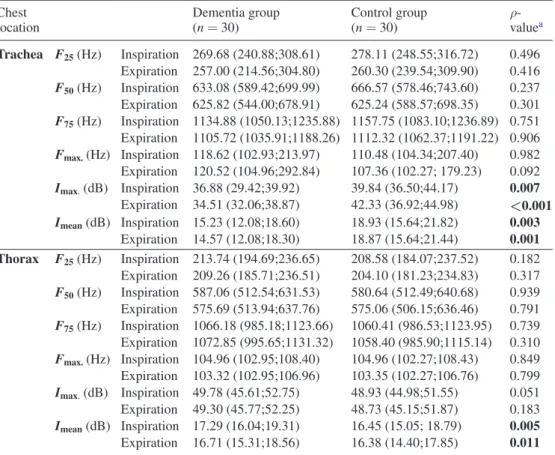
3.2.1.1. Frequency. There were no significant differences between the groups in frequency (i.e. F25, F50, F75 and Fmax.) during inspiration and expiration at both trachea and thorax (table 2).
3.2.1.2. Intensity. Subjects without dementia presented significantly higher values in Imax
dur-ing inspiration (ρ = 0.007) and expiration (ρ < 0.001) at trachea, than subjects with demen-tia (table 2). Significant differences were also found in Imean during inspiration (ρ = 0.003)
and expiration (ρ = 0.001) at trachea and during inspiration (ρ = 0.005) and expiration (ρ = 0.011) at thorax. Higher values were found at trachea in people without dementia and at thorax in people with dementia (table 2).
3.2.2. Adventitious respiratory sounds.
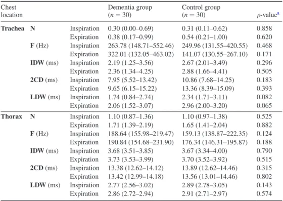
3.2.2.1. Crackles. There were no significant differences between groups in crackles’ mean number, frequency, IDW, 2CDs and LDW during inspiration and expiration in both trachea and thorax (table 3).
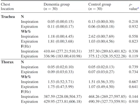
3.2.2.2. Wheezes. Groups were not significantly different in the mean number, frequency and Wh% of wheezes during inspiration and expiration in both trachea and thorax (table 4).
Low frequency wheezes were found in both groups during inspiration and expiration.
4. Discussion
This study characterized computerized respiratory sounds in people with and without dementia, confirming the applicability of computerized auscultation in a non-collaborative population.
Both, people with and without dementia presented similar characteristics of normal and adventitious respiratory sounds with the exception of NRS intensity at trachea and thorax. People with dementia presented significantly lower intensity values of NRS at trachea,
Table 1. Sample characteristics.
Variables Dementia group (n = 30)
Control group (n = 30) ρ-value Age 84.8 ± 7.5 81.7 ± 7.1 0.109a Gender 0.139b Female 22 73.3 17 56.7 Male 8 26.7 13 43.3 Marital Status 0.110b Widowed 16 53.3 16 53.3 Single 7 23.3 2 6.7
Married/living with a partner 7 23.3 9 30.0
Divorced/separated 0 0.0 3 10.0 Years of education 0.083b Illiterate 8 26.7 2 6.7 1–4 20 66.7 27 90.0 5–9 2 6.7 1 3.3 Waist circumference 92.30 ± 14.75 101.30 ± 11.19 0.010a Skin folds Triceps 7.4 ± 3.4 9.4 ± 3.9 0.044a Biceps 4.1 ± 2.3 6.1 ± 2.7 0.004a Suprailiac 7.1 ± 2.9 10.4 ± 4.1 0.001a Subscapular 6.9 ± 3.2 10.8 ± 3.4 < 0.001a
Cognitive status (MMSE) (0–30) 7.7 ± 7.9 24.6 ± 5.0 < 0.001a
Type of dementia N/A N/A
Unspecified dementia 17 56.7
Alzheimer’s disease 8 26.7
Vascular dementia 4 13.3
Dementia associated with PD 1 3.3
Severity of dementia (GDS) (0–7) 6.3 ± 0.9 N/A N/A
Legend: Values are presented as mean ± standard deviation or prevalence and percentage. a Independent samples t-test;
b Chi-square test; MMSE: mini mental state examination; GDS: global deterioration scale; N/A: not applicable; PD: Parkinson Disease; In bold statistically significant p-values: α < 0.005.
possibly explained by a decrease in sound generation resulting from the drop in inspiratory airflow. This decrease could be caused by their poor cooperation, as cognitive impairment in some cases lead to misunderstanding of the request to breathe deeply and due to the com-mon use of medicines to the central nervous system (Marques et al 2015). These drugs could cause depression of the movements of intercostal muscles, alteration of the shape and motion of chest wall and decrease of the rib cage excursion affecting lung compliance mechanics. Therefore, in contrast with people with dementia, people without dementia presented higher intensity values of NRS at trachea suggesting higher airflows, which is in agreement with a recent study from Jácome and Marques (2015). Their study found that in people with chronic obstructive pulmonary disease the normal respiratory sound intensity also increased at higher airflows (Jácome and Marques 2015).
In thorax, people with dementia presented higher mean intensity values of NRS when compared with people without dementia. The authors hypothesized that these higher intensity could result from the higher effort associated with breathing in people with dementia, known as ‘puerile respiration’ a term introduced by Laënnec as an increased sound intensity heard in adults after exertion (Duffin 1991). However, most of the differences between DG and CG are strictly speaking in limits of individual variability (<10%). This study does not allow us to
Table 2. Description of the normal respiratory sound spectrum (100–2000 Hz) during inspiration and expiration at trachea and thorax.
Chest
location Dementia group (n = 30) Control group (n = 30) ρ- valuea
Trachea F25 (Hz) Inspiration 269.68 (240.88;308.61) 278.11 (248.55;316.72) 0.496 Expiration 257.00 (214.56;304.80) 260.30 (239.54;309.90) 0.416 F50 (Hz) Inspiration 633.08 (589.42;699.99) 666.57 (578.46;743.60) 0.237 Expiration 625.82 (544.00;678.91) 625.24 (588.57;698.35) 0.301 F75 (Hz) Inspiration 1134.88 (1050.13;1235.88) 1157.75 (1083.10;1236.89) 0.751 Expiration 1105.72 (1035.91;1188.26) 1112.32 (1062.37;1191.22) 0.906 Fmax. (Hz) Inspiration 118.62 (102.93;213.97) 110.48 (104.34;207.40) 0.982 Expiration 120.52 (104.96;292.84) 107.36 (102.27; 179.23) 0.092 Imax. (dB) Inspiration 36.88 (29.42;39.92) 39.84 (36.50;44.17) 0.007 Expiration 34.51 (32.06;38.87) 42.33 (36.92;44.98) <0.001 Imean (dB) Inspiration 15.23 (12.08;18.60) 18.93 (15.64;21.82) 0.003 Expiration 14.57 (12.08;18.30) 18.87 (15.64;21.44) 0.001 Thorax F25 (Hz) Inspiration 213.74 (194.69;236.65) 208.58 (184.07;237.52) 0.182 Expiration 209.26 (185.71;236.51) 204.10 (181.23;234.83) 0.317 F50 (Hz) Inspiration 587.06 (512.54;631.53) 580.64 (512.49;640.68) 0.939 Expiration 575.69 (513.94;637.76) 575.06 (506.15;636.46) 0.791 F75 (Hz) Inspiration 1066.18 (985.18;1123.66) 1060.41 (986.53;1123.95) 0.739 Expiration 1072.85 (995.65;1131.32) 1058.40 (985.90;1115.14) 0.310 Fmax. (Hz) Inspiration 104.96 (102.95;108.40) 104.96 (102.27;108.43) 0.849 Expiration 103.32 (102.95;106.96) 103.35 (102.27;106.76) 0.799 Imax. (dB) Inspiration 49.78 (45.61;52.75) 48.93 (44.98;51.55) 0.051 Expiration 49.30 (45.77;52.25) 48.73 (45.15;51.87) 0.183 Imean (dB) Inspiration 17.29 (16.04;19.31) 16.45 (15.05; 18.79) 0.005 Expiration 16.71 (15.31;18.56) 16.38 (14.40;17.85) 0.011
Legend: Values are shown as median (interquartile range). F25: frequency at percentile 25; F50: frequency at percentile 50; F75: frequency at percentile 75; Fmax.: frequency at maximum intensity; Imax.: maximum intensity; Imean: mean intensity;
determine if the small differences found between the groups are due to the lack of real differ-ence or due to the small sample, which may not be sufficient to detect truly significant changes between people with and without dementia. Therefore, more studies are needed to compare findings, as very few information on standardized description and evaluation methods for nor-mal respiratory sounds is available (Duffin 1991, Pasterkamp et al 1997).
Similar frequency values of normal respiratory sounds at trachea and thorax were found in both groups, confirming similar airflow turbulence in people with and without demen-tia, which was expected due to the clinical stability of participants. Moreover, the frequency values of NRS found did not suggest the presence of respiratory disease, since they were in accordance with the standard clinical characteristics of NRS, which reference values were from 100 to 5000 Hz at trachea and from 100 to1000 Hz at thorax (Pasterkamp et al 1997, Bohadana et al 2014). Characterizing NRS constitute an important step in the establishment of the normal respiratory sound parameters in stable older people with and without demen-tia, which will allow future comparisons with people presenting respiratory tract infections. Further studies are needed to investigate this issue.
People with and without dementia presented ARS (crackles and wheezes) with similar characteristics during inspiration and expiration at trachea and thorax. Therefore, crackles’ number was similar in both groups, suggesting that crackles did not indicate lung pathol-ogy, agreeing with NRS findings. Two mechanisms could explain its genesis, i.e. air bubbling through secretions or the sudden opening of collapsed airways during inspiration or closing during expiration, as a result of fast pressure equalization of lung compartments (Forgacs
1967, Piirila and Sovijarvi 1995, Vyshedskiy et al 2009). Crackles’ occurrence depends on
Table 3. Crackles’ parameters during inspiration and expiration at trachea and thorax. Chest
location Dementia group (n = 30) Control group (n = 30) ρ-valuea
Trachea N Inspiration 0.30 (0.00–0.69) 0.31 (0.11–0.62) 0.858 Expiration 0.38 (0.17–0.99) 0.54 (0.21–1.00) 0.620 F (Hz) Inspiration 263.78 (148.71–552.46) 249.96 (131.55–420.55) 0.468 Expiration 322.01 (132.05–463.02) 141.07 (130.55–267.10) 0.171 IDW (ms) Inspiration 2.19 (1.25–3.56) 2.67 (2.01–3.49) 0.296 Expiration 2.36 (1.34–4.25) 2.88 (1.66–4.41) 0.505 2CD (ms) Inspiration 7.95 (5.52–13.42) 10.86 (7.68–14.25) 0.183 Expiration 9.65 (6.15–15.22) 13.36 (8.39–15.09) 0.393 LDW (ms) Inspiration 1.74 (0.84–2.74) 2.34 (1.71–3.11) 0.082 Expiration 2.06 (1.52–3.07) 2.96 (2.00–3.20) 0.065 Thorax N Inspiration 1.10 (0.87–1.36) 1.10 (0.97–1.38) 0.525 Expiration 1.71 (1.39–2.19) 1.65 (1.41–2.04) 0.882 F (Hz) Inspiration 188.64 (155.98–219.47) 159.13 (138.87–222.35) 0.124 Expiration 190.84 (154.68–231.90) 176.34 (146.31–195.87) 0.188 IDW (ms) Inspiration 3.68 (3.51–3.85) 3.67 (3.34–4.00) 0.790 Expiration 3.73 (3.53–3.99) 3.70 (3.52–3.92) 0.515 2CD (ms) Inspiration 13.38 (12.62–14.12) 13.89 (12.62–14.46) 0.315 Expiration 13.42 (12.99–14.18) 13.56 (13.01–14.46) 0.802 LDW (ms) Inspiration 2.77 (2.56–3.02) 2.89 (2.78–3.05) 0.143 Expiration 2.86 (2.72–2.94) 2.91 (2.71–2.97) 0.574
Legend: Values are shown as median (interquartile range); N: number; F: frequency; IDW: initial deflection width; 2CD: two cycle duration; LDW: largest deflection width;
the lung volumes achieved during auscultation (Piirila and Sovijarvi 1995) and on properties of the lungs. Both groups were elderly and lung properties change with age, i.e. the lung elas-tic recoil pressure decreases at the same time that residual volumes increases, explaining the crackling sounds (Connolly et al 1992) of both groups.
Most crackles presented longer durations and low frequencies suggesting the presence of coarse crackles in both groups (American Thoracic Society 1977, Sovijarvi et al 2000a), that are consistent with the presence of sputum in proximal airways (Postiaux 2004, Marques et al 2009). This finding could be explained by the common low forced expiratory flow rates and lower lung elastic recoil at an advanced age (Piirila and Sovijarvi 1995), which reduce the efficacy of air-way secretions clearance by coughing (Janssens and Krause 2004, Sharma and Goodwin 2006) and leads to an accumulation in proximal airways. Therefore, crackles assessment is essential, to contribute for estimating the presence and location of secretions (Reichert et al 2008). It can also contribute for the differential diagnosis of respiratory diseases, as their number relates with the severity of the disease and their waveform and positioning within the respiratory cycle are characteristics to differentiate lung pathological cases (Reichert et al 2008).
Following our previous findings of NRS and crackles, no significant differences in the number and frequency of wheezes were found between groups, which suggests that partici-pants from both groups did not have airway obstruction or presented a flow limitation that interferes with the flutter mechanism required to produce wheezes (Pasterkamp et al 1997, Janssens and Krause 2004). Additionally, a low occupation rate was found in both people with and without dementia, meaning that a small percentage of the respiratory cycle was occupied by wheezes. This suggests a reduced airway obstruction at proximal airways possibly due to the secretions movement (Pasterkamp et al 1997), but consistent with the absence of respira-tory disease. This absence among the two groups is an important clinical finding, as wheezes
Table 4. Wheezes parameters during inspiration and expiration at trachea and thorax. Chest
location Dementia group (n = 30) Control group (n = 30) ρ-valuea
Trachea N Inspiration 0.05 (0.00;0.15) 0.13 (0.00;0.30) 0.218 Expiration 0.11 (0.00;0.17) 0.06 (0.00;0.18) 0.932 Wh% Inspiration 1.18 (0.00;4.45) 2.62 (0.00;7.69) 0.558 Expiration 1.81 (0.00;3.68) 1.03 (0.00;4.56) 0.823 F(Hz) Inspiration 410.44 (277.21;510.31) 357.30 (289.63;401.82) 0.338 Expiration 336.96 (183.00;410.98) 375.12 (328.35;522.28) 0.119 Thorax N Inspiration 0.05 (0.02;0.10) 0.05 (0.02;0.13) 0.739 Expiration 0.09 (0.03;0.33) 0.07 (0.03;0.27) 0.734 Wh% Inspiration 1.53 (0.52;3.71) 1.51 (0.56;3.35) 0.667 Expiration 1.75 (0.47;5.99) 1.07 (0.49;4.50) 0.641 F(Hz) Inspiration 387.59 (228.08;504.37) 468.26 (269.27;597.85) 0.144 Expiration 429.95 (273.81;606.18) 490.39 (327.73;559.91) 0.910
Legend: Values are shown as median (interquartile range) or N sum: sum of mean number; min.: minimum; max.: maximum; Wh%: occupation rate—duration of wheeze/duration of phase;
F: frequency; N/A: not applicable;
are considered one of the most easily recognized adventitious respiratory sound (Bohadana et al2014) and their presence is an important indicator of the respiratory system status which also complements the crackles’ assessment.
Although previous studies stated that differences in anthropometric values influenced adventitious respiratory sounds characteristics (Sanchez and Pasterkamp 1993, Ellington et al
2014), this was not supported by our findings, which deserves further investigation.
Finally, it is known that age affects lung volumes and capacities, due to some degree of physi-ological degeneration of the respiratory system, reduced mucociliary function and lower flow rates (Sharma and Goodwin 2006). This study suggested a decrease in airflow, accompanied by a great effort to breathe in people with dementia, based on the characteristics of NRS. These alterations along with the low anthropometric values and poor mobility found in subjects with dementia may represent great difficulty in overcoming future respiratory disease, explaining their higher rates of hospitalization (Muder 1998), longer periods in the hospital (Draper et al
2011) and higher mortality (Steen et al 2006). However, these findings should be considered with caution, due to the exploratory nature of this study. Therefore, NRS and ARS routine assessment and analysis through computerized auscultation demonstrate potential to obtain relevant clini-cal information about the respiratory system. This could allow prevention, early diagnosis and continuous monitoring of LRTIs, mainly in non-collaborative populations, in different settings.
4.1. Limitations and future research
The relative small sample size included in this study limits the generalization of the findings. Larger samples will strengthen these results. Moreover, the sample size used may not be sufficient to detect truly significant changes between older people and people with dementia (type II error). Studies with sample size estimations are also needed. Therefore, this exploratory study is a first step towards the use of computerized respiratory sounds in the objective assessment of people with dementia and could be used as a pilot study to compute sample sizes in future studies.
In this study only one recording per chest location was performed as people with dementia are extremely restless and agitated. The inability to collect 3 measurements at each respiratory system site did not allow reliability assessment, which would have strengthened our findings. However, previous studies (Elphick et al 2004, Marques et al 2009) have demonstrated excel-lent intra-subject reliability and validity of computerized respiratory sounds.
The lack of airflow assessment simultaneously with respiratory sounds also limited our findings, as respiratory sound generation is affected by lung volume and airflow (Gavriely and Cugell 1996, Kiyokawa and Pasterkamp 2002). However the cognitive impairment pre-sented by people with dementia broadly restricted their collaboration in data collection, and in most cases it will be nearly impossible to take them to breath by a mouth piece, while the researcher/clinician perform auscultation.
Finally, studies including 3 groups (people with dementia versus people with dementia presenting a respiratory infection versus matched people without dementia) could be interest-ing to further enhance knowledge on the respiratory system and inform health promotion and prevention of respiratory infections.
5. Conclusion
People with and without dementia had similar characteristics of normal and adventitious respiratory sounds, with the exception of NRS intensity. People with dementia presented lower intensity values at trachea and higher intensity values at thorax.
Although people with dementia have extreme difficulties in participating in the diagnosis and treatment of respiratory diseases, it was possible to collect and study their respiratory sounds, due to the non-invasive nature of computerized auscultation and the minimal need for collaboration. Moreover, the recording of computerized respiratory sounds in people with stable dementia, without respiratory disease, could be the step towards prevention, early diag-nosis and continuous monitoring of respiratory diseases or exacerbations states in different settings.
Acknowledgments
The authors would like to acknowledge to Cátia Pinho and João Dinis for their engineering contributions and to Ana Oliveira for her advices in data analysis. The authors are also grateful to all institutions, staff and older people involved in this research.
The authors also report no conflicts of interest.
References
American Psychiatric Association 1994 Diagnostic and Statistical Manual of Mental Disorders: DSM-IV 4th edn (Washington, DC)
American Thoracic Society 1977 Update nomenclature for membership relation ATS News 3 5–6 Baughman R P and Loudon R G 1984 Quantitation of wheezing in acute asthma Chest 86 718–22
Bohadana A, Izbicki G and Kraman S S 2014 Fundamentals of lung auscultation New Engl. J. Med. 370 744–51
Carusone S B C, Walter S D, Brazil K and Loeb M B 2007 Pneumonia and lower respiratory infections in nursing home residents: predictors of hospitalization and mortality J. Am. Geriatrics Soc. 55 414–9
Carvalhaes-Neto N, Lorino H, Gallinari C, Escolano S, Mallet A, Zerah F, Harf A and Macquin-Mavier I 1995 Cognitive function and assessment of lung function in the elderly Am. J. Respir. Crit. Care
Med.152 1611–5
Cheetham B M G, Charbonneau G, Giordano A, Helistö P and Vanderschoot J 2000 Digitization of data for respiratory sound recordings Eur. Respir. Rev. 10 621–4
Connolly M J, Crowley J J and Vestal R E 1992 Clinical significance of crepitations in elderly patients following acute hospital admission: a prospective study Age Ageing 21 43–8
Davey R J and Jamieson S 2004 The validity of using the mini mental state examination in NICE dementia guidelines J. Neurol. Neurosurg. Psychiatry 75 343–4
Draper B, Karmel R, Gibson D, Peut A and Anderson P 2011 The hospital dementia services project: age differences in hospital stays for older people with and without dementia Int. Psychogeriatr. 1 1–10
Duffin J M 1991 Puerile respiration: Laënnec’s stethoscope and the physiology of breathing Trans. Stud.
Coll. Physicians Phil. 13 125–45
Ellington L, Emmanouilidou D, Elhilali M, Gilman R, Tielsch J, Chavez M, Marin-Concha J, Figueroa D, West J and Checkley W 2014 Developing a reference of normal lung sounds in healthy peruvian children Lung 192 765–73
Elphick H E, Lancaster G A, Solis A, Majumdar A, Gupta R and Smyth R L 2004 Validity and reliability of acoustic analysis of respiratory sounds in infants Arch. Dis. Child. 89 1059–63
Folstein M S, Folstein S E and McHugh P R 1975 ‘Mini-mental state’. A pratical method for grading the mental state of patients for the clinician J. Psychiatric Res. 12 189–98
Forgacs P 1967 Crackles and wheezes Lancet 290 203–5
Gavriely N and Cugell D W 1996 Airflow effects on amplitude and spectral content of normal breath sounds J. Appl. Physiol. 80 5–13
Gavriely N, Palti Y and Alroy G 1981 Spectral characteristics of normal breath sounds J. Appl. Physiol.
Respir. Environ. Exerc. Physiol. 50 307–14
Guerreiro M, Silva A P, Botelho M A, Leitão O, Caldas A C and Garcia C 1994 Adaptação à população portuguesa do ‘Mini Mental State Examination’ (MMSE) Revista Portuguesa de Neurologia
Hadjileontiadis L J and Rekanos I T 2003 Detection of explosive lung and bowel sounds by means of fractal dimension IEEE Signal Process. Lett. 10 311–4
Hoevers J and London R G 1990 Measuring crackles Chest 98 1240–3
Jácome C and Marques A 2015 Computerized respiratory sounds are a reliable marker in subjects with COPD Respir. Care 60 1264–75
Janssens J and Krause K 2004 Pneumonia in the very old Lancet Infectious Dis. 4 112–24
Joseph M M 2007 Pneumonia and Bronchitis Infection Management for Geriatrics in Long-Term Care
Facilities 2nd edn, ed T T Yoshikawa and J G Ouslander (New York: Informa Health Care) pp 215–29
Katz M J 1988 Fractals and the analysis of waveforms Comput. Biol. Med. 18 145–56
Kirkwood B R and Sterne J A C 2006 Essential Medical Statistics 2nd edn (Oxford: Blackwell) Kiyokawa H and Pasterkamp H 2002 Volume-dependent variations of regional lung sound, amplitude,
and phase J. Appl. Physiol. 93 1030–8
Lev S, Glickman Y A, Kagan I, Shapiro M, Moreh-Rahav O, Dahan D, Cohen J, Grinev M and Singer P 2010 Computerized lung acoustic monitoring can help to differentiate between various chest radiographic densities in critically ill patients Respiration 80 509–16
Marini J J, Pierson D J, Hudson L D and Lakshminarayan S 1979 The significance of wheezing in chronic airflow obstruction Am. Rev. Respir. Dis. 120 1069–72
Marques A, Bruton A and Barney A 2006 Clinically useful outcome measures for physiotherapy airway clearance techniques: a review Phys. Ther. Rev. 11 299–307
Marques A, Bruton A and Barney A 2009 The reliability of lung crackle characteristics in cystic fibrosis and bronchiectasis patients in a clinical setting Physiol. Meas. 30 903–12
Marques A, Oliveira A and Jácome C 2014 Computerized adventitious respiratory sounds as outcome measures for respiratory therapy: a systematic review Respir. Care 59 765–76
Marques A, Rocha V, Pinto M, Sousa L and Figueiredo D 2015 Comorbidities and medication intake among people with dementia living in long-term care facilities Rev. Portuguesa de Saúde Pública 33 42–8
Monteiro I and Reisberg B 2000 Global Deterioration Scale: Validation to the Portuguese Population (Portugal)
Muder R R 1998 Pneumonia in residents of long-term care facilities: epidemiology, etiology, management, and prevention Am. J. Med. 105 319–30
Murphy R L Jr, Del Bono E A and Davidson F 1989 Validation of an automatic crackle (rale) counter
Am. Rev. Respir. Dis.140 1017–20
Murphy R L H, Holford S K and Knowler W C 1977 Visual lung sound characterization by time expanded wave form analysis New Engl. J. Med. 296 968–71
Ntoumenopoulos G and Glickman Y 2012 Computerised lung sound monitoring to assess effectiveness of chest physiotherapy and secretion removal: a feasibility study Physiotherapy 98 250–5
Oliveira A and Marques A 2014 Respiratory sounds in healthy people: a systematic review Respir. Med. 108 550–70
Oliveira A, Pinho C, Dinis J, Figueiredo D and Marques A 2013 Automatic Wheeze detection and lung function Proc. 6th Int. Conf. on Health Informatics (Barcelona, Spain, 11–14 February 2013) ed S Deborah et al pp 233–8
Oliveira D, Pinho C, Marques A and Dinis J 2011 Validation of a time frequency wheeze detector in cystic fibrosis: a pilot study Eur. Respir. J. 38 1280
Pasterkamp H, Kraman S S and Wodicka G R 1997 Respiratory sounds Am. J. Respir. Crit. Care Med. 156 974–87
Pasterkamp H, Powell R E and Sanchez I 1996 Lung sound spectra at standardized air flow in normal infants, children, and adults Am. J. Respir. Crit. Care Med. 154 424–30
Piirila P 1992 Changes in crackle characteristics during the clinical course of pneumonia Chest 102 176–83
Piirila P and Sovijarvi A R A 1995 Crackles: recording, analysis and clinical significance Eur. Respir.
J.8 2139–48
Piirila P, Sovijarvi A R A, Kaisla T, Rajala H M and Katila T 1991 Crackles in patients with fibrosing alveolitis, bronchiectasis,COPD, and heart failure Chest 99 1076–83
Pinho C, Oliveira A, Jácome C, Rodrigues J and Marques A 2015 Integrated approach for automatic crackle detection based on fractal dimension and box filtering Proc. Int. Conf. on Health and Social
Care Information Systems and Technologies (HCIST) (Vilamoura, Portugal, 2015)
Pinho C, Oliveira D, Oliveira A, Dinis J and Marques A 2012 LungSounds@UA interface and multimedia database Proc. Technol. 5 803–11
Ponte D F, Moraes R, Hizume D C and Alencar A M 2012 Characterization of crackles from patients with fibrosis, heart failure and pneumonia Med. Eng. Phys. 1 1–9
Postiaux G 2004 Fisioterapia Respiratória Pediátrica 2nd edn, ed A Médicas (Brussels: Artmed) p 302 Prince M, Bryce R, Albanese E, Wimo A, Ribeiro W and Ferri C P 2013 The global prevalence of
dementia: a systematic review and meta-analysis Alzheimer’s Dementia 9 63–75
Reichert S, Gass R, Brandt C and Andres E 2008 Analysis of respiratory sounds: state of the art
Clin. Med. 2 45–58
Reisberg B, Ferris S H, Leon M J d and Crook T 1982 The global deterioration scale for assessment of primary degenerative dementia Am. J. Psychiatry 139 1136–9
Sanchez I and Pasterkamp H 1993 Tracheal sound spectra depend on body height Am. Rev. Respir. Dis. 148 1083–7
Sands L P, Yaffe K, Lui L, Stewart A, Eng C and Covinsky K 2002 The effects of acute illness on ADL decline over 1 year in frail older adults with and without cognitive impairment J. Gerontol. A 57 449–54
Sevcik C 2010 A procedure to estimate the fractal dimension of waveforms Complexity Int. 5 1–19 Sharma G and Goodwin J 2006 Effect of aging on respiratory system physiology and immunology
Clin. Interv. Aging.1 253–60
Shen J, Shen W, Castan S and Zhang T 2002 Sum-box technique for fast linear filtering Signal Process. 82 1109–26
Shulman K I, Herrmann N, Brodaty H, Chiu H, Lawlor B, Ritchie K and Scanlan J M 2006 IPA survey of brief cognitive screening instruments Int Psychogeriatr. 18 281–94
Sovijarvi A R A, Dalmasso F, Vanderschoot J, Malmberg L P, Righini G and Stoneman S A T 2000a Definition of terms for application of respiratory sounds Eur. Respir. Rev. 10 597–610
Sovijarvi A R A, Malmberg L P, Charbonneau G, Vanderschoot J, Dalmasso F and Sacco C 2000b Characteristics of breath sounds and adventitious respiratory sounds Eur. Respir. Rev. 10 591–6 Sovijärvi A R A, Vanderschoot J and Earis J E 2000c Computerised respiratory sound analysis
(CORSA): recommended standards for terms and techniques. ERS task force report Eur. Respir.
Rev. 10 595–649
Steen J T Vd, Mehr D R, Kruse R L, Sherman A K, Madsen R W, D’Agostino R B, Ooms M E, Gvd W and Ribbe M W 2006 Predictors of mortality for lower respiratory infections in nursing home residents with dementia were validated transnationally J. Clin. Epidemiol. 59 970–9
Taplidou S A and Hadjileontiadis L J 2007 Wheeze detection based on time-frequency analysis of breath sounds Comput. Biol. Med. 37 1073–83
Volicer L 2005 End-of-life Care for People with Dementia in Residential Care Settings (Tampa, FL: University of South Florida)
Vyshedskiy A, Alhashem R M, Paciej R, Ebril M, Rudman I, Fredberg J J and Murphy R 2009 Mechanism of inspiratory and expiratory crackles Chest 135 156–64
Woodhead M et al 2011 Guidelines for the management of adult lower respiratory tract infections— summary Clin. Microbiol. Infection 17 1–24