FACULDADE DE FARMÁCIA
PROBING ENTRY INHIBITORS’ ACTIVITY ON HIV AND
DEVELOPMENT OF NEW FUSION INHIBITORS:
INTEGRATING EVOLUTIONARY BIOLOGY WITH VIROLOGY
Pedro José Vieira Borga Martins Borrego
DOUTORAMENTO EM FARMÁCIA
(MICROBIOLOGIA)
FACULDADE DE FARMÁCIA
PROBING ENTRY INHIBITORS’ ACTIVITY ON HIV AND
DEVELOPMENT OF NEW FUSION INHIBITORS:
INTEGRATING EVOLUTIONARY BIOLOGY WITH VIROLOGY
Pedro José Vieira Borga Martins Borrego
DOUTORAMENTO EM FARMÁCIA
(MICROBIOLOGIA)
2011
A presente dissertação foi realizada na Unidade de Retrovírus e Infecções Associadas da Faculdade de Farmácia, Universidade de Lisboa, sob a orientação do Professor Doutor Nuno Eduardo Moura dos Santos da Costa Taveira e co-orientação da Professora Doutora Maria Helena de Sousa Barroso e do Professor Doutor José Moniz Pereira.
Este trabalho foi desenvolvido no âmbito de uma Bolsa de Doutoramento atribuída ao autor pela Fundação para a Ciência e Tecnologia.
Todas as afirmações efectuadas no presente documento são da exclusiva responsabilidade do seu autor, não cabendo qualquer responsabilidade à Faculdade de Farmácia, Universidade de Lisboa, pelos conteúdos nele apresentados.
Many people have contributed to this work and therefore I would like to acknowledge them for making this project possible.
Thank you Professor Nuno Taveira, for all your guidance through this journey and for all the knowledge and expertise you shared with me. Thank you for showing me the passion for research and for all the straightforward motivation, reminding me that there are no unreachable boundaries. You will always be a reference to me. Thank you for your friendship.
Thank you Professor Helena Barroso, for all the support given during my years in the lab. You have been amazingly available for questions and thoughts about my work and influenced my learning in a most incredible positive way. Thank you for your friendship.
Professors Nuno and Helena, I am very grateful for the opportunities you have given me to work in several projects in the lab, which have been so important in my scientific learning process and helped me find new research interests.
I would like to thank Professor José Moniz Pereira, director of the Unidade de Retrovírus e
Infecções Associadas – Centro de Patogénese Molecular, for allowing me to developed my
work at this unit. In addition, I would like to thank all the teachers that work in this unit, particularly: Prof. José Azevedo Pereira, Prof. João Gonçalves, Prof. Isabel Portugal, Prof. Madalena Pimentel, Prof. Elsa Anes and Prof. Aida Duarte.
To my laboratory colleagues Rita, Cheila, Marcelino, Inês, Andreia and Claudia Palladino, thank you for making it such a great working environment, for always being there and helping me whenever I needed. Importantly, thank you for standing all my crazy talk, music taste and singing! Thank you for all the lab meetings that were not only an incentive to conviviality but also a stimulation to my belly! I will always be available for as many Italian lunches you want me to join! Thank you for your friendship.
Especially to Rita, thank you for your collaboration and endless devotion that has contributed to my work in a most precious way.
I would also like to thank the good spirit of other colleagues with whom I’ve shared the working bench in the past: Vânia, Filipa, Sofia, Daniela Moutinho and Hugo.
This work wouldn’t be possible without the great collaboration provided by Prof. Alexandre Quintas. Thank you for your expertise and for keeping the doors of your lab always open
Thank you to all the patients and doctors that provided the samples needed for analysis, without your participation none of this work could have been done.
In the backstage of science there are always indispensable people that work hard every day so that students and teachers are provided with all they need. Thank you Lena Brás, Teresa and Lídia for being there whenever we need.
My daily work in the laboratory would not be possible without the help of D. Fátima, Sr. Augusto, Kleida and D. Noémia, providing us proper working conditions.
To Ofélia and Vera thank you for taking care of all the paperwork, for your availability and contagious good mood.
To all the other lab colleagues at URIA-CPM (Quirina, João, Cláudia, Carla, Marta Calado, Maria, Paula, Daniela, Marta, Camila, Emilie, Sylvie, Paula Brito, Andreia, Ana Catarina, Catarina, Mariana, Acilino, Inês, Sara, Luís, André, Patrícia, Leonor, Diana, Joana, David, Paulo, Nuno), with whom I’ve shared not only the P3 but also good companionship and positive vibes, many thanks!!
Thank you Prof. Jorge Vítor and Joana Vital for your availability and good will.
I’m also very fortunate to have contacted with several people along the past few years who have so kindly given me the opportunity to collaborate in other activities outside the lab. For different reasons, thank you Luís Proença, Jorge Félix and Filipa Duarte Ramos.
Finally, and most importantly, I would like to thank all my family, godparents and close friends for believing in me, for always supporting my decisions and for all the effort in fulfilling my dreams.
Obrigado mãe, pai, mano e avós, por serem uma fonte de inspiração e orientação inesgotáveis.
Raquel, obrigado pela ajuda, pelo carinho e compreensão, sem os quais teria sido impossível realizar este trabalho. A vida é melhor contigo ao meu lado.
This thesis is based on the following publications:
Manuscripts in international journals
2011 – Borrego P, Calado R, Marcelino J, Oliveira L, Pereira P, Quinta A, Barroso H, Taveira N. Design and evaluation of an ancestral peptide with potent and broad HIV fusion inhibitor activity. Manuscript submitted, July 2011.
2011 – Borrego P, Calado R, Marcelino JM, Bártolo I, Rocha C, Cavaco-Silva P, Doroana M, Antunes F, Maltez F, Caixas U, Barroso H, Taveira N. Baseline susceptibility of primary Human Immunodeficiency Virus Type 2 to entry inhibitors. Manuscript submitted, Antiviral Therapy, June 2011.
2011 – Barroso H, Borrego P, Bártolo I, Marcelino J, Família C, Quintas A, Taveira N. Evolutionary and structural features of the C2, V3 and C3 envelope regions underlying the differences in HIV-1 and HIV-2 biology and infection. PLoS One. 2011 Jan;6(1):e14548.
Book Chapters
2008 – Pedro Borrego, Helena Barroso, Nuno Taveira. Mecanismo Molecular de Entrada do VIH nas Células e sua Inibição Farmacológica [The molecular mechanism of HIV entry into
cells and its pharmacologic inhibition]. In “8th HIV-AIDS Virtual Congress”. 2008. pages
279-295.
Oral communications in international conferences
2010 – Borrego P, Calado R, Quintas, A, Barroso H, Taveira N. Design and evaluation of a new HIV-1 and HIV-2 fusion inhibitor peptide. XII Iberian Peptide Meeting, February 10 – 12, 2010, Faculty of Medicine, University of Lisbon, Lisbon, Portugal.
Oral communications in national conferences
2010 – Borrego P, Calado R, Quintas, A, Barroso H, Taveira N. Design and evaluation of a new HIV-1 and HIV-2 fusion inhibitor peptide. Centre for Pharmaceutical Studies 2010, September 28th 2010, Faculty of Pharmacy, University of Coimbra, Coimbra, Portugal.
Poster communications in international conferences
2011 – Borrego P, Barroso H, Bártolo I, Marcelino JM, Família C, Quintas A, Taveira N. Evolutionary and structural features of the C2, V3 and C3 envelope regions underlying the differences in HIV-1 and HIV-2 biology and infection. In: Keystone Symposia - HIV Evolution, Genomics and Pathogenesis, March 20 – 25, 2010, Whistler Conference Centre, Whistler, British Colombia, Canada (Abstract no. 115).
2011 – Borrego P, Calado R, Marcelino JM, Rocha C, Barroso H, Taveira N. Susceptibility of HIV-2 primary isolates to fusion and entry inhibitors. In: Keystone Symposia - Protection
2011 – Borrego P, Calado R, Marcelino JM, Bártolo I, Cavaco-Silva P, Quintas A, Barroso H, Taveira N. Design and evaluation of a new HIV-1 and HIV-2 fusion inhibitor peptide. In: Keystone Symposia - Protection from HIV: Targeted Intervention Strategies, March 20 – 25, 2010, Whistler Conference Centre, Whistler, British Colombia, Canada (Abstract no. 115). 2009 – Borrego P, Marcelino JM, Rocha C, Doroana M, Maltez F, Barroso H, Taveira N. Evolutionary features underlying the differences in HIV-1 and HIV-2 clinical outcome In: 15th International BioInformatics Workshop on Virus Evolution and Molecular Epidemiology, September 7 – 11, 2009, Erasmus Postgraduate School of Molecular Medicine, Rotterdam, Netherlands. (Abstract no. 26)
Other Publications:
Manuscripts in international journals
2011 – Bártolo I, Abecasis AB, Borrego P, Barroso H, McCutchan F, Camacho F, Taveira N. Origin and epidemiologic history of HIV-1 CRF14_BG. Submitted, PLoS One, March 2011. 2011 – Félix J, Andreozzi V, Soares M, Borrego P, Gervásio H, Moreira A, Costa L, Marcelo F, Peralta F, Furtado I, Pina F, Albuquerque C, Santos A, Passos-Coelho JL; Portuguese Group for the Study of Bone Metastases. Hospital resource utilization and treatment cost of skeletal-related events in patients with metastatic breast or prostate cancer: estimation for the portuguese national health system. Value Health. 2011 Jun;14(4):499-505.
2010 - Marcelino JM, Borrego P, Nilsson C, Barroso H, Doroana M, Antunes F, Novo C, Taveira N. Escape from neutralization is a frequent event in HIV-2 infection and is strongly associated with X4 tropism. AIDS Res Hum Retroviruses. 2010 26: A6.
2010 – Marcelino J, Borrego P, Rocha C, Barroso H, Quintas A, Novo C, Taveira N. Potent and broadly reactive HIV-2 neutralizing antibodies elicited by a Vaccinia virus vector-prime C2V3C3 polypeptide-boost immunization strategy. J Virol. 2010 Sep;84(23):12429-12436. 2010 – Skar H, Borrego P, Wallstrom TC, Mild M, Marcelino JM, Barroso H, Taveira N, Leitner T, Albert J. HIV-2 genetic evolution in patients with advanced disease is faster than that in matched HIV-1 patients. J Virol. 2010 Jul;84(14):7412-5.
2008 – Borrego P, Marcelino JM, Rocha C, Doroana M, Antunes F, Maltez F, Gomes P, Novo C, Barroso H, Taveira N. The role of the humoral immune response in the molecular evolution of the envelope C2, V3 and C3 regions in chronically HIV-2 infected patients. Retrovirology. 2008 Sep 8;5(1):78.
2008 – Marcelino JM, Nilsson C, Barroso B, Gomes P, Borrego P, Maltez F, Rosado L, Doroana M, Antunes F, Taveira N. Envelope-specific antibody response in HIV-2 infection: C2V3C3-specific IgG response is associated with disease progression. AIDS. 2008 Nov 12;22(17):2257-65.
2008 – Nuno Taveira, Pedro Borrego and Inês Bártolo. Biologia molecular do VIH [The
Molecular Biology of HIV]. Manual de VIH/SIDA. 3rd Edition. Pernmayer Portugal. 2009.
pages 27-50.
Oral communications in international conferences
2010 – Marcelino JM, Borrego P, Nilsson C, Barroso H, Doroana M, Antunes F, Novo C, Taveira N. Escape from neutralization is a frequent event in HIV-2 infection and is strongly associated with X4 tropism. AIDS Vaccines 2010, September 28th – October 1st, 2010,
Atlanta, Georgia, USA.
Oral communications in national conferences
2007 - Borrego P, Marcelino J, Rocha C, Doroana M, Maltez F, Barroso H, Taveira N. Interplay between humoral response and viral evolution in HIV-2 infection. URIA-CPM Seminars, July 6th 2007, Faculty of Pharmacy, University of Lisbon, Lisbon, Portugal
2005 – Borrego P, Carneiro C, Félix J. “Willingness-to-pay of diabetic and/or hypertensive patients for Pharmaceutical Care in the Community Pharmacy”. 10th Anniversary
Conference of the Centre for Pharmacoepidemiology Studies, February 25 – 26, 2005, Lisbon, Portugal.
Poster communications in international conferences
2010 – Marcelino JM, Borrego P, Nilsson C, Barroso H, Doroana M, Antunes F, Novo C, Taveira N. 2010. Escape from neutralization is a frequent event in HIV-2 infection and is strongly associated with X4 tropism. IN: AIDS Vaccines 2010, Atlanta, USA. Abstract book p. 62. Published in: AIDS Res. Hum. Retroviruses, 2010, 26: A6.
2010 – Taveira N, Borrego P, Novo C, Barroso H, Marcelino JM. Potent and broad spectrum anti-HIV-2 neutralizing antibodies induced by C2V3C3-directed vaccination. In: Keystone Symposia HIV Vaccines, March 21 – 26, 2010, Fairmont Banff Springs, Banff, Alberta, Canada.
2008 – Skar H, Borrego P, Marcelino JM, Wilbe K, Alaeus A, Rocha C, Barroso H, Taveira N, Leitner T, Albert J. The rate of HIV-2 evolution. In: 15th International HIV Dynamics &
Evolution, April 27 – 30, 2008, Hotel Santa Fe, Santa Fe, New Mexico, USA. (Abstract no. 115)
2008 - Borrego P, Marcelino JM, Rocha C, Doroana M, Maltez F, Barroso H, Taveira N. Interplay between the immune response to the envelope glycoproteins and viral evolution in chronic HIV-2 infection. In: Keystone Symposia HIV Vaccines: Progress and Prospects, March 27th –April 1st, 2008, Fairmont Banff Springs, Banff, Alberta, Canada. (Abstract no.
115)
2007 – Borrego P, Marcelino JM, Rocha C, Doroana M, Maltez F, Barroso H, Taveira N. Intrapatient molecular evolution of the env gene in HIV-2 infection. In: 13th International
2006 – Félix J, Ferreira JM, Borrego P, Duarte-Ramos F, Andreozzi V, Urano Study Group. Utilization of Granulocyte Colony Stimulating Factors in Chemotherapy Induced Neutropenia in Patients with Breast or Lung Cancer: Portugal. In: 22nd International Conference on Pharmacoepidemiology & Therapeutic Risk Management, 2006, Lisbon, Portugal. Pharmacoepidemiology and Drug Safety, 2006. v. 15. p. S166.
O Vírus da Imunodeficiência Humana do tipo 1 e do tipo 2 (VIH-1 e VIH-2) são os agentes etiológicos do Síndrome de Imunodeficiência Adquirida (SIDA). Embora sejam semelhantes na sua organização estrutural e genómica, estes lentivírus humanos apresentam características antigénicas distintas e partilham uma semelhança genética de apenas 50%. Enquanto o VIH-1 é responsável pela pandemia mundial, a infecção pelo VIH-2 localiza-se sobretudo na África Ocidental, em alguns países europeus como Portugal e França, e na Índia. A infecção pelo VIH-2 tem melhor prognóstico, a progressão para a doença é mais lenta e há melhor controlo imunológico do que na infecção pelo VIH-1.
Ao contrário do VIH-1, o arsenal terapêutico actualmente disponível para tratar a infecção por VIH-2 é reduzido. Os fármacos antiretrovirais em uso foram especificamente desenvolvidos para o VIH-1 e, consequentemente, a sua actividade pode ser reduzida ou nula no VIH-2. Este é o caso concreto dos inibidores não nucleosídicos da transcriptase reversa e de alguns inibidores da protease. Neste contexto, os inibidores de entrada poderão ser úteis para tratar a infecção por VIH-2. Contudo, a susceptibilidade dos isolados primários de VIH-2 aos inibidores de entrada é actualmente desconhecida.
A susceptibilidade do VIH aos inibidores de entrada é determinada pela qualidade da interacção do vírus com os receptores celulares. O VIH-1 e VIH-2 são substancialmente diferentes a este nível. Por exemplo, o VIH-2 pode ligar-se ao co-receptor CCR5 independentemente do receptor CD4 e da região V3 do invólucro. Por outro lado, as regiões C2, V3 e C3 do VIH-2 são substancialmente diferentes do VIH-1 a nível antigénico. Colectivamente, estes dados indicam que a estrutura e conformação das glicoproteínas de superfície do VIH-1 e VIH-2 são substancialmente diferentes e sugerem que a susceptibilidade e resistência dos dois tipos de vírus aos inibidores de entrada podem também ser diferentes.
Os principais objectivos desta tese foram: 1) analisar as características moleculares, estruturais e evolutivas das regiões C2, V3 e C3 no VIH-1 e VIH-2; 2) comparar a susceptibilidade do VIH-1 e VIH-2 aos inibidores de entrada e avaliar o seu potencial terapêutico na infecção por VIH-2; 3) produzir um novo inibidor de fusão para o VIH-2.
Para melhor compreender as potenciais diferenças destes dois vírus na resposta aos inibidores de entrada começámos por analisar as características moleculares, estruturais e evolutivas da região V3 e as regiões circundantes C2 e C3, num número significativo de vírus VIH-1 e VIH-2 isolados em Portugal e noutras regiões do globo, com recurso a diferentes metodologias de biologia evolutiva e computacional (Capitulo 2). Apesar da
região poderá constituir um domínio neutralizante. No entanto, ao contrário do VIH-1, a maioria das mutações adaptativas no VIH-2 são prejudiciais e levam à extinção das linhagens virais pelo que o efeito final é um forte constrangimento à variabilidade das regiões analisadas. Ao contrário do VIH-1, verificámos que a ansa V3 do VIH-2 se encontra oclusa no complexo glicoproteico do invólucro, numa conformação que parece ser estabilizada por interacções que mantém com alguns resíduos da regiões C2 e C3. Estes resultados são consistentes com o facto de a V3 não ser imunodominante no VIH-2, ficando assim mais protegida da resposta imunitária e das eventuais mutações que dela resultam. A forte conservação da V3, da C2 e da C3 também é consistente com a sua potencialmente importante actividade imunosupressora. Em conclusão, este primeiro estudo permitiu caracterizar algumas das características estruturais e funcionais que distinguem as glicoproteínas do invólucro do VIH-1 e do VIH-2 e que estão associadas às diferentes características biológicas e fenotípicas destes dois vírus. Estes dados podem ter impacto na resposta dos dois vírus aos inibidores de entrada (analisado no Capítulo 3) e no desenvolvimento de novas vacinas.
No segundo estudo (Capítulo 3) comparámos a actividade antiviral dos antagonistas dos co-receptores (AMD3100, TAK-779 e maraviroc) e dos inibidores de fusão (T-20 e T-1249) entre um grupo de 20 isolados de VIH-2 (19 isolados primários + um isolado laboratorial) e nove isolados de VIH-1 (sete isolados primários + dois isolados laboratoriais). Verificámos que a sensibilidade ao AMD3100 e ao TAK-779 é semelhante no VIH-1 e o VIH-2. No entanto, o perfil da curva dose-resposta do maraviroc (MVC) obtido para os isolados R5 foi diferente nos dois tipos de vírus. No VIH-2 os valores de IC90 foram significativamente mais elevados
do que no VIH-1; por outro lado, os declives da curva dose-resposta foram mais baixos no VIH-2 do que no VIH-1. Colectivamente, estes resultados sugerem que poderão ser necessárias concentrações mais elevadas de MVC para tratar os doentes infectados pelo VIH-2. Adicionalmente, encontrámos uma correlação forte e de sentido inverso entre as susceptibilidade do VIH-2 ao MVC e o número de células T CD4+ dos doentes quando os vírus
foram isolados. Vírus isolados em doentes em fase de SIDA foram menos susceptíveis ao MVC do que os vírus isolados em doentes com uma contagem de células T CD4+ superior a
200 células/l. Ao contrário do VIH-1 não encontrámos qualquer correlação entre a carga da V3 e a susceptibilidade dos isolados R5 de VIH-2 ao MVC. De um modo geral, os nossos resultados sugerem que são necessários ensaios clínicos para avaliar a efectividade do MVC na infecção pelo VIH-2, determinar a dose terapêutica mais adequada e esclarecer se é
resistentes ao MVC, é de extrema importância o desenvolvimento de um ensaio de tropismo (genotípico e/ou fenotípico) para o VIH-2 de modo a determinar o tropismo antes do início da terapia com MVC. Sem o conhecimento prévio do tropismo viral, o tratamento com MVC poderá seleccionar espécies X4 minoritárias que estão associadas a maior resistência à neutralização e uma progressão mais rápida da doença.
No que diz respeito aos inibidores de fusão, verificámos que o T-20 tem actividade reduzida no VIH-2, confirmando estudos anteriores realizados com dois isolados laboratoriais. Por outro lado, observámos uma elevada susceptibilidade deste vírus ao T-1249, indicando que os inibidores de fusão são potencialmente eficazes na infecção pelo VIH-2. Assim, o desenvolvimento de um novo inibidor de fusão do VIH-2 foi o objectivo do último estudo desta tese (Capítulo 4).
No Capítulo 4, desenvolvemos novos péptidos inibidores de fusão a partir da reconstrução de sequências ancestrais da glicoproteína gp36 do invólucro de VIH-2 e de Vírus de Imunodeficiência dos Símios (VIS). Com esta abordagem inovadora pretendemos incorporar a história evolutiva dos vírus na sequência dos péptidos e desta forma melhorar a tolerância destas moléculas aos polimorfismos naturais da sua região alvo bem como às mutações de resistência seleccionadas na sua presença. Obteve-se um péptido ancestral (P3) constituído por 34 aminoácidos, cuja sequência corresponde às posições homólogas 628 – 661 da proteína Env do isolado VIH-1 HXB2 (ou 623 – 656 do isolado VIH-2 ROD). A sequência do P3 difere em 21 aminoácidos da sequência consenso de VIH-1, 14 aminoácidos da sequência do T-20 e 6 aminoácidos da sequência consenso de VIH-2. Ao contrário da natureza não-estruturada do T-20, o P3 tem uma conformação típica em hélice-, o que lhe poderá conferir maior a estabilidade contra a degradação proteolítica, bem como maior afinidade para a região alvo. Por outro lado, o P3 foi facilmente solúvel em soluções aquosas o que é uma vantagem num futuro desenvolvimento de uma fórmula farmacêutica. O P3 demonstrou ter uma forte actividade antiviral contra isolados primários e laboratoriais de VIH-1 e VIH-2 (IC50 médio, 11 nM para o HIV-1 e 63.8 nM para o HIV-2),
incluindo variantes resistentes ao T-20 (IC50, 0.15 – 11.8 nM). Através da passagem
consecutiva de vírus em cultura na presença do péptido, foi seleccionada uma mutação de resistência na região HR1 da gp41 (VIH-1), a qual é responsável pela redução da susceptibilidade do VIH-1 ao P3 em 120x. Nas mesmas condições, e após 60 dias em cultura, não foi possível seleccionar mutações de resistência ao P3 no VIH-2. Estes resultado, em conjugação com a sua forte ligação à glicoproteína transmembranar de um
que a barreira genética para a resistência ao P3 é significativamente superior no VIH-2 do que no VIH-1. Neste estudo demonstrámos ainda que o P3 é significativamente menos antigénico do que o T-20 nos doentes infectados pelo VIH-1 o que poderá traduzir-se numa maior duração da eficácia clínica do P3 em comparação com o T-20. Os resultados obtidos com o P3 demonstram pela primeira vez que é possível desenvolver péptidos com actividade antiviral significativa utilizando metodologias de biologia evolutiva, pelo que esta abordagem poderá ser explorada no futuro para a produção de medicamentos peptídicos e, eventualmente, de vacinas.
The general aims of this thesis were: 1) to examine the C2, V3 and C3 envelope regions of HIV-1 and HIV-2 at the molecular, evolutionary and structural levels; 2) to compare HIV-1 and HIV-2 susceptibility to entry inhibitors and assess their potential value in HIV-2 therapy; 3) to produce a new fusion inhibitor peptide using evolutionary biology based strategies.
In the first study (Chapter 2), HIV-1 and HIV-2 were compared at the molecular, evolutionary and structural levels in the C2, V3 and C3 envelope regions. We identified significant structural and functional constrains to the diversification and evolution of C2, V3 and C3 in the HIV-2 envelope but not in HIV-1. In particular, we found that V3 in HIV-2 is less exposed and more conserved than in HIV-1, suggesting fundamental differences in the biology and infection of these viruses as well as in their susceptibility to entry inhibitors.
In the second study (Chapter 3) we measured the baseline susceptibility of HIV-1 and HIV-2 primary isolates to different fusion inhibitors and coreceptor antagonists, including enfuvirtide (T-20) and maraviroc (MVC). MVC inhibited HIV-2 R5 variants at significantly higher IC90 concentrations than HIV-1 variants. Moreover, as previously found in HIV-1,
susceptibility of HIV-2 R5 variants to MVC was inversely related with CD4+ T cell counts at time of virus isolation. These results suggest that the structure of the envelope complex of R5 variants changes along the course of infection. More importantly, the results call for new clinical studies to evaluate the efficacy of MVC in HIV-2 infection and to determine its best therapeutic dosage in early and late stage disease. We also provide definitive evidence demonstrating that T-20 is not useful for HIV-2 therapy.
In the final study (Chapter 4), we designed a new HIV fusion inhibitor peptide (P3) based on the ancestral sequences of the HIV-2 and SIV envelope genes. P3 has an -helix structure as demonstrated by circular dichroism. It has broad antiviral activity at the nanomolar range against HIV-1 and HIV-2 primary isolates, including HIV-1 variants resistant to T-20. Binding ELISA assays and selection of resistant mutants suggest that P3 prevents viral fusion by binding to the transmembrane protein in the HR1 region. These studies provide proof of concept that viable antiviral peptides can be constructed using evolutionary biology strategies. Such strategies should be explored to enhance the production of peptide drugs and vaccines.
95%CI confidence interval at 95 percent
AIC Akaike’s information criterion
AIDS Acquired Immunodeficiency Syndrome
BSA bovine serum albumin
ºC Celsius degree
CA conic shaped viral capsid
CD circular dichroism spectroscopy CDC Centres for Disease Control
CO2 carbon dioxide
COT center-of-the-tree
CRF circulating recombinant form
CTL cytotoxic T lymphocyte
DC dendritic cells
D/M dual/mixed population
DMEM Dulbecco’s minimal essential medium
dN rate of non-synonymous substitutions
DNA deoxyribonucleic acid
dS rate of synonymous substitutions
ECLs extracellular loops
ECL2 second extracellular loop
ELISA Enzyme-Linked Immunosorbent Assay
FEL fixed effects likelihood
FP fusion peptide
GARD Genetic Algorithm Recombination Detection
GM growth medium
GTR General Time Reversible model
h hour
HAART highly active anti-retroviral therapy
HIV human immunodeficiency virus
HIV-1 HIV type 1
HIV-2 HIV type 2
hLRT hierarchical likelihood ratio test
HPLC high-pressure liquid chromatography
HR heptad repeat
HTLV human T-cell leukaemia viruses
IC50 50% inhibitory concentration
IC90 90% inhibitory concentration
ICAM1 intercellular adhesion molecule 1
IFEL internal fixed effects likelihood method
IN integrase
kDa kilodalton
LBD lipid-binding domain
LFA1 lymphocyte function-associate antigen 1
mg micrograms µl microliters µM micromolar ml milliliters mm millimeters MP maximum parsimony
MRCA most recent common ancestor
mRNA messenger RNA
ML maximum likelihood method
MSM men who have sex with men
MVC maraviroc
NC nucleocapsid proteins
NK natural killer cells
nM nanomolar
nm nanometers
NNI nearest-neighbor interchange
NNRTIs non-nucleoside reverse transcriptase inhibitors
NRTIs nucleoside reverse transcriptase inhibitors
NtRTIs nucleotide reverse transcriptase inhibitors
OD optical density
PBD pocket-binding domain
PBMC peripheral blood mononuclear cell
PCR polymerase chain reaction
PDB protein data bank
PFD pocket-forming domain
PHA phytohemagglutinin
PIC pre-integration complex
PIs protease inhibitors
PR protease
PS positively selected
PT Portuguese patients
RAL raltegravir
REL random effects likelihood method
RNA ribonucleic acid
RRE rev responsive element
rsCD4 recombinant soluble CD4 molecule
RT reverse transcriptase
SBP Simple Breakpoint Recombination
SCH-D vicriviroc
SD standard deviation
SIV simian immunodeficiency virus
SIVcpz SIV from Pan troglodytes troglodytes chimpanzees
SIVgor SIV from Western lowland gorillas
SIVsmm SIV from Cercocebus torgnatus atys sooty mangabeys
STIs sexually transmitted infections
SU surface glycoprotein
T-20 enfuvirtide
TAR transactivation response region
TBR tree bisection and reconnection
TCID50 50% tissue culture infectious dose
TM transmembrane glycoprotein
TNX-355 ibalizumab
TVM transversion model
U units
USA United States of America
ACKNOWLEDGEMENTS i LIST OF PUBLICATIONS v RESUMO ix ABSTRACT xiii LIST OF ACRONYMS xv CHAPTER I INTRODUCTION 1 GENERAL INTRODUCTION 3
The discovery of HIV 3
The HIV/AIDS pandemic 3
The origin and genetic diversity of HIV 4
HIV Genome and structure 7
HIV Life cycle 8
HIV Transmission 11
HIV Pathogenesis 12
HIV ENVELOPE 14
Molecular and structural organization of the viral envelope 14
Mechanism of HIV entry 15
Interaction with the CD4 receptor 16
Interaction with the coreceptor 16
Fusion 17
Cell-to-cell viral entry 18
Coreceptor usage, pathogenesis and disease progression 19
HIV entry inhibitors 21
Inhibitors of gp120-CD4 interaction 21
CCR5 antagonists 22
Resistance to CCR5 antagonists 23
CXCR4 antagonists 24
Fusion inhibitors 25
Resistance to fusion inhibitors 29
HIV EVOLUTION 30
Mechanisms of viral evolution 30
An introduction to phylogeny reconstruction 32
Multiple alignment of sequences 32
Estimating selection pressure 36
Ancestral state reconstruction 37
Homology modelling 39
AIMS AND WORK PLAN 41
REFERENCES 43
CHAPTER II
Evolution in HIV-1/HIV-2 Env 59
TITLE PAGE 61
ABSTRACT 63
INTRODUCTION 63
MATERIALS AND METHODS 65
RESULTS 68 DISCUSSION 76 ACKNOWLEDGEMENTS 78 REFERENCES 78 SUPPLEMENTARY MATERIAL 83 CHAPTER III
HIV-2 susceptibility to entry inhibitors 95
TITLE PAGE 97 ABSTRACT 99 INTRODUCTION 99 METHODS 100 RESULTS 100 DISCUSSION 106 ACKNOWLEDGEMENTS 107 REFERENCES 107 SUPPLEMENTARY MATERIAL 109 CHAPTER IV
Design and evaluation of an ancestral HIV fusion inhibitor peptide 113
TITLE PAGE 115
ABSTRACT 117
INTRODUCTION 118
MATERIALS AND METHODS 119
REFERENCES 135
SUPPLEMENTARY MATERIAL 139
CHAPTER V
General Discussion and Conclusions 143
GENERAL DISCUSSION AND CONCLUSIONS 145
FUTURE PERSPECTIVES 148
Introduction
GENERAL INTRODUCTION
The discovery of HIV
The Acquired Immunodeficiency Syndrome (AIDS) was first described in 1981. Symptoms of immune suppression were observed in young homosexual men developing Kaposi´s sarcoma and Pneumocystis carinii pneumonia [1,2]. These cases were initially reported in individuals from the United States of America (USA), but shortly after similar observations were made in patients from Haiti [3], Europe [4] and Africa [5]. In 1982, the Centres for Disease Control (CDC), USA, coined the term ―acquired immunodeficiency syndrome‖ [6], and by 1983 the risk groups for contracting AIDS already included homosexuals, injection drug users, haemophiliacs [7], women maintaining sexual contacts with infected men [8,9], and infants (vertical transmission) [10].
Luc Montagnier and Françoise Barré-Sinoussi at Pasteur Institute (France) isolated the first virus from a patient with AIDS in 1983 [11]. It was reported to be a retrovirus belonging to the family of the human T-cell leukaemia viruses (HTLV), but distinct from each previous isolate. In the following year, a similar retrovirus (HTLV-III) was isolated by a group of American investigators [12]. The evidence produced confirmed that this retrovirus, later classified as Human Immunodeficiency Virus type 1 (HIV-1), was the causative agent of AIDS [13].
In 1986, a new retrovirus distinct from HIV-1 was isolated in patients from Guinea-Bissau and Cape Verde Islands (West Africa) interned at a Lisbon (Portugal) hospital. They presented a clinical syndrome similar to AIDS [14,15]. The isolation and characterization of the second HIV virus, HIV type 2 (HIV-2), resulted from a successful collaboration between Pasteur Institute and the pioneer work of Maria Odette Santos Ferreira at Faculty of Pharmacy of Lisbon.
The Nobel Foundation has recently acknowledged the discovery of HIV by rewarding Luc Montagnier and Françoise Barré-Sinoussi with the 2008 Nobel Prize for Medicine.
The HIV/AIDS pandemic
Since the beginning of the epidemic, more than 60 million people have been infected with HIV worldwide and almost 30 million people have died of AIDS-related causes [16]. At the end of 2009, there were an estimated 33 million people living with HIV, including 2.5 million children with less than 15 years of age. Indeed, the number of people living with HIV tended to rise since the late 1990s due to high rates of HIV transmission, but also to the significant scale up of successful antiretroviral therapy. Nonetheless, the latest reports
indicate that the overall growth of the epidemic has now stabilized and the number of new infections and AIDS-related deaths are decreasing [17].
Over the last decade, the incidence of HIV infection has decreased by more than 25%, even in countries from sub-Saharan Africa. This region represents 68% of the global HIV prevalence, has the highest number of new infections and is still the only region, besides the Caribbean, where girls and women are significantly more affected than male individuals. There are, however, a few selected countries in Eastern Europe and Central Asia that escape this global trend. In these countries the incidence has increased by 25% and the HIV epidemics involves a complex association between injection drug users, sex workers, their sexual partners and men who have sex with men (MSM). Noticeably, there is also evidence of a re-emergence of HIV infection among MSM in North America and Western Europe [17].
In contrast to HIV-1 pandemic, HIV-2 infection is mainly restricted to West African countries, such as Guinea-Bissau [14,18], Gambia [19], Senegal [20] and Ivory Coast [21]. Notably, an increasing number of dual infections of HIV-1 and HIV-2 have been documented in HIV-2 endemic countries, and no evidence has been found of a protective effect of HIV-2 against HIV-1 infection [22,23,24]. However, recent data indicates that HIV-2 prevalence is now decreasing, particularly in regions where the number of cases used to be particularly high [23,25,26].
In Portugal, there were a total of 37201 notified cases of HIV/AIDS infection by the end of 2009, the majority of which are associated to injection drug usage and heterosexual transmission. Over the last five years, the number of new infections is decreasing and heterosexual transmission is becoming the most frequent route of infection. Portugal is one of the few countries outside West Africa with a significant number of HIV-2 infection cases. Indeed, it represents 3.2% of the total notified cases of HIV/AIDS in Portugal [27].
The origin and genetic diversity of HIV
Despite being considered human lentiviruses, humans are not the natural hosts of either HIV-1 or HIV-2. Compelling evidence has demonstrated that both viruses were introduced in human population by the zoonotic transmission of distinct lentiviruses naturally infecting non-human primates. While HIV-1 descends from the Simian Immunodeficiency Virus (SIV) infecting Pan troglodytes troglodytes chimpanzees (SIVcpz) [28], HIV-2 descends from SIVs endemic in Cercocebus torgnatus atys sooty mangabeys (SIVsmm) [29,30]. In fact, it seems that SIVs have entered the human population on 12 separate occasions, resulting in 12 distinct phylogenetic (evolutionary) lineages (groups) of HIV.
So far, HIV-1 has been classified into four groups (M, N, O and P). HIV-1 groups M and N descend from SIVcpz infecting West Central African chimpanzees communities, particularly the ones from Cameroon [31], while HIV-1 groups O and P are more closely related to SIVs infecting western lowland gorillas (SIVgor) from the same region [32,33]. Concerning HIV-2, a total of 8 groups (A to H) have been described [30,34,35,36]. They all descend from SIVsmm endemic in sooty mangabeys inhabiting the West African region [37,38].
The development of sophisticated phylogenetic analysis together with the availability of increasing number of well-characterized viral sequences, have allowed the possibility to estimate, with a reasonable level of confidence, the time when SIV zoonoses occurred [39]. Several studies point to the early 20th century as the time for the origin and initial
spread of epidemic HIV strains [40,41,42,43]. Still, the first documented case of HIV-1 infection was identified in a blood sample from 1959 stored in the Democratic Republic of Congo [44]. Noticeably, a recent study indicates that SIVs have been present in African primates for more than 32 000 years, suggesting that SIV transmission to humans may have occurred repeatedly over the ages [45]. Exposure to primate blood by bushmeat trade of wild animals (hunting, consumption as food source or other related activities) is one plausible route for the cross-species transmission [39], and even nowadays constitutes a risk for potentially new transmissions [46].
Changes in human behaviour like social disruption, urbanization and prostitution, or the use of non-sterilized needles might have significantly contributed for the establishment of the nascent HIV epidemics [39,43]. The epicentres for the initial spread of HIV-1 and HIV-2 were probably the West Equatorial Africa and West Africa (respectively), due to the greatest diversity of HIV strains that have been co-circulating in these regions over the years [39].
The dissemination of HIV in humans has resulted in the emergence of highly genetic diverse HIV strains. Indeed, extensive genetic heterogeneity is one of the key characteristics of HIV. Apart from the epidemiological patterns described above, the major mechanisms contributing for such variability are the lack of proofreading activity of the reverse transcriptase, high rate of replication, host selective immune pressures and recombination events during replication. Notably, these variants are unevenly distributed around the globe [47,48,49,50].
The majority of HIV-1 strains found worldwide and responsible for the pandemic belong to group M [50]. These strains seem to have efficiently adapted to the new host, spreading around the world and generating multiple subtypes. In contrast, HIV-1 group O variants are restricted to the West Central African region, particularly in Cameroon, the country where also a limited number of group N and group P viruses have been identified [33,47].
HIV-1 group M can be divided into 9 subtypes or clades (A, B, C, D, F, G, H, J and K), representing phylogenetically linked strains of HIV-1 that are approximately at the same genetic distance from each other [48,49] and have arisen from just one cross-species transmission event [28,39]. Subtypes A and F can be further separated into sub-subtypes A1 – A5, and F1 – F2, respectively [48,49,51]. There are also numerous recombinant forms of HIV-1, which have a mosaic genome composed of regions from different subtypes. A recombinant form is classified as circulating recombinant form (CRF) if it is documented in at least three people without direct epidemiologic linkage. Otherwise it is classified as a unique recombinant form (URF) [48]. So far, there were already identified 49 CRFs and multiple URFs (Los Alamos Sequence Database, http://www.hiv.lanl.gov).
The identification of subtypes and CRF is a useful strategy to track the dissemination of HIV in the worldwide pandemic [48,49]. Globally, the most prevalent HIV-1 genetic forms are subtypes A, B, C, CRF01_AE and CRF02_AG. Subtype A is primarily found in Central and Eastern Africa and in Eastern Europe, and subtype B is the main genetic form in Western and Central Europe, the Americas and Australia. Subtype C is responsible for 50% of the global prevalence and is predominant in India, China, Eastern and Southern Asia. Regarding the CRF01_AE and CRF_AG, each account for 5% of all HIV-1 infections worldwide and while the former circulates mainly in Southeast Asia, the latter is found in West Africa [47,48,49]. In Portugal, the most prevalent HIV-1 genetic forms are subtypes B and G and CRF14_BG [52,53,54].
As mentioned above, HIV-2 infection is primarily restricted to West Africa. Of the 8 phylogenetic clades, only HIV-2 groups A and B are considered as endemic [36,39,55], with group A being frequent in the western part of West Africa (Senegal and Guine-Bissau and Cape Verde) and group B in Ivory Coast [21,56,57,58]. For all other HIV-2 clades, only a few cases have been documented, mostly in Sierra Leone and Liberia (groups C–F) [30,34] or Ivory Coast (groups G and H) [35,36]. The first recombinant form identified for HIV-2 was an A/B recombinant isolated in a patient from Ivory Coast [30]. More recently, three additional HIV-2 A/B recombinants were identified in Japan [59]. Altogether, these findings culminated in the determination of the first CRF for HIV-2, the HIV-2 CRF01_AB [59]. HIV-2 group A infection have also been documented in countries sharing socio-historical links to West Africa, such as, for example, Portugal and France [55,56,60]. It has been proposed that the independence war of Guinea-Bissau against Portugal (1963-1974), and the associated blood transfusions and sexual activities at that time, might have facilitated the spread of the virus out of West Africa [41,61]. HIV-2 group A is also found in other countries with historical and socio-economical ties to Portugal, like Brazil and India [26,55,62].
HIV Genome and structure
HIV-1 and HIV-2 are classified as belonging to the Retroviridae family, the
Orthoretrovirinae subfamily and the Lentivirus genus [63].
HIV is a spherical enveloped virus with a diameter of approximately 110 nm (Figure 1). The envelope consists of a lipid bilayer spanned by the transmembrane glycoprotein (TM), which is anchored to the outer surface glycoprotein (SU). In the mature virion these heterodimers are associated as trimers. The envelope is surrounding internally by a matrix protein (MA). Inside the conic shaped viral capsid (CA) there are two identical copies of a positive sense single stranded RNA bound to nucleocapsid proteins (NC). The CA also encloses the viral enzymes reverse transcriptase (RT), integrase (IN), and protease (PR) and the four accessory proteins Nef, Vif, Vpr and Vpu (HIV-1) or Vpx (HIV-2) [64].
Figure 1. Schematic structure of the HIV particle. (Adapted from Taveira N, Borrego P, Bártolo I (2008) Biologia molecular de VIH. In: Antunes F, editor. Manual sobre SIDA. 3th ed. Lisbon: Permanyer Portugal. pp. 27-50.)
Each RNA molecule is about 9800 nucleotides long and is delimited by long terminal repeats (LTR) at both 5’ and 3’ ends (Figure 2). It combines nine genes by the use of all three open reading frames. Three genes encode for structural or enzymatic proteins (gag,
pol and env), two for regulatory proteins (tat and rev) and four for accessory proteins (nef, vif, vpr and vpu/vpx). The gag gene encodes the polyprotein precursor Pr55Gag that is then
cleaved by the PR enzyme into the MA (with a molecular weight of 17 kDa, p17), CA (p24), NC (p7) and p6 proteins. This process generates the additional p1 and p2 spacer peptides. The pol gene encodes for the RT (p66 and p51 subunits), IN (p31), and PR (p15) enzymes. They are produced after PR processes the Pr160GagPol, a polyprotein precursor that is
synthesized when the reading frame is shifted during the transcription of Pr55Gag. The env
which is cleaved by a cellular protease into the SU (gp120 in HIV-1 or gp125 in HIV-2) and TM (gp41 or gp36) glycoproteins [64].
Figure 2. Genomic organization of HIV-1 and HIV-2. (Adapted from Taveira N, Borrego P, Bártolo I (2008) Biologia molecular de VIH. In: Antunes F, editor. Manual sobre SIDA. 3th ed. Lisbon: Permanyer Portugal. pp. 27-50.)
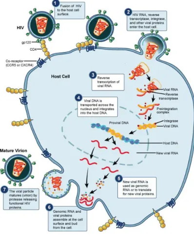
HIV Life cycle
Generally, the viral life cycle of HIV starts when the SU glycoprotein binds to the main receptor, the CD4, present in the cellular surface of the host cell (T-lymphocytes, monocytes, macrophages and dendritic cells). This interaction induces conformational changes in the SU, whereby the site for binding to a second receptor (co-receptor) becomes exposed. In vivo, the major co-receptors of HIV are the CCR5 and CXCR4 chemokine receptors. Both CD4 and co-receptor binding leads to conformational changes in TM glycoprotein that result in the insertion of the fusion peptide of TM into the host cellular membrane and, consequently, on the fusion of the viral envelope with the host cell. Thereafter, the viral capsid is release into the cytoplasm (reviewed in [64]) (Figure 3).
After HIV uncoating, the RT enzyme starts the reverse transcription of viral RNA. In the first stage, a single DNA strand is synthesised using one of the two RNA molecules as a template and the tRNAlys molecule as a primer. Once the first complementary DNA strand (negative strand) is transcribed, the ARNase H subunit of RT enzyme (p51) degrades the
RNA template. A new positive DNA strand, complementary to the negative one, is then synthesized by the p61 subunit of RT. The double stranded DNA, together with the MA, NC, IN, RT and Vpr proteins (plus the Vpx in HIV-2), make the pre-integration complex (PIC), which is transported to the nucleus using the cytoplasmatic microtubules network. This process is mediated by the IN and Vpr (and Vpx in HIV-2). Still outside the nucleus, the IN enzyme digests the 3’ LTR of both DNA strands creating two recessive ends. The IN will later use these ends to unite (integrate) the viral DNA into an open region of the host chromosomal genome, thus generating a provirus. Proviral DNA can either remain latent (silent) in the host cell or be transcribed by the cellular machinery, progressing with the viral life cycle [64].
Figure 3. The life cycle of HIV. (Adapted from http://www.niaid.nih.gov/topics/HIVAIDS/)
The promoter region within the 5’ LTR mediates the transcription of the proviral DNA. Three classes of RNA are obtained: (1) completely spliced mensager RNA (mRNA) translating for Rev, Tat and Nef (early transcripts); (2) incomplete spliced mRNA encoding for Env, Vif, Vpr and Vpu/Vpx (late transcripts); (3) unspliced and complete mRNA molecules that translate for polyprotein precursors Pr55Gag and Pr160GagPol (late transcripts)
and will be incorporated in the nascent viral particles as genomic RNA. Indeed, proteins from early transcripts (Tat and Rev) are required to complete the expression of the later
transcripts. Binding of Tat protein to the transactivation response region (TAR), a secondary structure downstream the LTR of the nascent RNA, is important for stable and efficient elongation of mRNA. The transport of unspliced and incompletely spliced mRNA outside the nucleus is dependent on Rev, which binds to the Rev responsive element (RRE) in the RNA env region before carrying them to the cytoplasm to be translated [64].
Once the Env precursor poliproteins are translated, they are glycosylated in the Golgi apparatus before they oligomerize in trimers. The polyproteins are, then, cleaved into the SU and TM glycoproteins and transported to the cytoplasmatic membrane, where the assembly of the viral particles takes place. These particles include the genomic RNA and the polyprotein precursors Pr55Gag and Pr160GagPol. They bud from the cell by gemulation of
the cytoplasmatic membrane, thus acquiring the lipid envelope already containing the TM/SU trimers (and some cellular membrane proteins). Finally, the Pr55Gag and Pr160GagPol
polyproteins are processed into the functional proteins by the PR enzyme [64]. This final maturation of the viral particle (virion) occurs outside the host cell (Figure 4).
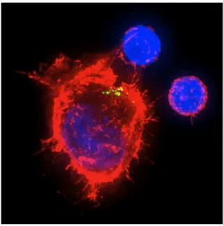
Figure 4. Maturation of the virus particle. False-colored image of two HIV virus particles budding from a
human T cell: (left) the CA protein is still associated with the viral membrane in the immature particle, whereas (right) the mature particle has a condensed core inside the virus shell. (Image by Klaus Boller, Paul-Ehrlich-Institute, Germany; http://www.cell.com/Cell_Picture_Show-hiv)
Several host restriction factors can hinder the retroviral replicative cycle [65]. Among these factors are APOBEC3G, TRIM5- and tetherin proteins. APOBEC3G, a member of the family of cytidine deaminases that is packaged within viral particles, induces G-to-A hipermutation and degradation of the nascent proviral DNA [65,66]. However, the viral protein Vif impairs the activity of this enzyme [65,66,67]. TRIM5- is a member of the tripartite motif protein family [65,68]. TRIM5- interacts with the viral capsid and blocks
uncoating, but its activity is highly dependent on species-specific compatibility [66,68,69]. It has been reported that, when compared to HIV-1, HIV-2 is more susceptible to TRIM5-
but more resistant to APOBEC3G [70]. Tetherin is a recently identified host restriction factor that inhibits the release of new viral particles [71]. Vpu and Env proteins can neutralize tetherin’s activity in HIV-1 and HIV-2, respectively [71,72].
HIV Transmission
The most common routes of HIV transmission include sexual contacts, contaminated blood or blood products (medical injections, blood transfusions, injection drug usage) and mother-to-child transmission (before, during and after birth or through breast feeding) [69]. Still, heterosexual transmission is the most frequent route of HIV-1 infection worldwide [17,66]. Several human- and HIV-specific determinants are required for efficient viral transmission.
There is evidence that HIV-1 transmission is directly correlated with the level of virus in circulation [73,74]. Moreover, the concentration of HIV-1 in blood and genital secretions varies depending on the stage of disease [69,75]. Indeed, increasing rates of HIV-1 transmission occur during the very early (acute) and later stages of infection (advanced disease), the periods when intense viral replication is observed and the highest levels of viral load are detected [66,69,75]. Notably, up to 50% of new HIV-1 infections are acquired from recently infected patients [76].
The risk of HIV infection is also influenced by the presence of other sexually transmitted diseases, such as syphilis and herpes simplex virus-2. The erosion of skin or mucosa resulting from genital inflammation and ulceration can enhance HIV-1 sexual transmission [66,69,77], or even increase the concentration of HIV-1 in the genital tract of the infecting partner [69]. On the other hand, male circumcision offers a degree of protection against HIV-1 acquisition, probably because removing the penile foreskin eliminates an easily breached entry portal containing many cellular targets of HIV [66,69,76]. Successful antiretroviral treatment also has the potential to prevent HIV transmission, by reducing the levels of HIV in blood and genital secretions [78,79]. In addition, research for new HIV prevention strategies led to the development of microbicides as topical agents to be applied on the vagina or rectum in order to protect from sexually transmitted infections (STIs) [80,81]. The impact of this approach in the prevention of HIV transmission has been highlighted by the results from the recent CAPRISA trial, which reported the use of tenofovir (an antiretroviral agent) in a vaginal gel formulation as a safe and effective method that can reduce HIV acquisition by 54% [81]. Nevertheless, on a global perspective, better access to healthcare services and behaviour changes (like adoption of safer sex
practices), are key strategies to reduce the risk of HIV infection and have a significant impact in the shape of the current epidemics [17].
Despite using similar routes of transmission, the prevalence rates of HIV-2 are much lower than HIV-1 [26,55,62]. As in HIV-1, heterosexual transmission is the most common route of HIV-2 infection [27,82], but at significantly lower rates [83]. Mother-to-child transmission is a rare event in HIV-2 with rates below 5% when compared to almost 25% in HIV-1, in the same untreated population [26,84]. The reduced transmissibility of HIV-2 is probably linked to the markedly lower plasma viremia [25,84] and reduced viral shedding in the genital tract [62,79].
A number of genotypic and phenotypic evidence support the active selection of specific variants during HIV-1 transmission [76]. Newly infected individuals acquire only a limited number of variants (1-10) circulating in the source donor (bottleneck effect), the majority of which are only able to use the CCR5 coreceptor [85]. This is observed either in sexual or percutaneous routes of infection. Although the mechanisms underlying these observations are not totally clear, it seems that the availability, infectability and spatial distribution of early target cells might severely limit the variability of the initial viral population (reviewed in [76]). This should be particularly true in mucosal transmission where a small, focal infected founder population of cells expands locally before posterior dissemination and systemic infection [86]. HIV then evolves away (diverge) from the founder virus as soon as anti-HIV humoral and cellular immune responses arise after exposure (usually takes several weeks) [66].
HIV Pathogenesis
The course of HIV infection can be divided into four stages: the acute phase (primary infection), the chronic asymptomatic phase, the early symptomatic phase and AIDS [87]. The acute phase is characterized by intense viral replication and massive loss of CD4+ T
cells that takes place mainly in mucosal tissues, particularly in the gut [88]. At the early stages of infection, HIV transmission across the mucosal epithelial layers is enhanced by dendritic cells (DC) present at the lamina propria. This is where productive viral replication initially occurs mostly in memory CD4+ T cells. DCs also seem to contribute for HIV dissemination to draining lymph nodes and secondary lymphoid tissue throughout the organism (e.g. the gut-associated lymphoid tissue), where high levels of activated CD4+ T
cells are present (reviewed in [66,89,90]). CD4+ T cell depletion is a combination of direct
viral infection, activation-induced cell death and host-derived cytotoxic responses [91]. The integrin 47 mediates the migration of T cells to the gut-associated lymphoid tissue
CD4+ T cells, in which it appears in a complex with CD4 [92,93]. Notably, this integrin is
also an HIV-1 receptor [93]. Binding of HIV-1 gp120 to integrin 47 seems to facilitate
cell-to-cell spread of HIV-1 and may enhance viral propagation following mucosal transmission [92,93].
The majority of HIV-infected individuals develop flu-like symptoms (acute HIV syndrome), approximately two to four weeks following the transmission of the virus. Seroconversion, with detection of specific anti-HIV antibodies, usually occurs within 3 to 12 weeks after exposure. Among the first antibodies detected are those directed against the viral capsid (p24) [87,94]. Plasma viremia (or viral load) typically peaks at three to four weeks after infection and then decreases to a steady state (viral set-point) [66], due to HIV-specific cytotoxic T lymphocyte (CTL) responses and humoral responses (neutralizing antibodies) [89]. The viral set-point marks the beginning of the chronic stage and is an important determinant on the rate of disease progression in untreated patients [66,95] (Figure 5).
Figure 5. The clinical and laboratorial course of untreated HIV-1 infection. (Adapted from Daskalakis D (2011) HIV Diagnostic Testing: Evolving Technology and Testing Strategies. Top Antivir Med. 2011;19(1):18-22)
The chronic phase is the asymptomatic stage of HIV infection that lasts on average between 8 to 10 years in HIV-1 (it can be much longer in HIV-2) [70,96]. It is a period of clinical latency (silent infection) characterized by low levels of viral replication in the lymphoid tissue (viral reservoir) and constant antigen stimulation of the host immune system (immune activation) [96]. Persistent immune activation is manifested by increased turnover of T cells, monocytes and natural killer (NK) cells, high levels of CD4+ and CD8+ T
cell apoptosis and polyclonal B cell activation which leads to generalized hipergammaglobulinemia (reviewed in [91]). It should be noted that in HIV-2 patients IgA
levels are not increased suggesting a selective B cell activation [97]. Chronic immune activation, which is a strong predictor of HIV disease progression, will eventually lead to the exhaustion of the immune system and occurrence or reactivation of opportunistic infections (e.g. candidiasis, pneumonia and tuberculosis) and development of neoplasic diseases (Epstein-Barr virus-related lymphomas, Kaposi’s sarcoma, etc) [66,87,96]. Clinical manifestations of these co-infections mark the onset of the early symptomatic phase [87]. In untreated patients, progression to AIDS occur by continuous loss of CD4+ T cells and
rising viremia, as a consequence of intensifying viral replication from viral reservoirs and latently infected CD4+ T cells [66,96]. Ultimately, the level of CD4+ T lymphocytes drops
below 200 cells/ml, defining the beginning of the AIDS stage [87,96].
Despite having similar proviral loads (n. of proviral DNA copies in PBMCs), at the same disease stage [70,98,99], HIV-1 and HIV-2 infections lead to very different immunological and clinical outcomes. Compared to HIV-1 infected patients, the majority of HIV-2 infected individuals have reduced general immune activation, normal CD4+ T cell counts, low or
absent plasma viremia and absence of clinical disease [55,70,99,100,101,102]. Indeed, HIV-2 infection is characterized by slow disease progression, long survival and reduced mortality rates [55,70,100,103,104,105,106]. These observations might be a consequence of the lower replication capacity of HIV-2 [107,108] and more effective immune response produced against HIV-2. In fact, most HIV-2 infected individuals have strong cytotoxic responses to Env and Gag proteins and raise autologous and heterologous neutralizing antibodies [55,109,110,111,112,113]. The lower state of immune activation in HIV-2 patients may be related with the immunosuppressive activity of the C2, V3, and C3 envelope regions of HIV-2 [19,20,21]. Nevertheless, with disease progression CD4+ T cell
depletion becomes similar in HIV-1 and HIV-2 infections [102,114], most of the immunological differences are lost and the mortality risk is equivalent [55,70,105,106].
HIV ENVELOPE
Molecular and structural organization of the viral envelope
Viral entry into host cells is mediated by the envelope SU and TM glycoproteins, which are encoded by the env gene. These glycoproteins are attached by a noncovalent association and are assembled as trimers [3x(SU/TM)], representing up to 14 functional spikes on the surface of the mature virion [115,116].
The SU glycoprotein is composed by five hypervariable regions, V1 to V5, separated by five more conserved regions, C1 to C5 (Figure 6). Hypervariable regions tend to form loops, stabilized by disulfide bridges. In its native trimeric conformation, SU has two domains,
one internal, hydrophobic in nature, and one external. After binding to the CD4 receptor, a major structural change occurs and a bridging sheet is formed between the V1/V2 stem and 20/21 in C4. While both the external domain and bridging sheet are involved in the interaction between the SU and the cellular receptors (CD4, CCR5 and/or CXCR4), the internal domain is essential for SU–TM association [64,115,117,118,119]. Also, interaction between SU and the integrin 47 gut-homing receptor is mediated by a conserved motif in
the V2 loop of the bridging sheet [93]. Numerous glycosylation sites as well as major antigenic determinants, including neutralizing epitopes, can be found on the external domain [64,115,118,120].
The TM glycoprotein consists of one extracellular ectodomain, one transmembrane region and one intracytoplasmatic domain (Figure 6). The fusion peptide, at the hydrophobic N-terminal end of the ectodomain, is followed by two -helices containing leucine zippers-like motifs: heptad repeats 1 and 2 (HR1 and HR2, respectively). Separating these heptad repeats, there is a small loop defined by cysteine residues (CC, cysteine bridge). HR1 and HR2 contain repeated patterns of seven residues and are arranged as trimers. The fusion peptide and both HR1 and HR2 play a significant role on the fusion of the viral envelope with the host cellular membrane. On the other hand, the intracytoplasmatic domain mediates the binding of the envelope to the MA protein, during the maturation of new viral particles [64,115,117,121].
Figure 6. Schematic representation of SU and TM envelope glycoproteins. The SU glycoprotein is composed
by five conserved (C1 to C5) and five variable (V1 to V5) domains. The TM glycoprotein contains the N-terminal fusion peptide (FP), two heptad repeats (HR1 and HR2), one transmembrane region (TM) and the intracytoplasmic domain. The figure is numbered according to the HIV-1 JR-FL isolate. (Adapted from Taveira N, Borrego P, Bártolo I (2008) Biologia molecular de VIH. In: Antunes F, editor. Manual sobre SIDA. 3th ed. Lisbon: Permanyer Portugal. pp. 27-50.)
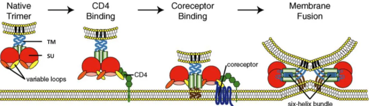
Mechanism of HIV entry
The process of HIV entry generally involves three sequential steps occurring on the surface of the target cell: (1) binding of the SU glycoprotein to the CD4 receptor, (2) binding of the SU to the CCR5 and/or CXCR4 coreceptor and, finally, (3) fusion of the viral envelope with the cellular membrane (Figure 7). The mechanisms underlying these stages will be described in the next sections, and they characterize viral spread driven by cell-free
virions. Alternatively, HIV can disseminate through cell-to-cell contact using either viral synapses or membrane nanotubes [122,123].
Figure 7. Model of the multi-step process of HIV entry. (Adapted from Tilton JC, Doms RW (2010) Entry inhibitors in the treatment of HIV-1 infection. Antiviral Res 85: 91-100.)
Interaction with the CD4 receptor
The CD4 receptor is a transmembrane protein with 58 kDa that exists on the surface of several cell lines, like T cells, monocytes, macrophages and DCs [64]. As mentioned above, it is often found in a complex with the integrin 47 in activated CCR5high/CD4+ T cells in
the gut compartment [92,93]. Four domains compose the extracellular region of CD4, D1 to D4. Attachment to the viral SU glycoprotein occurs at the CDR2 sub-region, one of the three sub-regions of D1 domain [64].
Electrostatic forces are responsible for the interaction between CD4 (positive charge) and the SU (negative charge), which is stabilized by Van der Walls forces and hydrogen bonds [117]. This interaction promotes conformational changes in the SU, leading, as previously stated, to the formation of the bridging sheet and increasing the exposure of V1, V2, V3 and C4. This results in the approximation of the viral envelope and the cellular membrane and the subsequent interaction of V3 with the coreceptor [64,115,121,124,125].
Interaction with the coreceptor
In vivo, the major coreceptors for HIV entry are the CCR5 and CXCR4 G-protein coupled
receptors that function as the natural receptors for and chemokines [64,118]. These receptors are integral membrane proteins with seven transmembrane helices, an extracellular N-terminus and three extracellular loops (ECLs) that form a small pocket [118]. CCR5 is predominantly expressed on the surface of memory T lymphocytes, activated T lymphocytes and macrophages, while CXCR4 is mainly found in T lymphocytes, monocytes, DCs and B lymphocytes [126].
Upon SU – CD4 binding, the viral V3 loop is projected into closed proximity to the cellular membrane where it can interact with the coreceptor [125]. Interaction with the viral SU involves two coreceptor regions. Initially, the N-terminal region binds to the SU core and the base of the V3 loop, and then the second extracellular loop (ECL2) binds to the V3 tip [125,127,128]. While both coreceptor regions are necessary for successful cell entry by variants using the CCR5 coreceptor, only ECL2 seems to be critical for CXCR4 usage [129,130,131].
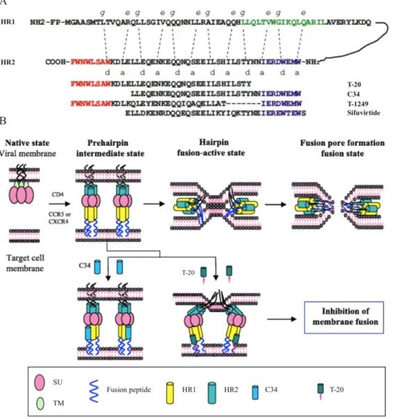
Fusion
Attachment of SU to CD4 and coreceptor promotes the approximation of the viral envelope and the cellular membrane and structural rearrangements of the TM glycoprotein. As a result, the fusion peptide becomes exposed and is inserted into the cytoplasmatic membrane, thus creating a prehairpin intermediate configuration of TM [132,133,134,135,136]. Notably, this intermediate state can be initiated by CD4 binding alone, but binding of a coreceptor enhances the process [137]. Then, the HR2 trimer folds back on an anti-parallel fashion towards the HR1 trimer, forming a six-helix bundle structure (6HB; final hairpin state) stabilised by the hydrophobic interactions between the HR1 domains in the center (central coiled-coil) and the HR2 domains outside. During this process, the viral envelope and the cellular membrane are brought together, leading to the formation of the fusion pore, through which the viral capsid enters the target cell [132,133,134,135,136] (Figure 8).
An alternative model of cell free HIV-1 cell entry is via the endocytic pathway [138]. Time-resolved imaging of single viruses and differential blocking of fusion by site-specific and universal inhibitors revealed that fusion with the cytoplasmatic membrane at the cell surface did not progress beyond the lipid mixing step [139]. Instead, HIV-1 was internalised upon CD4 and coreceptor interaction and complete fusion occurred only in endossomal compartments, leading to productive infection. Nonetheless, further studies are still needed to confirm the incidence and biological relevance of this pathway in HIV infection.
Figure 8. Model of the envelope glycoprotein-mediated membrane fusion. (Adapted from Weiss CD (2003) HIV-1 gp41: mediator of fusion and target for inhibition. AIDS Rev. 2003 Oct-Dec;5(4):214-21.)