To My Grandmother
and my Father
Acknowledgments
I want to address my first words of gratitude to my supervisors, who turned this challenge into a wonderful journey and inspired me as a researcher but most of all, as a person. First and foremost, I would like thank Professor Fernanda Borges for her unrivalled guidance, enthusiasm, kindness, and most importantly for the unconditional friendship. I also would like to thanks Professor Eugenio Uriarte for his support, encouragement and friendliness.
I would also like to thanks the FCT for its financial support, to Faculty of Sciences of the University of Porto and CIQ-(―Centro de Investigação em Química‖), for providing me the means for the execution of the experimental work.
I want to express my gratitude to all the research groups that collaborated in the execution of the work described in this thesis, namely the Department of Pharmacology of Faculty of the Pharmacy, University of Santiago de Compostela, Spain, the Institute of Pharmacology and Toxicology, University of Wuzburg, Germany , Department of Pharmacology of the Faculty of Pharmacy, University ―Magna Græcia‖, Catanzaro, Italy and the Molecular Modeling Section (MMS) of the Pharmaceutical Science Department, university of Padua, Italy.
To Professor Nuno Milhazes, my profound thanks for the invaluable brain storming sessions, encouragement and his one of a kind humour, but above all for his precious friendship.
Thank you to Professor Jorge Garrido and Professor Manuela Garrido, for their support, companionship and for believing in me.
To all of my colleagues in the lab, whose names I will not discriminate to avoid of forgetting someone, a genuine thank you for all the good times shared in the lab.
I would also like to thank all my friends outside of the research group with whom I will forever share great memories and, undoubtedly, create new ones in the future.
Abstract
The last few decades have provided stunning progresses in the understanding of physiopathology of several diseases. The remarkable progress in research fields, like genetics, immunology, neurobiology, among others, as well as the advent of more powerful tools, have made it possible to characterize, monitor, and understand far more of the basis of physiology and disease. However, for some diseases the treatment remains a problematic issue as the efforts performed so far were not translated into therapeutic solutions. Notwithstanding the steady increase in the total amount of money spent on pharmaceutical research and development over the past decade, the number of new drug approvals has declined in recent years. Despite the advances in technology, drug discovery is still a lengthy, expensive, difficult, and inefficient process, with a low rate of success. Therefore, the search for new chemical entities is still an unmet need for the drug discovery process.
Application of new drug discovery approaches/concepts for multifactorial diseases, such as cancer and neurodegenerative diseases, have prompted a switch on the strategy of one-molecule one-target to a new one, the multi-target approach, where a single chemical entity may be able to modulate multiple targets simultaneously.
In the present project, the main challenges were the validation of chromones as a privileged structure for the design of new drug candidates for Parkinson disease and the development dual-target lead compounds. Accordingly, the design of a chromone library to attain structure-activity relationships and the development of concise and diversity-oriented synthetic strategies has been carried out along this project. The small but innovative chromone libraries were screened to ascertain their potential as MAO-B inhibitors as well as A2A adenosine receptor ligands. The output of the biological screening assays gave rise to preliminary structure-activity relationship regarding both of the targets. The overall data showed that chromones are privileged structures for drug discovery and development processes in the field of Parkinson disease and that chromone-3-(3‘-hydroxy-4‘-methoxyphenyl)carboxamide and chromone-3-(4‘-chlorophenyl)carboxamide, exhibiting a IC50 for hMAO- B in a nanomolar range and affinity towards A2A ARs, can be regarded as putative leads for further optimisation. In addition, during the drug discovery process interesting A3 AR ligands based on chromone scaffold have been found, namely chromone-2-carboxamides such as the chromone-2-(4‘-methoxyphenyl)carboxamide which presented a Ki value for hA3 AR of 9,580 nM and reasonable to good selective indexes regarding the other ARs ligands.
Complementarily, the work described along this thesis was sustained on molecular docking studies performed over MAOs isoforms and ARs, for the most active chromone derivatives.
Resumo
As últimas décadas têm sido pautadas por um impressionante progresso em áreas como a genética, imunologia, neurobiologia, entre outras. O crescente conhecimento científico, bem como o desenvolvimento de novas e mais potentes tecnologias, tornaram possível não só a caracterização e monitorização dos processos fisiológicos mas também uma maior compreensão dos processos patológicos de inúmeras doenças. No entanto, para algumas enfermidades os esforços até agora realizados não conseguiram ser traduzidos em soluções terapêuticas.
O processo de descoberta e desenvolvimento de novos fármacos é dispendioso, demorado e com uma baixa taxa de sucesso De facto, e apesar do crescente investimento económico por parte da indústria farmacêutica, o número de novos fármacos tem diminuído ao longo das últimas décadas. Neste contexto, a descoberta de novas entidades químicas (NCEs), candidatas a novos fármacos, continua a ser um tópico da maior relevância na química medicinal.
No caso das doenças de natureza multifatorial, como cancro e eventos neurodegenerativos, o processo de descoberta e desenvolvimento de novos fármacos é ainda um desafio. Numa tentativa de acelerar o processo assiste-se actualmente a uma modificação da estratégia do desenho racional de fármacos com passagem do paradigma ―uma molécula-um alvo terapêutico‖ para uma abordagem multi-alvo, onde uma única entidade química tem ser capaz de modular, simultaneamente, mais do que um alvo.
O presente projeto consistiu na validação da cromona como uma estrutura privilegiada para a descoberta de novos candidatos a fármacos para a doença de Parkinson e no desenvolvimento de compostos líder com um mecanismo de acção em dois alvos terapêuticos. Neste contexto procedeu-se à obtenção e desenvolvimento de estratégias sintéticas para a construção de pequenas e inovadoras bibliotecas baseadas no núcleo da cromona. Adicionalmente, foram efetuados estudos para avaliação do seu potencial como inibidores da MAO-B e ligandos do recetor A2A da adenosina. Os resultados obtidos foram interpretados através do estabelecimento de relações de estrutura-atividade e em estudos de modelização molecular, envolvendo as isoformas da MAO e os recetores da adenosina. os quais foram realizados para os derivados de cromonas mais potentes.
Os dados obtidos até à data permitiram concluir que a cromona pode ser uma estrutura privilegiada para a descoberta e desenvolvimento de novos fármacos para doença de Parkinson. Do estudo efectuado salienta-se os resultados obtidos para dois derivados da cromona sintetizados, nomeadamente a cromona-3-(3'-hidroxi-4'-
metoxifenil)carboxamida e a cromona-3-(4'-clorofenil)carboxamida, os quais exibiram um CI50 para hMAO-B na ordem dos nanomolar e afinidade relevante para o recetor A2A da adenosina. Estes compostos, após uma etapa subsequente de otimização, podem produzir candidatos inovadores, com um mecanismo de ação duplo-alvo, e uma nova solução terapêutica para a doença de Parkinson. Salienta-se ainda os resultados obtidos para uma biblioteca de cromonas (cromona-2-carboxamidas), as quais evidenciaram, de uma foram geral, uma afinidade relevante para os recetores A3 da adenosina, nomedamente a cromona-2-(4'-metoxifenil)carboxamida a qual apresentou um valor de Ki = 9,580 nM para o referido recetor. Os dados obtidos, assim como a seletividade apresentada, leva a considerar este tipo de derivados de cromonas compostos passíveis de ser otimizados para o desenvolvimento de novos anticancerígenos.
Abbreviations
Ach – Acetylcholine AChE – Acethylcolinesterase Amino acids: Asn – Asparagine Cys – Cysteine Gln – Glutamine Glu – Glutamic acid His – Histidine Ile – Isoleucine Leu – Leucine Phe – Phenylalanine Ser – Serine Thr – Threonine Trp – tryotophan Tyr – Tyrosine Val – Valine ARs – Adenosine receptorsBOP – O-Benzotriazol-1-yloxytris(dimethylamino)phosphonium hexafluorophosphate CNS – Central nervous system
COMT – Catechol-O-methyltransferase COSY – Correlation spectroscopy DA – Dopamine
DBU – Diazabicyclo[5.4.0]undec-7-ene DOPAC – Dihydroxyphenylacetic acid EI/MS – Electron Impact Mass Spectrometry EL – Extra loop
GABA – Gamma aminobutyric acid GP – Globus pallidus,
GPCRs – G protein coupled receptors GPe – Globus pallidus pars externa GPi – Globus pallidus pars interna
hA1 AR – Human adenosine subtype A1 receptor
hA2A AR – Human adenosine subtype A2A receptor
hA3 AR – Human adenosine subtype A3 receptor
hMAO-A – Human monoamine oxidase type A isoform hMAO-B – Human monoamine oxidase type B isoform
HMBC – Heteronuclear multiple bond correlation HMPA – Hexamethylphosphoramide
HMQC – Heteronuclear multiple quantum coherence HTS – High-throughput screening
HVA – Homovanillic acid IL – Intra loop
L-AAD – L-Aromatic amino acid decarboxylase or dopa decarboxylase
L-dopa– L-Dihydroxyphenylalanine
MAO-A – Monoamine oxidase type A isoform MAO-B – Monoamine oxidase type B isoform MPP+ – 1-Methyl-4-phenylpyridinium ion
MPTP – 1-Methyl-4-phenyl-1,2,3,6-tetrahydropyridine NCEs – New chemical entities
NMR – Nuclear magnetic resonance PD – Parkinson‘s disease
PyBOP – O-Benzotriazol-1-yloxytris(pyrrolidino)phosphonium hexafluorophosphate PyBrOP – Bromotripyrrolidinophosphonium hexafluorophosphate
SARs – Structure–activity relationships SN – Substantia nigra
SNr – Substantia nigra pars reticulata STN – Subthalamic nucleus TH – Tyrosine hydroxylase
Metric units
µg – Microgram µL – Microliter µM – Micromolar g – Gram K – Degrees Kelvin L – Litre M – Molar mg – Milligram min – Minute mL – MililitermM – Milimolar ng – Nanogram nL – Nanoliter nM – Nanomolar ºC – Degrees Celsius ppm – Parts per million
Symbols
δ – Chemical shift in ppm
d – Duplet
dd – Double duplet
ddd – Double double duplet J – Coupling constant m – Multiplet
m/z – mass charge ratio
s – Singulet t – Triplet
Index
ACKNOWLEDGMENTS ... II
ABSTRACT ... III
RESUMO ... V
ABBREVIATIONS ... VII
METRIC UNITS ... VIII
SYMBOLS ... IX
INDEX ... X
FIGURE INDEX ... XIII
SCHEME INDEX ... XV
TABLE INDEX... XVI
... 17
INTRODUCTION ... 17
1.1 INTRODUCTION ... 18
1.2 SCOPE OF THE THESIS ... 20
... 21
LITERATURE REVIEW ... 21
2.1 PARKINSON‘S DISEASE ... 22
2.2 DOPAMINE BIOSYNTHESIS AND NIGROSTRIATAL PATHWAYS ... 23
2.3 PARKINSON'S DISEASE THERAPY THE CURRENT STATUS ... 26
2.3.1 Dopaminergic treatments for Parkinson´s disease ... 26
2.3.1.1 L-Dopa... 26
2.3.1.2 Dopamine agonists ... 27
2.3.1.3 Catechol-O-methyltransferase inhibitors ... 28
2.3.1.4 Monoamine oxidase-B inhibitors ... 31
2.3.2 Non-dopaminergic treatments for Parkinson´s disease ... 33
2.3.2.1 Anticholinergic drugs ... 33
2.3.2.2 Glutamate antagonists ... 34
2.4 LOOKING FOR NEW TARGETS: ADENOSINE A2A RECEPTORS AND PARKINSON´S DISEASE 35 2.4.1 G Protein-coupled receptors and adenosine receptors: a general outlook . 35 2.4.2 Adenosine, adenosine receptors and neurodegenerative diseases ... 37
2.4.2.1 A2A AR antagonists... 38
2.5 CHROMONE AS A PRIVILEGED SCAFFOLD ... 39
2.5.2 Simple chromones ... 40
2.5.3 Chromones in therapy ... 41
2.5.4 Biological activity of simple chromones and derivatives ... 41
2.5.4.1 Anti-inflammatory activity ... 42
2.5.4.2 Antimicrobial activity ... 44
2.5.4.3 Anticancer activity ... 45
2.5.4.4 Drugs for neurodegenerative diseases ... 47
2.6 METHODS OF SYNTHESIS OF SIMPLE CHROMONES ... 48
2.6.1 Chromones from 2-hydroxyarylalkyl ketones ... 48
2.6.2 Synthesis of chromones from phenols ... 52
2.6.3 Synthesis of chromones from salicylic acids and derivatives ... 53
2.6.4 Synthesis of chromones via C-C cross coupling reactions ... 54
2.6.5 Synthesis of chromones from chromanones ... 56
2.6.6 Synthesis of chromones from other chromones ... 56
... 60
RESULTS ... 60
3.1 CHROMONE-2- AND -3-CARBOXYLIC ACIDS INHIBIT DIFFERENTLY MONOAMINE OXIDASES A AND B 61 3.2 CHROMONE, A PRIVILEGED SCAFFOLD FOR THE DEVELOPMENT OF MONOAMINE OXIDASE INHIBITORS. ... 66
3.3 TOWARDS THE DISCOVERY OF A NOVEL CLASS OF MONOAMINE OXIDASE INHIBITORS: STRUCTURE–PROPERTY–ACTIVITY AND DOCKING STUDIES ON CHROMONE AMIDES. ... 76
3.4 CHROMONE 3-PHENYLCARBOXAMIDES AS POTENT AND SELECTIVE MAO-B INHIBITORS 82 3.5 IN SEARCH FOR NEW CHEMICAL ENTITIES AS ADENOSINE RECEPTOR LIGANDS: DEVELOPMENT OF AGENTS BASED ON BENZO-Γ-PYRONE SKELETON. ... 86
3.6 DISCOVERY OF NOVEL A3 ADENOSINE RECEPTOR LIGANDS BASED ON CHROMONE SCAFFOLD 92 3.7 COMBINING QSAR CLASSIFICATION MODELS FOR PREDICTIVE MODELING OF HUMAN MONOAMINE OXIDASE INHIBITORS. ... 102
3.8 SYNTHESIS AND NMR STUDIES OF NOVEL CHROMONE-2-CARBOXAMIDE DERIVATIVES. 119 ... 124
INTEGRATED OVERVIEW OF THE PERFORMED STUDIES ... 124
4.1 DESIGN, SYNTHESIS AND STRUCTURAL CHARACTERISATION OF THE DIVERSE FUNCTIONALIZED CHROMONE LIBRARIES ... 125
4.1.1 Synthesis of ester, carboxylic acid and formylchromone derivatives ... 129
4.1.3 Synthesis of amide chromone derivatives ... 131
4.1.4 Structural characterization of the chromone derivatives ... 133
4.2 CHROMONE A VALID SCAFFOLD FOR THE DEVELOPMENT OF MAO-B INHIBITORS ... 133
4.3 CHROMONE A VALID SCAFFOLD FOR THE DEVELOPMENT OF ADENOSINE RECEPTORS LIGANDS 138 4.4 CHROMONE-2-CARBOXAMIDE A VALID SCAFFOLD FOR THE DEVELOPMENT OF A3AR LIGANDS 143 ... 151
CONCLUDING REMARKS AND FUTURE PERSPECTIVES ... 151
5.1 CONCLUDING REMARKS AND FUTURE PERSPECTIVES ... 152
Figure Index
FIGURE 2.1 SCHEMATIC REPRESENTATION OF THE BASAL GANGLIA ANATOMY ... 22
FIGURE 2.2 SCHEMATIC REPRESENTATION OF DOPAMINE METABOLISM. L-AAD: L-AROMATIC AMINO ACID DECARBOXYLASE AD: ALDEHYDE DEHYDROGENASE, COMT: CATECHOL-O-METHYLTRANSFERASE, DOPAC: DIHYDROXYPHENYLACETIC ACID, DOPAL:3,4-DIHYDROXYPHENYLACETALDEHYDE, HVA: HOMOVANILLIC ACID, MAO-B: MONOAMINE OXIDASE-B, TH: TYROSINE HYDROXYLASE. ... 24
FIGURE 2.3 SCHEMATIC REPRESENTATION OF BASAL GANGLIA DIRECT AND INDIRECT PATHWAYS. GPE: GLOBUS PALLIDUS PARS EXTERNA, GPI: GLOBUS PALLIDUS PARS INTERNA, SNC: SUBSTANTIA NIGRA PARS COMPACTA, SNR: SUBSTANTIA NIGRA PARS RETICULATA STN: SUBTHALAMIC NUCLEUS. ... 25
FIGURE 2.4 CHEMICAL STRUCTURE OF L-DOPA. ... 26
FIGURE 2.5 CHEMICAL STRUCTURES OF L-AAD INHIBITORS. ... 27
FIGURE 2.6 CHEMICAL STRUCTURES OF ERGOT AND NON-ERGOT DOPAMINE AGONISTS. ... 28
FIGURE 2.7 PERIPHERAL BIOTRANSFORMATION OF L-DOPA BY ACTION OF COMT. COMT: CATECHOL-O -METHYLTRANSFERASE AND 3-OMD:3-O-METHYLDOPA. ... 29
FIGURE 2.8 CHEMICAL STRUCTURES OF COMT INHIBITORS. ... 30
FIGURE 2.9 MAOS CATALYSED REACTIONS ... 31
FIGURE 2.10 CHEMICAL STRUCTURES OF MAO-B INHIBITORS IN CLINICAL PRACTICE ... 32
FIGURE 2.11 CHEMICAL STRUCTURES OF MULTITARGET DRUGS IN CLINICAL TRIALS. ... 33
FIGURE 2.12 CHEMICAL STRUCTURES OF ANTICHOLINERGIC DRUGS ... 34
FIGURE 2.13 CHEMICAL STRUCTURE OF AMANTADINE. ... 34
FIGURE 2.14 SCHEMATIC REPRESENTATION OF G PROTEIN-COUPLED ADENOSINE RECEPTORS. ATP: ADENOSINE-TRIPHOSPHATE; CA2+: ION CALCIUM (II); CAMP: CYCLIC ADENOSINE MONOPHOSPHATE; GI/GO: INHIBITORY G PROTEIN; GS/GQ STIMULATORY G PROTEIN; K: POTASSIUM; P: PHOSPHORUS; PLC: PHOSPHOLIPASE C. ... 36
FIGURE 2.15 CO-LOCALIZATION OF A2A AR AND D2 RECEPTORS ... 38
FIGURE 2.16 CHEMICAL STRUCTURES OF ISTRADEFYLLINE AND PRELADENANT. ... 38
FIGURE 2.17 CHROMONE AND (ISO)FLAVONOIDS BACKBONE. ... 40
FIGURE 2.18 EXAMPLES OF CHROMONE DERIVATIVES IN THERAPY ... 41
FIGURE 2.19 CHEMICAL STRUCTURES OF (1):7-METHANESULFONYLAMINO-6-PHENOXYCHROMONE AND (2): T-614. ... 42
FIGURE 2.20 CHEMICAL STRUCTURES OF STELLATIN AND ITS DERIVATIVES. ... 43
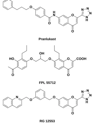
FIGURE 2.21 CHEMICAL STRUCTURES OF PRANLUKAST, FPL 55712 AND RG 12553... 43
FIGURE 2.22 LT RECEPTOR ANTAGONISTS BASED ON (PIPERIDINYLALKOXY)CHROMONE SCAFFOLD. .... 44
FIGURE 2.23 CHEMICAL STRUCTURES OF A: SULFONAMIDE CHROMONE DERIVATIVE AND B: DITHIAZOLE CHROMONE DERIVATIVE. ... 45
FIGURE 2.24 CHROMONE DERIVATIVES WITH ANTIVIRAL ACTIVITY. A)2-STYRYLCHROMONES; B) 5-HYDROXY CHROMONE SCAFFOLD. ... 45
FIGURE 2.262-STYRYLCHROMONES WITH ANTIPROLIFERATIVE ACTIVITY ON A PANEL OF CARCINOMA CELLS. ... 46
FIGURE 2.27 CHEMICAL CORE OF CHROMONE DERIVATIVES TESTED AS A: ACHE AND MAO INHIBITORS AND B: DOPAMINE D2 AGONISTS. ... 48 FIGURE 4.1. PHOSPHONIUM COUPLING AGENTS... 132
FIGURE 4.2. VIRTUAL DOCKING OF CHROMONE-3-CARBOXYLIC ACID INTO THE HMAO-B CATALYTIC SITE
(FIGURE EXTRACTED FROM ARTICLE IN SECTION 3.1). ... 135
FIGURE 4.3. CHEMICAL STRUCTURES OF FIVE OF THE BEST CHROMONE-3 CARBOXAMIDES AND THEIR INHIBITORY ACTIVITY TOWARDS HMAO-B. ... 136
FIGURE 4.4 CHEMICAL STRUCTURES OF N-PHENYL (COMPOUND 20), N-CYCLOHEXYL (COMPOUND 91)
AND N-PROPYL (COMPOUND 105) CHROMONE-3-CARBOXAMIDES AND THEIR INHIBITORY ACTIVITY TOWARDS HMAO-B. ... 137
FIGURE 4.5 VIRTUAL DOCKING OF N-(4-(CHLOROPHENYL)-CHROMONE-3-CARBOXAMIDE INTO THE HMAO
-B CATALYTIC SITE (FIGURE EXTRACTED FROM ARTICLE IN SECTION 3.2). ... 137
FIGURE 4.6. CHEMICAL STRUCTURES OF THREE OF THE BEST CHROMONE-3 CARBOXAMIDES AS A2A AR
LIGANDS. ... 140 FIGURE 4.7. HYPOTHETICAL BINDING MODES OF ONE OF OUR MOST ACTIVE COMPOUND OBTAINED AFTER DOCKING SIMULATIONS:(A) INSIDE THE HA2A AR BINDING SITE;(B) INSIDE THE HA3 AR BINDING SITE.
(FIGURE EXTRACTED FROM ARTICLE IN SECTION 3.6) ... 141
FIGURE 4.8. CHEMICAL STRUCTURE OF ZM241385... 141
FIGURE 4.9. CHEMICAL STRUCTURES OF MONO SUBSTITUTED CHROMONE-2-CARBOXAMIDES, THEIR CORRESPONDING HA3 KI AND SELECTIVE INDEX (SI). ... 144 FIGURE 4.10. CHEMICAL STRUCTURES OF DI-EXOCYCLIC SUBSTITUTED CHROMONE-2-CARBOXAMIDES,
THEIR CORRESPONDING HA3 KI AND SELECTIVE INDEX (SI). ... 146 FIGURE 5.1. CHEMICAL STRUCTURES OF TWO OF THE BEST CHROMONE-3-CARBOXAMIDES AS
scheme index
SCHEME 2.1 SYNTHESIS OF CHROMONES VIA BAKER-VENKATAMARAN REARRANGEMENT. ... 49
SCHEME 2.2 SYNTHESIS OF CHROMONES VIA CLAISEN CONDENSATION. ... 49
SCHEME 2.3 PROPOSAL MECHANISM OF KOSTANECKI- ROBINSON REACTION ADAPTED FROM [248]. ... 51
SCHEME 2.4 SYNTHESIS OF CHROMONES VIA ... 51
SCHEME 2.5 SYNTHESIS OF CHROMONES VIA BENZOPYRYLIUM SALT INTERMEDIATES. ... 52
SCHEME 2.6 SYNTHESIS OF CHROMONES VIA SIMONIS AND RUHEMANN REACTIONS. ... 53
SCHEME 2.7 SYNTHESIS OF CHROMONES FROM SALICYLIC ACIDS AND DERIVATIVES. ... 54
SCHEME 2.8 SYNTHESIS OF CHROMONES VIA C-C CROSS COUPLING REACTIONS. ... 54
SCHEME 2.9 SYNTHESIS OF CHROMONES CATALYSED BY NHCS. ... 56
SCHEME 2.10 SYNTHESIS OF CHROMONES FROM HYDROXYL OR THIOCHROMONES. ... 57
SCHEME 2.11 SYNTHESIS OF CHROMONES FROM CHROMONE CARBOXYLIC ACIDS. ... 57
SCHEME 2.12 SYNTHESIS OF CHROMONES FROM 3-FORMYLCHROMONES. ... 58
SCHEME 2.13 SYNTHESIS OF HETEROCYCLIC AND VINYL SUBSTITUTED CHROMONES. ... 58
SCHEME 2.14 SYNTHESIS OF STYRYLCHROMONES FROM OTHER CHROMONES. ... 59
SCHEME 4.1 SYNTHESIS OF ESTER CHROMONE DERIVATIVES BY FISHER ESTERIFICATION. ... 129
SCHEME 4.2 SYNTHESIS OF ESTER CHROMONE DERIVATIVES BY CLAISEN CONDENSATION. ... 130
SCHEME 4.3. SYNTHESIS OF 6-CHLORO-3-CHROMONE CARBOXYLIC ACID. ... 130
SCHEME 4.4. SYNTHESIS OF HYDROXYMETHYLCHROMONE DERIVATIVES. ... 131
table index
TABLE 4.1. CHROMONE LIBRARY I: ESTER, CARBOXYLIC ACID, HYDROXYMETHYL AND FORMYLCHROMONE DERIVATIVES. ... 125
TABLE 4.2 CHROMONE LIBRARY II: PHENYLCARBOXAMIDE CHROMONE DERIVATIVES. ... 126
TABLE 4.3: CHROMONE LIBRARY III: ALKYL, PHENYL AND HETEROCYCLIC CHROMONE CARBOXAMIDE DERIVATIVES. ... 128
TABLE 4.4. RADIOLIGANDS BINDING ASSAYS CONDITIONS. ... 139
TABLE 4.5 AFFINITY (KI, NM) OF MONOSUBSTITUTED CHROMONE-2-CARBOXAMIDES IN RADIOLIGAND BINDING ASSAYS AT HARS. ... 144
TABLE 4.5(CONT.) AFFINITY (KI, NM) OF MONOSUBSTITUTED CHROMONE-2-CARBOXAMIDES IN
RADIOLIGAND BINDING ASSAYS AT HARS. ... ERROR!BOOKMARK NOT DEFINED. TABLE 4.6. AFFINITY (KI, NM) OF DISUBSTITUTED CHROMONE-2-CARBOXAMIDES IN RADIOLIGAND BINDING ASSAYS AT HARS. ... 146
TABLE 4.7. AFFINITY (KI, NM) OF MONOSUBSTITUTED CHROMONE-2-CARBOXAMIDES WITH WITHDRAWING GROUPS IN RADIOLIGAND BINDING ASSAYS AT HARS. ... 147
TABLE 4.8. AFFINITY (KI, NM) OF MONOSUBSTITUTED CHROMONE-2-CARBOXAMIDES WITH DONATING GROUPS IN RADIOLIGAND BINDING ASSAYS AT HARS. ... 148
TABLE 4.9. AFFINITY (KI, NM) OF TERTIARY CHROMONE-2-CARBOXAMIDES IN RADIOLIGAND BINDING ASSAYS AT HARS. ... 148
TABLE 4.10. AFFINITY (KI, NM) OF HETEROCYCLIC CHROMONE-2-CARBOXAMIDES IN RADIOLIGAND BINDING ASSAYS AT HARS. ... 149
1.1 Introduction
The Committee of the International Union of Pure and Applied Chemistry (IUPAC) defines medicinal chemistry as ―a chemistry-based discipline, also involving
aspects of biological, medical and pharmaceutical sciences. It is concerned with the invention, discovery, design, identification and preparation of biologically active compounds, the interpretation of their mode of interaction at the molecular level, the construction of their structure-activity relationships, and the study of their metabolism" [1]. The interdisciplinarity between Chemistry and Biology, essential for medicinal chemistry, was brilliantly recognized by Arthur Kornberg, a Nobel laureate, in 1959 who wrote: ―We have the paradox of the two cultures, chemistry and biology,
growing further apart even as they discover more common ground.” [2]. Due to the harmonization of these apparently distinct two sciences, conditions have been created for the growth of medicinal chemistry and consequently for drug discovery and development processes.
Nowadays, medicinal chemistry is seen as a science situated at the interface of organic chemistry and life sciences, encompassing the knowledge of biochemistry, pharmacology, molecular biology, genetics, immunology, pharmacokinetics and toxicology on one side, and chemistry based disciplines, such as organic and physical chemistry, crystallography, spectroscopy and computer-based techniques, data analysis and data imaging on the other side [3].
Medicinal chemistry is a dynamic science that changes as the drug discovery paradigms shifts. Before 1990 the lead generation in the drug discovery processes was based on natural ligands and massive synthesis [4-7]. Usually, the therapeutic purpose is fixed in advance and a large number (several thousands) of molecules is tested on a limited number of experimental models. This method, called as random screening, has been used for the discovery of several drugs, particularly antibiotics. The common criticism of this type of methodology is that it constitutes, by the absence of a rational lead, a sort of fishing.
The steep increase in the knowledge about biological processes, the factors leading to their misregulation and ultimately to disease as well as the development of new technologies, such as high-throughput screening (HTS), microwave assisted synthesis and parallel and combinatorial technology, have a tremendous impact on the methodologies of drug discovery, and correspond to the entrance of a new era - the so-called ―rational drug design‖. Classically, the drug discovery process embraces four stages of: i) target identification (enzyme, receptor, ion channel, signalling protein, transport protein or DNA.), ii) target validation, iii) (hit) lead identification and optimisation, iv) candidate(s) selection [8]. During the process it is essential to ensure
that the molecule is innovative (new chemical entities (NCEs)) and that it reaches its target effectively while also ensuring that it satisfies necessary safety requirements.
Drug design is a complex issue that still lacks a general approach that has proven reliability. There is still a need to select the appropriate structural series of compounds, to follow and pursue the SARs in order to identify suitable drug candidates for advancement to safety and clinical testing [5]. Furthermore, it has become increasingly clear that many effective drugs act by interactions with multiple receptors and/or enzymes, although they may have been designed to be highly selective.
For some time, scientists involved in drug discovery have been questioning the wisdom of the reductionist philosophy (one drug-one target.) of hitting single targets as the accurate approach to ameliorate complex disease states [9]. Although the strategy of one-molecule-one-target has led to the discovery of many successful drugs, this concept has been recently shifted to a new one, the multi-target approach, where a single chemical entity may be able to modulate simultaneously multiple targets. This new strategy seems to be of particular interest in areas that involve multiple pathogenic factors, like neurodegenerative diseases, diabetes, cardiovascular diseases, and cancer [9, 10].One of the main limitations with this approach is the ability to define the set of targets that is causative of a particular disease state and design compounds that will hit the key targets with a desirable ratio of potencies. This is certainly a daunting challenge.
However, and despite the remarkable conceptual and technique differences of the earlier era of drug discovery, medicinal chemistry faces today many of the challenges that it confronted in the past. The accepted paradigm for drug discovery in the last 25 years has failed to provide a sufficient number of innovative and effective new drugs to continue to fuel the research and development (R&D) engine. Drug approvals by the US Food and Drug Administration (FDA) have continued to fall from the levels of the 1990‘s. The challenge to select the most druggable targets and to find the suitable drug-like molecules, substances that not only interact with the target but also have specific pharmacokinetic and toxicological properties, has generated more attrition in the process [11].
Now and again a remembrance of the past appears as a way out. Although natural products have been marginalized by major pharmaceutical companies over the last 20–30 years, the changing landscape of drug discovery now favours a greatly enhanced role for nature‘s privileged structures [12]. Advances in total synthesis, especially function-oriented syntheses and biosynthetic technologies offer new avenues to the identification and use of privileged structures and molecular fragments that are able to interact with more than one target. For nearly 20 years, privileged
structures have offered an optimal source of core scaffolds and fragments for the design of chemical libraries directed at a broad spectrum of targets [13].
In summary, although the drug discovery approaches have changed significantly over the past 50 years as the workflows have been reinvented, the same goals remain: find and test novel molecules that can reach and act on disease targets.
1.2 Scope of the thesis
The research program of the thesis was aimed at the discovery and development of new chemical entities skilled to be a therapeutic solution for Parkinson‘s disease. The main challenges were by one side the validation of the chromone scaffold as a privileged structure for the design of new drug candidates for Parkinson´s disease and by the other the development of dual-target lead structures.
Therefore, the specific goals of this thesis were:
i. Design of a library of structural related chromone derivatives suitable to achieve proper structure-activity relationships;
ii. Establishment of concise and diversity-oriented synthetic strategies embedded with the benzopyran motif and development of efficient and less time consuming synthetic strategies;
iii. Biological screening towards MAO-B;
2.1 Parkinson’s disease
Parkinson‘s disease (PD) is the second most prevalent neurodegenerative disorder, affecting up to 10 million people worldwide[14, 15]. The cardinal neuropathological feature of PD is a dopaminergic cell loss within the substantia nigra (SN). Despite the enormous amount of research in the field, that reveal the implication of some molecular mechanisms such as mitochondrial dysfunction, production of free radicals and oxidative stress [16, 17], protein aggregation [18, 19], like α-synuclein aggregation [20], neuroinflammation [21], and impaired protein degradation [22],the features and sequence of the events that generate the exact causes of cell death in PD remain unknown[23]. It is also important to point out that some etiological factors have been associated with the disease, namely genetics [24], aging [15] and environmental toxins [25, 26].
Classically, PD is considered a motor system pathological condition with an extensive degeneration of the dopaminergic neurons and subsequent dopamine (DA) depletion [27]. The histopathological features of PD occur mainly on basal ganglia (Fig. 2.1), which comprises a group of interconnected deep brain nuclei, namely the striatum, formed by the caudate nucleus and putamen, the globus pallidus (GP), the substantia nigra and the
subthalamic nucleus (STN). The dopaminergic nigrostriatal projections, by modulation of the thalamus and the cortex output, influence the generation and
execution of voluntary
movements. DA also plays a crucial role in modulation of
other basal ganglia
neurotransmitters associated with motor control, like γ-aminobutyric acid (GABA), acetylcholine (ACh), glutamate, enkephalin, and substance P [27-29].
The degeneration of the nigrostriatal dopaminergic
neurons with the disruption of the complex circuitry mentioned above can culminate on movement disorders, such as those implied on PD [30, 31]. This disease is clinical
Figure 2.1 Schematic representation of the basal ganglia anatomy
translated into motor manifestations, such as bradykinesia, rigidity tremor and postural instability [27]. In addition to the degeneration and loss of the dopaminergic neurons, the neurodegenerative process is also associated with the loss of serotonergic, noradrenergic and cholinergic neurons and, consequently, with the depletion of the corresponding neurotransmitters [27, 32]. The latter described pathological processes are linked with the non-motor manifestations of PD, including cognitive decline, sleep abnormalities and depression as well as gastrointestinal and genitourinary disturbances [27, 32]. The advent of non-motor complications on PD is well documented although often under-recognised in clinical practice and diagnosis, which still relies on detection and evaluation of the cardinal motor symptoms [29].
2.2 Dopamine biosynthesis and nigrostriatal pathways
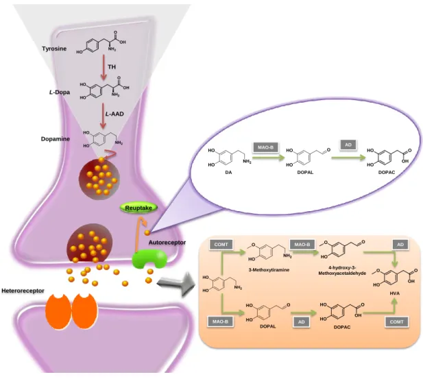
The biosynthesis of DA occurs in the dopaminergic neurons and starts with the conversion of the L-tyrosine to L-dihydroxyphenylalanine (L-dopa) by the action of the enzyme tyrosine hydroxylase (TH). L-Dopa is then converted into DA by the enzyme L -aromatic amino acid decarboxylase (L-AAD), also called dopa decarboxylase (Fig. 2.2) [33-36]. Once synthesised, DA is sequestered into vesicles and released into the synaptic cleft, where it exerts its role by interacting with specific receptors. The DA receptors are a class of G protein-coupled receptors (GPCRs), commonly divided in two major classes: D1-like receptors, comprising D1 and D5 receptors, and D2 like-receptors class formed by the D2, D3, and D4 receptors subtypes [37]. The DA metabolism encompasses either its reuptake by the dopaminergic neurons or its uptake by glial cells. Briefly, after its reuptake by the neurons, DA can be recycled and stored in vesicles, or converted to dihydroxyphenylacetic acid (DOPAC) by the action of monoamine oxidase-B (MAO-B) and aldehyde dehydrogenase (AD) (Fig 2.2). On the other hand, within glial cells, DA is converted into homovanillic acid (HVA) by the action of catechol-O-methyltransferase (COMT), MAO-B and AD (Fig. 2.2) [33-36].
Through its innervation systems, DA acts as a relatively slow modulator of a faster neurotransmission mediated by glutamate and GABA, and it has been implied in several vital functions of the central nervous system (CNS) such as voluntary movement, appetite, affection, reward, sleep, attention, working memory and learning [38]
. Accordingly, the typical movement disorders observed in PD have been linked with the degeneration and loss of basal ganglia dopaminergic projections, specially of the dopaminergic nigrostriatal neurons [28, 39, 40].
Figure 2.2 Schematic representation of dopamine metabolism. L-AAD: L-aromatic amino acid decarboxylase AD: aldehyde dehydrogenase, COMT: catechol-O-methyltransferase, DOPAC: dihydroxyphenylacetic acid,
DOPAL: 3,4-Dihydroxyphenylacetaldehyde, HVA: homovanillic acid, MAO-B: monoamine oxidase-B, TH: tyrosine hydroxylase.
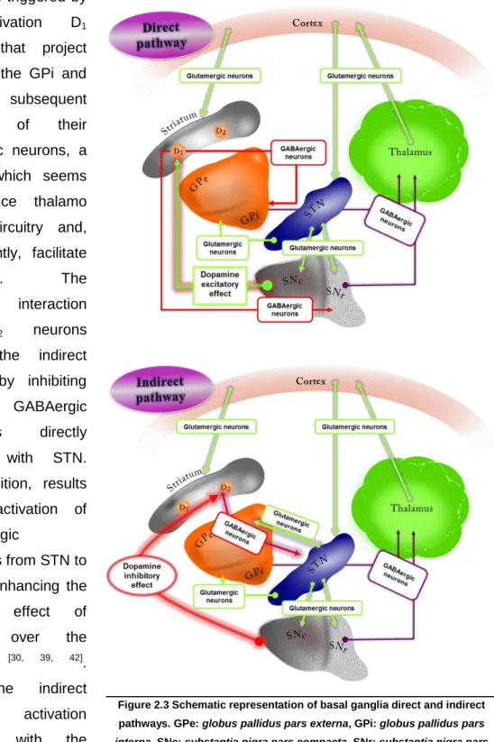
The actual model of the basal ganglia circuitry is based in the existence of two major projection systems: the direct and indirect pathways. Concisely, the direct and indirect pathways arise from different populations of striatal projection neurons, whose activity is regulated by striatal dopamine, supplied via nigrostriatal projection from the
substantia nigra compacta (SNc). The direct pathway (Fig.2.3) encloses D1-like receptors, involving dopaminergic projections from the striatum to the globus pallidus
pars interna (GPi) and substantia nigra pars reticulata (SNr). The indirect pathway
(Fig.2.3) selectively expresses D2-like receptors and involves the projections of the
striatum to globus pallidus pars externa (GPe) [37, 39, 41, 42]. It has been postulated that movement control by basal ganglia involves both direct and indirect pathways, by modulation of the cortical afferent activity through thalamus output.
TH Tyrosine L-Dopa Dopamine L-AAD Autoreceptor Reuptake Heteroreceptor MAO-B DOPAC AD DOPAL DA DOPAL 3-Methoxytiramine 4-hydroxy-3-Methoxyacetaldehyde DOPAC HVA MAO-B COMT MAO-B AD COMT AD
The direct pathway is triggered by the activation D1 neurons that project mainly to the GPi and SNr with subsequent inhibition of their GABAergic neurons, a process which seems to enhance thalamo cortical circuitry and, consequently, facilitate
movement. The
dopamine interaction
with D2 neurons
triggers the indirect pathway by inhibiting the GPe GABAergic projections directly attached with STN. This inhibition, results in the activation of glutamatergic
projections from STN to GPi/SNr enhancing the inhibitory effect of
GPi/SNr over the
thalamus [30, 39, 42]. Thus, the indirect pathway activation interferes with the thalamocortical circuitry
resulting in a movement inhibition [43].
More recently, controversial experimental data, led to questioning the consistency of the referred model. Although some of its features and hypotheses should be clearly reviewed [39, 44], significant assumptions of the basal ganglia circuitry model are still widely accepted (e.g. the basic connections between the basal ganglia nuclei and the
Figure 2.3 Schematic representation of basal ganglia direct and indirect pathways. GPe: globus pallidus pars externa, GPi: globus pallidus pars interna, SNc: substantia nigra pars compacta, SNr: substantia nigra pars
role of dopaminergic system in the striatum output) [39]. It is also important to point out its contribution to the study of the pathophysiology of movement disorders such as the ones observed in Parkinson‘s disease. In this neurodegenerative disease the regulatory role of DA in striatrum is compromised [27, 29], which leads to a reduced activity of the direct nigrostriatal pathway, whereas the indirect nigrostriatal pathway activity is increased, resulting in an excessive GPi inhibitory output to the thalamus with a consequent movement disorders [28].
2.3 Parkinson's disease therapy the current status
Nowadays the pharmacological therapy available for Parkinson's disease (PD) aims to improve patients‘ life quality, slowing down the progression of symptoms, either through the delay of the physical and psychological morbidity inherent to of the disease or by diminishing the long-term complications associated with the therapy [45]. The main strategic developments for symptomatic treatment and discovery of new compounds to modify the course of PD were focused on the improvement of dopaminergic therapies and in the identification of non-dopaminergic drugs.
Despite all the recent research in the field, there are only a few classes of drugs approved for the treatment of the motor related symptoms of PD, namely the ones that act primarily on the dopaminergic system, such as L-dopa, dopamine agonists, MAO-B and COMT inhibitors, and the non-dopaminergic drugs like anticholinergic and glutamate antagonists [45, 46].
In the fowling sections it will be succinctly presented the current status of the available chemotherapy for the treatment of the motor afflictions of Parkinson‘s disease will be briefly discussed, as well as the importance of the discovery and development of new drugs for PD.
2.3.1 Dopaminergic treatments for Parkinson´s disease
2.3.1.1 L-Dopa
The observation that dopamine levels in the basal ganglia of patients suffering from PD were much lower than those found in the brains of healthy persons, was the key information that boosted the drug discovery for
PD that lead to the introduction of L-dopa (Fig.2.4) in the market from the late 1960‘s [47]
.
Almost 50 years later, L-dopa remains the most useful oral drug in PD therapy, showing real effectiveness in the reversibility of motor limitations of the disease.
Figure 2.4 Chemical structure of L -dopa.
L-Dopa is a natural dopamine precursor that can cross the blood-brain barrier but has a relatively short half-life period of 60 to 90 min [48]. Once in the brain, exogenous
L-dopa is taken up by dopaminergic neurons, thus mimetizing the endogenous one, and converted to DA by the dopa-decarboxylase and stored in vesicles with subsequent release into the synaptic cleft.
The PD progressive loss of dopaminergic neurons, which are required to the biosynthesis, metabolism, storage and release of dopamine, influence the beneficial effect of each dose of L-dopa by shorting the time of its effectiveness and so the daily scheduled dose must be adjusted [47]. The long-term usage of this drug can lead to a narrow therapeutic window as well as several complications [49]. Standard treatment of PD with L-dopa causes a number of typical peripheral
and central dopaminergic adverse events, including nausea, vomiting, hypotension, hallucinosis, paranoid psychosis and particularly iatrogenic dyskinesias [50]. Some of the side effects are mainly due the peripheral metabolism of exogenous L-Dopa to DA in the liver and intestinal mucosa via peripheral L-AAD. In fact, less than 1% of administered L-dopa reaches the brain. To overcome this drawback a combination of L-dopa with
L-AAD inhibitors such as carbidopa and benserazide (Fig. 2.5) is commonly prescribed. Thus, the L-dopa bioavailability is increased and the necessary dose to obtain effect reduced as well as the peripheral side effects [51].
2.3.1.2 Dopamine agonists
One of the strategies employed to minimize the side effects of L-dopa is related to the use of dopamine agonists. This drugs were launched as first-line effective drugs for monotherapy in the early stages of the disease, in order to postpone L-dopa introduction and its associated dyskinesia [52]. Furthermore, due to the fact that this class of compounds can delay the onset of dyskinesias and motor fluctuations, by one to three years, after L-dopa introduction DA agonist are also used as adjuvants to L -dopa in the later stages of the disease [53] .
The main advantage associated with the use of dopamine agonists is the fact that they can act directly on dopamine receptors, bypassing the metabolic pathways in the degenerated nigrostriatal neurons [54-56]. It was also postulated that the antioxidant/free radical-scavenging activity demonstrated by this type of compounds, makes them good candidates for neuroprotection of the dopaminergic and
non-Benserazide Carbidopa
Figure 2.5 Chemical structures of L -AAD inhibitors.
dopaminergic neurons [56-59]. These additional effects, although very important to the management of PD, still remain unproven and may not be significant for clinical practice as suggested for some authors [60, 61].
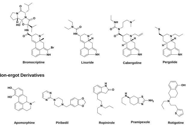
DA agonists are a heterogeneous group of drugs classically divided into ergot or non-ergot agonists. The ergot DA agonists (Fig. 2.6) are based on the structure of ergoline and include: bromocriptine, cabergoline, lisuride, and pergolide. The non-ergot DA agonists (Fig. 2.6) are a more diverse chemical group comprising apomorphine, piribedil, ropinirole, pramipexole, and rotigotine [54]. Due to several side effects, such as impulse control disorders (e.g. pathological gambling, hyper sexuality, or binge eating) the treatment with dopamine agonists must be initiated at low dosage and the titration period may be extended for months in order to improve tolerance to the peripheral side effects [62]. This dose escalation process may have direct implications in the patient´s compliance to the treatment due to the time needed for beneficial effects to manifest.
Figure 2.6 Chemical structures of ergot and non-ergot dopamine agonists.
2.3.1.3 Catechol-O-methyltransferase inhibitors
Catechol-O-methyltransferase (COMT) is an intracellular enzyme widely distributed in human tissues and highly active in liver, kidneys and gastrointestinal tract
Apomorphine Piribedil Ropinirole Pramipexole Rotigotine
Bromocriptine Lisuride Cabergoline Pergolide
Ergot Derivatives
[63]
. This enzyme in presence of Mg2+ promotes the O-methylation of one of the hydroxyl groups present on the catechol substrate [63].
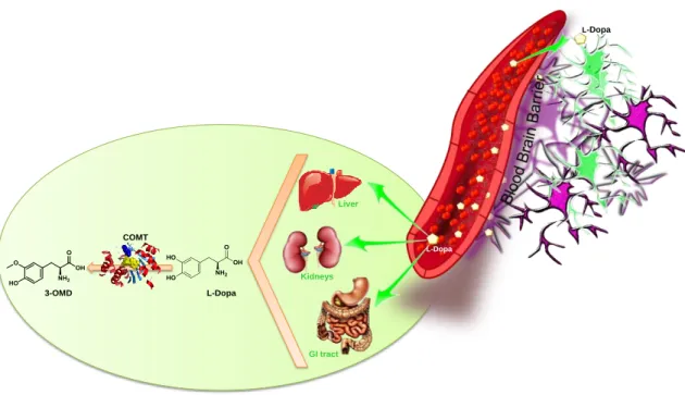
To understand the role of COMT inhibitors, as a valid resource for PD, it is important to highlight the role of this enzyme in the peripheral metabolism of L-dopa. Besides the peripheral conversion of L-dopa to dopamine via L-AAD, L-dopa is also peripherally metabolized to its inactive metabolite 3-O-methyldopa (3-OMD) (Fig. 2.7), by the action of COMT [63, 64]. The use of COMT inhibitors allows a prolonged maintenance of L-dopa serum levels, by increasing its half-tlife and boosting the central bioavailability of the drug. These drugs have the additional advantage of low motor fluctuations and less dependence on daily L-dopa [64].
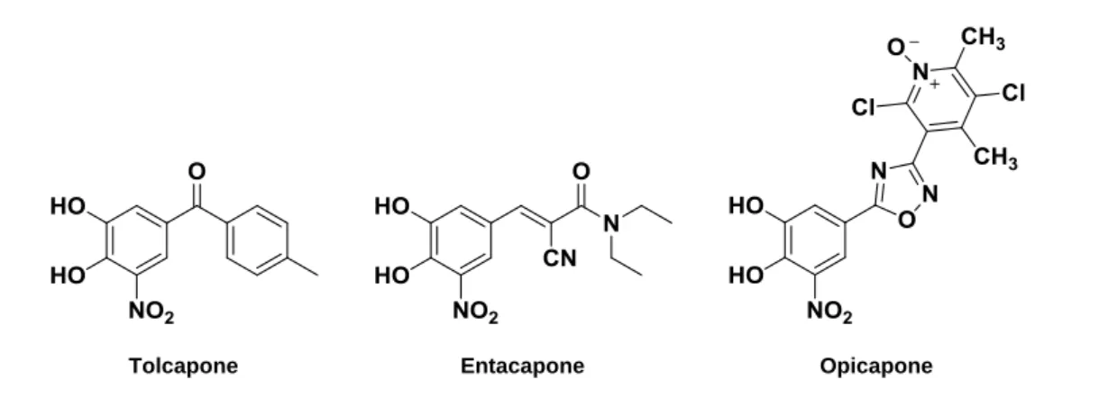
There are two COMT inhibitors, available since the mid-1990s, for clinical practice: tolcapone and entacapone (Fig. 2.8) [65]. Entacapone is a selective and reversible peripheral COMT inhibitor that does not cross the blood brain barrier (BBB) acting primarily in the increase and maintenance of plasmatic half-life of L-dopa [66]. Tolcapone has a half-life similar to entacapone but, as it has a greater bioavailability and smaller volume of distribution, thus resulting in higher potency for COMT inhibition [54]. Furthermore, tolcapone, unlike entacapone, may also inhibit central COMT brain [67, 68], increasing peripherally and central L-dopa availability, thus enhancing striatum dopamine neurotransmission [68]. However, the appearance of fatal acute hepatitis in some patients receiving tolcapone, raised several concerns about this
3-OMD L-Dopa COMT L-Dopa L-Dopa Liver Kidneys GI tract
Figure 2.7 Peripheral biotransformation of L-dopa by action of COMT. COMT: Catechol-O-methyltransferase and 3-OMD: 3-O-methyldopa.
drug and the European Medicines Agency (EMEA) has temporary suspended tolcapone in the European Union (EU) [67, 69]. In 2004, EMEA lift the suspension but its use was substantially curtailed [67]. Comparatively, entacapone does not show the same tendency to cause hepatotoxicity, being the most common side effects related with the dopaminergic system. As reflex of the increased central dopaminergic activity, nausea, vomiting, orthostatic hypotension, and dyskinesia, can be experienced by the patients undergoing COMT inhibitors therapy. Additionally, it was reported the occurrence of diarrhoea in about 10% of cases, and in some patients it may be intractable and discontinuation of the drug may be required [70].
Recently, a novel catechol-O-methyltransferase (COMT) inhibitor, opicapone (Fig.2.8), was developed by BIAL and has been proposed as adjunctive therapy for PD patients treated with L-dopa. Opicapone is currently in phase III clinical trials and a license agreement was signed with Ono Pharmaceutical Co., Ltd. (―ONO‖) for the development and commercialization in Japan [71].
Figure 2.8 Chemical structures of COMT inhibitors.
2.3.1.4 Monoamine oxidase-B inhibitors
Monoamine oxidases
(MAOs) are intracellular flavine-containing enzymes that play a major role in the in
vivo inactivation of biogenic
amines, both in peripheral and central neuronal tissues [72].
These enzymes are
ubiquitously distributed on the body and are mainly located in the outer mitochondrial membrane. The biological role of MAOs is the catalysis of the oxidative deamination of
endogenous monoamines
(Fig. 2.9) by a process that involves the oxidation of the amine function, with the formation of an imine
intermediate and
simultaneous reduction of the flavin cofactor. The imine
intermediate is then hydrolysed, yielding ammonia and the corresponding aldehyde. The cofactor is re-oxidized by molecular oxygen, with concomitant hydrogen peroxide release (Fig. 2.9) [73].
Two isoforms of MAOs are expressed in mammals: MAO-A and MAO-B, which can be distinguished based on their substrate preference for the substrate and their interaction with specific inhibitors [73, 74]. Within the CNS, the primary substrates of MAO are neurotransmitters, such as adrenaline, noradrenaline (NA), dopamine (DA), serotonin, also called 5-hydroxytryptamine (5-HT), and β-phenylethylamine (PEA). However, a variety of monoamines, like tyramine, octopamine, tryptamine and kynuramine, can be also metabolised by MAO. These substrates are deaminated by both isoforms of the enzyme, albeit with differing kinetic parameters, which are influenced by concentration, affinity and turnover rate of the substrate. Under normal physiologic conditions, NA and 5-HT are the preferred substrates of MAO-A, and PEA
FAD + H2O+O2 FAD + H2O2 + NH3
is preferentially metabolised by MAO-B. Both MAOs isoforms metabolise DA, although DA in human brain has higher affinity for MAO-B isoform [75].
These findings led to the development of selective inhibitors for each subtype of MAO, being MAO-A a target in the treatment of depression, whereas MAO-B inhibitors are used in therapy for neurodegenerative disorders, including PD [74]. MAO-B inhibition causes the blockade of the metabolism of dopamine and therefore could enhance both endogenous dopamine and the one produced from exogenously administered L-dopa.
1-Methyl-4-phenyl-1,2,3,6-tetrahydropyridine (MPTP), a chemical compound, which was linked with parkinsonism by serendipity, had a significant impact on PD research and it was used since then as a tool to investigate the cause and treatment of the disease namely in the development of suitable PD animal models [76]. The neurotoxic effects are not caused by MPTP by itself, but by its bioactive metabolite, the 1-methyl-4-phenylpyridinium ion (MPP+), a metabolic process which can be MAO-B mediated. The active metabolite exerts is toxicity by acting as mitochondrial complex I inhibitor inducing cell death [77]. There are several studies suggesting that MAO-B inhibitors can block the conversion of MPTP to its active metabolite MPP+, thus giving rise to the thought that they can possess some neuroprotective properties [75, 78].
The MAO-B inhibitors currently in the market, selegiline (L-deprenyl) and rasagiline (Fig. 2.10) are both selective and irreversible inhibitors of MAO-B isoform. Selegiline in combination with L-dopa presents very successful results in the treatment of PD. First synthesised in 1962, the low potential of selegiline to produce the so-called "cheese effect", a common reaction following the usage of MAO inhibitors, had attracted attention even before MAO subtypes have been discovered. Additionally, selegiline may also display neuroprotective
properties through the decrease of oxidative stress in neuronal striatal circuit [79]. A negative feature of selegiline is related to its biotransformation in amphetamine-like metabolites; two of them, L -amphetamine and L-methamphetamine, have shown to be potential neurotoxins. Despite the fact that the neurotoxicity mechanisms are not completely understood, it has been proposed that it involves damage in dopaminergic and serotonergic cells [80-82].
Rasagiline (Fig. 2.10) is a potent, selective and irreversible inhibitor of MAO-B. One of its main therapeutic advantages is related to its biotransformation, as no amphetamine-like metabolites are obtained [82-84]. Moreover, its major metabolite
((R)-1-Seligiline
Rasagiline
Figure 2.10 Chemical structures of MAO-B inhibitors in clinical practice
aminoindan) exhibits neuroprotective activity in animal PD models [85]. Studies with patients, with moderate to severe symptoms of PD treated with L-dopa, showed that rasagiline improves the typical symptoms and motor fluctuations [86]. The key to the success of rasagiline seems to be its ability to limit the process of neurodegeneration in the early stages of the disease [87].
The multitarget drug discovery approach recently proposed for multifactorial diseases has already brought out encouraging results regarding PD. The development of new chemical entities (NCEs) with a MAO-B inhibitory activity allied to another relevant mechanism of action, culminated in synthesis of two new compounds: ladostigil and safinamide (Fig. 2.11).
The identification of the propargylamine moiety as a key structure related with the neuroprotective activity of MAO-B inhibitors was the inspiration for the design and development of ladostigil [10]. Ladostigil, is a NCE that combines the neuroprotective effects of the propargylamine moiety, the MAO-B inhibition profile of rasagiline and retains the acetylcholinesterase (AChE) inhibitory activity of rivastigmine [88].
Safinamide is an aminoamide derivative, currently in Phase III clinical trial for Parkinson´s disease, which has been shown to be a reversible
MAO-B inhibitor with the capacity to reduce dopamine reuptake and with antiglutamatergic effects. This NCE may add a new dimension to PD treatment options, either as an adjuvant to current drugs for neuroprotection or motor symptomatic relief in PD, as it displays dopaminergic and non-dopaminergic effects [89].
2.3.2 Non-dopaminergic treatments for Parkinson´s disease
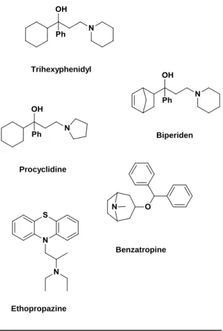
2.3.2.1 Anticholinergic drugs
Before the discovery of L-dopa the therapy of choice for PD relied on anticholinergic drugs. The first‘s reports involving the use of anticholinergic drugs for ameliorating the motor symptoms of PD remount to the nineteen century (late 1860‘s, early 1870‘s) with use of the belladonna alkaloids, atropine and scopolamine. The synthetic anticholinergic agents, namely, trihexyphenidyl, Biperiden, procyclidine, benzatropine and ethopropazine were only marketed for the treatment of PD in 1950‘s [90-92] (Fig. 2.12). Although the benefits were modest and inconsistent,
Ladostigil
Safinamide
Figure 2.11 Chemical Structures of multitarget drugs in clinical trials.
anticholinergic drugs remained the cornerstone of PD therapy for nearly one hundred years.
The rational use of anticholinergic agents in PD has been strengthened by
the clear demonstration of
dopaminergic-cholinergic antagonism in the striatal function [55]. The mechanism of action of the anticholinergic drugs in PD is not yet thoroughly known, but it is postulated that these drugs can antagonise overactive cholinergic
transmission via blockade of
postsynaptic muscarinic receptors, thereby restoring the balance between
DA and ACh in the
striatum [55, 93]. Although anticholinergics may be useful as monotherapy in early stages of PD, they are mainly used as adjuvants to L-dopa treatment. They are more effective in ameliorating the mild symptoms of tremor and rigidity, without
significant changes in bradykinesia signals [94]. Anticholinergic agents can also block muscarinic ACh receptors peripherally leading to a myriad of side effects like xerostomia, sweating inhibition, blurred vision, and urinary retention as the most common ones. CNS side effects, such as confusion, dementia and other psychiatric
symptoms are also
frequent [95].
2.3.2.2 Glutamate antagonists
Amantadine (Fig. 2.13) is a tricyclic amine that was firstly used as an antiviral agent. Its benefits in PD therapy were discovered by serendipity in 1969 by Schwab et al. [96]. Despite the lack of scientific evidence of its efficacy regarding the delay of PD progression, this drug has been used for the relief of PD motor since then [55]. Amantadine is
administered either in combination with L-dopa or in monotherapy, in patients that have less tolerance for L-dopa [97]. More recently, the interest on this drug has re-emerged
Figure 2.13 Chemical structure of amantadine. Trihexyphenidyl Biperiden Procyclidine Benzatropine Ethopropazine
Figure 2.12 Chemical structures of anticholinergic drugs
due to its putative beneficial role in the treatment of motor fluctuations and dyskinesias [98, 99]. The exact mechanism of action of amantadine remains unclear. The role of this drug in PD has been related to the antagonism exerted on N-methyl-D-aspartate (NMDA) receptor subtype, but other mechanisms have also been proposed, such as direct action over the dopaminergic system. None of the mechanisms so far proposed met the rigorous criteria for strength of evidence [55,100, 101].
However, the use of amantadine in therapy is limited due to its side effects, which are generally mild but may be severe, especially in elderly patients, and also due to the discovery of more effective therapeutic approaches. Adverse effects associated with amantadine usage are primarily central nervous system ones, like anxiety, hallucinations, depression, insomnia, somnolence/drowsiness, fatigue and impaired coordination, along with non-SNC effects, such as diarrhoea, nausea and vomiting, constipation, xerostomia, and livedo reticularis [98].
2.4 Looking for new targets: adenosine A2A receptors and
Parkinson´s disease
To date, most of the currently available therapies in PD target the dopaminergic system and none of these therapeutic approaches have been proven to modify the course of the disease. To various extents, these drugs can also cause motor and non-motor complications [102].
The need for potent and safe antiparkisonian agents has been the motor of an intensive research towards the development of new therapeutic approaches for PD, preferentially one that can operate beyond the damaged dopaminergic system. In fact, in the last years a large number of medicinal chemistry programs have been focused in looking for new non-dopaminergic modulators of basal ganglia motor circuits [103].
As mentioned previously in section 2.3.2, there are available medicines whose mechanisms of action operate via a non-dopaminergic pharmacological approach (e.g. anticholinergic drugs and amantadine) illustrating the viability and the problems of this type of therapeutic strategy for PD motor symptoms relief [104]. So, the development of novel antiparkinsonian agents whose mechanisms of action are mediated by other specific CNS receptors, preferentially restricted to basal ganglia neurons, may have an intrinsic therapeutic advantages while reducing the occurrence of adverse CNS events.
2.4.1 G Protein-coupled receptors and adenosine receptors: a general
outlook
Adenosine can act as neuromodulator and has been associated with the coordination responses to DA and other neurotransmitters, in areas of the brain that are responsible for motor function, mood, learning and memory [105, 106]. The
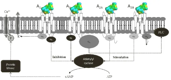
physiological functions of adenosine are mediated by adenosine receptors (ARs). Briefly, these receptors are G protein-coupled receptors (GPCRs) consisting of a single polypeptide chain that transverses the membrane from the extracellular side, beginning at the N terminus, to form seven transmembranar helices (TMs), which are connected by three intracellular and three extracellular loops (ILs and ELs, respectively), leaving the N-terminus in the extracellular milieu and the C-terminus in the cytoplasm [107] (Fig. 2.14).
There are four distinct ARs subtypes designated as A1, A2A, A2B and A3. Adenosine A1 and A3 receptors are coupled to inhibitory G proteins, while A2A and A2B receptors are coupled to stimulatory G proteins. The A2A and A2B ARs preferably interact with members of the Gs family of G proteins, with consequent activation of adenylyl cyclase and consequently the production of cyclic AMP (cAMP), and contrariwise, the A1 and A3 ARs inhibit the adenylyl cyclase activity by interaction with Gi proteins [107] (Fig. 2.14).
Figure 2.14 Schematic representation of G protein-coupled adenosine receptors. ATP: Adenosine-triphosphate; Ca2+: ion Calcium (II); cAMP: Cyclic adenosine monophosphate; Gi/Go: inhibitory G protein; Gs/Gq stimulatory G
protein; K: potassium; P: phosphorus; PLC: phospholipase C.
GPCRs have been implicated in a multitude of human disorders and numerous diseases as their signalling regulates an incredibly vast array of physiological functions and pathological conditions. Consequently, they have a high pharmaceutical appeal and constitute the target of a very large segment of the currently marketed drugs [108]. It is estimated that nearly half of all modern drugs somehow regulate GPCR activity [109].
Although GPCRs are among the most fruitful targets for marketed drugs, and while intense discovery efforts have been performed to find agents for GPCR subtypes, a selective drug candidate has not yet been reported. Particularly, ARs have long been
considered as targets for drug discovery programs of a variety of maladies, such as cerebral and cardiac ischaemia, immune and inflammatory disorders, and more recently for neurodegenerative and neoplastic events [108, 110, 111]. Consequently, numerous medicinal chemistry groups have made intense efforts to create selective agonists and antagonists for each AR subtype [112-115].
2.4.2 Adenosine, adenosine receptors and neurodegenerative diseases
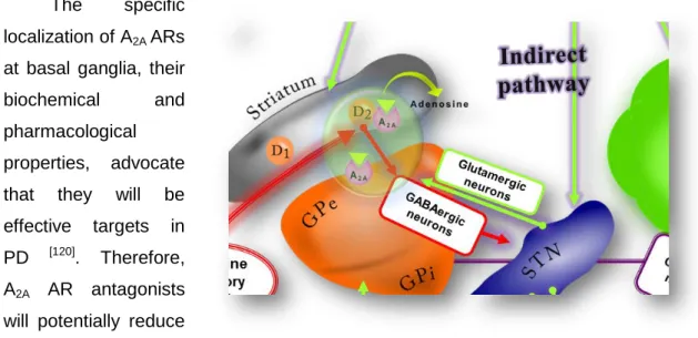
Adenosine is a neuromodulator with several functions in the CNS, such as inhibition of neuronal activity in many signalling pathways [105,106,116,117]. Adenosine plays a major role in a diverse array of neural phenomena, which includes regulation of sleep and the level of arousal, neuroprotection, seizure susceptibility, locomotor effects, neuroprotection and modulation of various neurotransmitters, namely dopamine. The existence of an interaction between adenosine and dopamine within the basal ganglia, which could be correlated with the modulation of motor function, has been demonstrated [105, 116, 117]. Adenosine can also counteract the excitotoxicity associated with excessive glutamate release in the brain [118]. The overall findings support the hypothesis that adenosine plays an important role in the modulation of glutamatergic and GABAergic neurotransmission [28, 119, 120] of the basal ganglia indirect pathway [121].
The neuromodulation effect exerted by adenosine occurs probably through the activation of the A1 and A2A ARs, which are highly expressed in brain [122]. The A2A AR in the CNS, distinguishes itself from the other adenosine receptors by its selective localization in striatum [107] (Fig. 2.15). In the striatum the A2A AR are co-localize and physically associated with DA D2 receptors. A2A AR and D2 receptors have opposing effects on adenylyl cyclase and cAMP production in cells. Interestingly, A2A AR activation reduces the affinity of striatal D2 receptor for dopamine and the blockade of A2A AR with specific antagonists facilitates function of the D2 receptor. Blockade of A2A AR signalling by selective A2A AR receptor antagonists was shown to be beneficial not only by enhancing the therapeutic effects of L-Dopa but also by reducing dyskinesia from long-term L-Dopa treatment.
The specific localization of A2A ARs at basal ganglia, their
biochemical and
pharmacological properties, advocate that they will be effective targets in PD [120]. Therefore, A2A AR antagonists will potentially reduce the effects associated
with dopamine depletion in PD. In fact this class of compounds have recently attracted considerable attention as they have shown potential effectiveness in counteracting motor dysfunctions, while displaying additional neuroprotective and anti-inflammatory effects in animal models of PD [60, 117, 118]. They have distinguished themselves over other antiparkinsonian agents in development, by the convergent epidemiological and preclinical evidence of their neuroprotective benefits as well as symptomatic relief [117, 118].
2.4.2.1 A2A AR antagonists
The knowledge acquired over the last decades regarding the involvement of adenosine on motor functions, mainly
through modulation of A2A receptor, makes A2A AR antagonists promising non-dopaminergic agents for the treatment of PD motor symptoms. A2A AR antagonists developed do far have been traditionally categorized as xanthine based (e.g. caffeine and styrylxanthines) or non-xanthine based derivatives (e.g. pyrazolotriazolo pyrimidines, arylindeno pyrimidines, thiazolotriazolo pyrimidines). Excellent reviews have been published on this topic in the last five years[123-126]. However, until now only two A2A AR antagonists have undergone clinical
evaluation, namely, istradefylline (KW-6002) and preladenant (SCH 420814) (Fig. Figure 2.15 Co-localization of A2A AR and D2 receptors
Istradefylline
Preladenant
Figure 2.16 Chemical structures of istradefylline and preladenant.
2.16). The preclinical data demonstrates that istradefylline possesses beneficial effects on PD symptoms, and in clinical trials it has shown the ability to reduce the ―off‖ time in patients with PD receiving dopaminergic therapy [117]. Istradefylline completed phase III clinical trials in 2009, but it was not approved by the FDA due to lack of efficacy in comparison with placebo [127]. Preladenant was being researched as a potential treatment for Parkinson's disease. Although positive results were reported in Phase II clinical trials it did not prove itself to be more effective than a placebo during Phase III trials, and so it was discontinued in May 2013.
2.5 Chromone as a privileged scaffold
2.5.1 Privileged structures for lead discovery
The term privileged structure was first introduced in 1988 by Evans and co-workers[128] who have recognised the potential of certain regularly occurring structural motifs as templates for structural modification to expand the discovery of novel ligands for binding to proteins. Since then, the original concept definition has evolved and nowadays privileged structures (scaffolds or motifs) can be defined as molecular frameworks which are able of providing useful ligands for more than one type of receptor or enzyme targets by judicious structural modifications[129]. Privileged structures typically exhibit drug-like properties and usually generate drug-like compound libraries and lead compounds. However, one should keep in mind that the presence of a privileged scaffold in a compound is no guarantee that all compounds in the library will be bioavailable and non-toxic.
Many of these frameworks have been used as templates for the design of libraries of molecules for drug discovery [130-134]. In fact, the privileged structure concept has emerged as a successful approach for the drug discovery process, e.g. as core structures for synthesis and optimal starting points for the library design of ligands with affinity to certain molecular targets [130-134]. The last decade has brought more popularity to this idea and numerous scaffolds have been claimed to be privileged: benzazepinone, diphenylmethane, piperidine, biphenyltetrazole, indole, biphenyl and benzopyrane [13, 130]. However, the prerequisites that make a structural motif a privileged one still remain unknown. Nevertheless, it is consensual that there are at least two important issues that can make a privileged structure suitable for fragment-based design: synthetic availability and ease of modifying its structure. The decoration of the privileged scaffolds using diversity-oriented synthesis has been proven to be an essential tool to rapidly discover biologically active small molecules. During the design of a synthetic library, it is advised to exploit simple one-pot approaches, e.g. condensation reactions that afford high yields. On the other hand, for building the