DAPTOMYCIN DELIVERY INTO THE EYE BY ENCAPSULATION INTO CHITOSAN COATED ALGINATE NANOPARTICLES
by
Joana Ribeiro Costa
DAPTOMYCIN DELIVERY INTO THE EYE BY ENCAPSULATION INTO CHITOSAN COATED ALGINATE NANOPARTICLES
Thesis presented to Escola Superior de Biotecnologia of the Universidade Católica Portuguesa to fulfill the requirements of Master of Science degree in Biomedical Engineering
by
Joana Ribeiro Costa
Places: Escola Superior de Biotecnologia da Universidade Católica Portuguesa Faculdade de Farmácia da Universidade do Porto
Instituto Superior de Ciências da Saúde - Norte
Supervision: Professor Manuela Pintado Professor Bruno Sarmento
iii
A endoftalmite bacteriana é uma inflamação ocular, resultante da introdução de um agente infeccioso no segmento posterior do olho. Grande parte das infeções são provocadas por bactérias Gram-positivas, tal como Staphylococcus aureus resistente à meticilina e
Staphylococcus epidermidis. Atualmente, o tratamento de infeções oculares é dificultado pelas
barreiras anatómicas e natureza delicada do interior do globo ocular. A aplicação de fármacos no próprio olho é uma solução não-invasiva, segura e menos dolorosa do que tratamentos cirúrgicos, a laser ou injeções oculares. A daptomicina é um novo péptido cíclico antimicrobiano, com atividade contra bactérias Gram-positivas, constituindo um poderoso agente no tratamento da endoftlamite bacteriana. Contudo, a aplicação tópica de daptomicina no olhoé limitada devido à rápida renovação do fluído ocular, requerendo formulações com propriedades mucoadesivas. Apesar da existência de nanopartículas de quitosano para encapsulamento de daptomicina, é necessária uma alternativa eficiente que possa melhorar as suas propriedades biológicas e farmacêuticas.
Neste trabalho, apresentam-se nanopartículas mucoadesivas de alginato revestidas com quitosano como possível sistema de libertação de daptomicina, uma vez que o alginato e o quitosano possuem diversas propriedades biológicas favoráveis ao prolongamento do tempo de residência pré-corneal do antibiótico, permitindo a acumulação e permeabilidade deste fármaco
e integrando um método promissor para o tratamento tópico da endoftalmite bacteriana.
As nanopartículas foram preparadas através de pré-gelificação ionotrópica do alginato, seguido de complexação polielectrónica do quitosano e caracterizadas pelo seu tamanho, polidispersão e potencial zeta. As suas eficiências de encapsulação foram determinadas e a actividade antimicrobiana foi testada. Foi também avaliada, in vitro, a permeabilidade da daptomicina em células oculares.
As nanopartículas obtidas apresentam uma carga negativa, com tamanhos entre 382 e 421 nm e as eficiências de encapsulação apresentam valores entre 79 e 91%. A atividade antimicrobiana da daptomicina não sofreu alterações com o encapsulamento em nanopartículas. A permeabilidade ocular da daptomicina, in vitro, alcançou os 6% para células da córnea e 5% para células da retina, após 4 horas da aplicação.
Em conclusão, as nanopartículas obtidas são apropriadas para a libertação ocular de daptomicina, constituindo um potencial tratamento da endoftalmite bacteriana.
v
Bacterial endophthalmitis is an ocular inflammation resultant from the introduction of an infectious agent into the posterior segment of the eye. Gram-positive bacteria such as methicillin-resistant Staphylococcus aureus and Staphylococcus epidermidis are the main cause of majority of endophthalmitis cases. Currently, treatment of bacterial infections and inflammation in the eye poses the dilemma of anatomic barriers and the delicate nature of the interior of the eye. Local drug applied to the eye represents a non-invasive, safe and less painful solution than surgery, laser treatments or eye injections. Daptomycin is a novel acidic cyclic lipopeptide antimicrobial agent with activity against Gram-positive organisms, including methicillin-resistant Staphylococcus aureus, becoming a powerful agent for treatment of bacterial endophthalmitis, though, topical administration of daptomycin directly into the eye is limited due to the rapid ocular fluid turn-over, requiring formulations with mucoadhesive properties. In spite of the existence of chitosan nanoparticles as daptomycin carrier, it is required an efficient alternative that may improve their biological and pharmaceutical properties. In the present project, mucoadhesive chitosan-coated alginate nanoparticles are proposed as effective delivery systems for daptomycin through ocular epithelium, taking advantage of the favourable biological properties of alginate and chitosan to prolong precorneal residence time of the antibiotic, enhancing drug accumulation and permeation.
Nanoparticles were prepared by ionotropic pre-gelation of an alginate core followed by chitosan polyelectrolyte complexation, characterized regarding particle size, polydispersity and zeta potential, encapsulation efficiency was determined, and antimicrobial activity was also tested after encapsulation of the antibiotic. Also, in vitro ocular permeability of
daptomycin-loaded nanoparticles was tested using cell culture models.
Formulated daptomycin-loaded chitosan coated alginate nanoparticles were negatively charged and sized between 380-420 nm, suitable for ocular application. Encapsulation efficiencies were between 79 and 92%. Antibacterial activity of daptomycin against major microorganisms responsible for bacterial endophthalmitis was not affected by encapsulation into nanoparticles. In vitro permeability was up to 6% for corneal cells and 5% for retinal cells after 4 hours.
In conclusion, obtained nanoparticles are suitable for daptomycin delivery in the eye and seem to be a promising vehicle for bacterial endophthalmitis treatment.
vii
Firstly, I would like to acknowledge my supervisors, Professor Manuela Pintado and Professor Bruno Sarmento, for all the help and guidance given throughout the course of this work, providing all the conditions to carry out this work.
I wish to express my sincere gratitude to Nádia Silva, thank you for all your precious assistance, patience and wise advises that helped me to overcome many obstacles during this work.
Also, I would like to offer my regards to my lab colleagues: to Eduardo Costa and Sara Silva, thank you for all the help in the laboratory; to Francisca Araújo, thank you for the assistance with the cell growing work; to Adriana Pereira and Inês Montenegro, thank you for gentleness and for receiving me so well.
To my dear classmates, Marta Godinho, Paulo Dias and Pedro Sousa, thank you for all the shared ideas and moments of joy through this master degree.
Moreover, I would like to thank to all my friends for the all their friendship, care and support given every day. Special thanks to Ana Serra, Daniela Rodrigues, Fernando Moreira, Liliana Barbosa, Lúcia Rios, Luís Adães and Sebastião Almeida for your interest and encouragement throughout this work.
Finally, this project would not have been possible without my family support, particularly my parents, who have always supported and understood me, a special thanks for all your love and care.
ix
Resumo iii
Abstract v
Acknowledgments vii
List of Figures xi
List of Tables xiii
List of Abbreviations xv
I. Introduction 1
1. Anatomy of the eyeball 1
1.1 Fibrous layer 2 1.2 Vascular layer 3 1.3 Neural Layer 4 1.4 Lens 5 1.5 Vitreous body 5 1.6 Conjunctiva 5 2. Bacterial endophthalmitis 5 2.1. Types of endophthalmitis 6 2.1.1 Exogenous endophthalmitis 6 2.1.2 Endogenous endophthalmitis 7 2.2 Treatment 7 3. Resistance to antibiotics 8 4. Daptomycin 9
4.1. Mechanism of action and pharmacology 9
4.2. Spectrum of activity and susceptibility 10
5. Routes of drug delivery to the eye 11
5.1. Topical delivery 12
5.2. Systemic delivery 12
5.3. Intravitreal delivery 13
5.4. Periocular delivery 13
6. Ocular barriers 13
6.1. Drug loss from the ocular surface 14
x
7.1. Nanoparticles for ocular drug delivery 16
7.1.1. Alginate 17
7.1.2. Chitosan 18
7.1.3. Alginate-Chitosan polyionic complexes 19
8. Objectives 20
II. Material and Methods 21
1. Materials 21
2. Development of chitosan coated sodium alginate nanoparticles 21
2.1. Unloaded nanoparticles 21
2.2. Daptomycin-loaded nanoparticles 23
3. Physico-chemical characterization of nanoparticles 25 4. Determination of Daptomycin encapsulation efficiency 25
4.1. Instrumentation 25
4.2. Chromatography method setup 26
4.3. Preparation of calibration solutions and calibration function 26
5. Determination of Minimal Inhibitory Concentrations 26
6. Determination of ocular cells permeability to Daptomycin 27
6.1. Cell culturing 28
6.2. Cell plating 28
6.3. Permeability experiment 29
7. Statistical analysis 29
III. Results 30
1. Characterization of chitosan coated sodium alginate nanoparticles 30
1.1. Unloaded nanoparticles 31
1.2. Daptomycin-loaded nanoparticles 35
2. Determination of encapsulation efficiency of daptomycin-loaded nanoparticles 36
3. Minimal Inhibitory Concentrations 38
4. Determination of ocular cells permeability to Daptomycin 39
IV. Conclusions 44
xi
Figure 1.1 – Diagramatic representation of the gross anatomy of the globe. Source: Malhotra
et al. (2011) 1
Figure 1.2 - Anterior segment of the human eyeball. Source: Presland (2007) 4 Figure 1.3 - Chemical Structure of Daptomycin. Source: Jeu and Fung (2004) 9 Figure 1.4 – Different routes of ocular drug delivery. Source: Gaudana et al. (2010) 12 Figure 2.1 – Schematic representation of the in vitro cell culture model, using transwell
inserts. Source : Barar et al. (2009) 28
Figure 3.1 – Permeability of daptomycin solution and daptomycin-loaded nanoparticles in
HCE cells 41
Figure 3.2 – Permeability of daptomycin solution and daptomycin-loaded nanoparticles in
xiii
Table 1.1 – MIC50 and MIC90 of daptomycin among different species isolated. Adapted from
Enoch et al. (2007). 11
Table 2.1 - Different concentrations of alginate, chitosan and CaCl2 used to prepare preliminary
unloaded chitosan coated sodium alginate nanoparticles. 22
Table 2.2 - Different concentrations of alginate, chitosan and CaCl2used to prepare final set of
unloaded chitosan coated sodium alginate nanoparticles, after preliminary study. 23
Table 2.3 - Different concentrations of alginate, chitosan and CaCl2and daptomycin mass in
the final solution used to prepare chitosan coated sodium alginate nanoparticles loaded with
daptomycin. 24
Table 3.1 - Particle size, polydispersity and zeta potential of preliminary experiment for
unloaded chitosan coated alginate nanoparticles. 32
Table 3.2 - Particle size, polydispersity and zeta potential of unloaded chitosan coated alginate
nanoparticles, after preliminary study. 34
Table 3.3 - Particle size, polydispersity and zeta potential of daptomycin loaded chitosan coated alginate nanoparticles, prepared at different alginate: daptomycin mass ratios. 35
Table 3.4 - Encapsulation efficiency of daptomycin loaded chitosan coated alginate nanoparticles, at different alginate: daptomycin mass ratios. 37
Table 3.5 – Minimum Inhibitory Concentrations for free daptomycin and daptomycin-loaded
xiv 5-FU - 5-Fluorouracil
DMEM – Dulbecco’s Modified Eagle Medium
EMA - European Medicines Agency
FDA - Food and Drug Administration
HBSS – Hanks Balanced Salt Solution
HCE – Human Corneal Epithelial
MIC – Minimum Inhibitory Concentration
MRSA – Methicillin-resistant Staphylococcus aureus
MRSE – Methicillin-resistant Staphylococcus epidermidis
MSSA – Methicillin-susceptible Staphylococcus aureus
Pen - Penicillin
PRSP – Penicillin-resistant Streptococcus pneumonia
RPE – Retinal Pigment Epithelium
RP-HPLC - Reverse-Phase High Performance Liquid Chromatography
TER – Transepithelial resistance
VA - Vancomycin
VRE – Vancomycin-resistant enterococci
1
I.
Introduction
1.
Anatomy of the eyeball
The human eye is a small organ that provides sense of sight, allowing the perception of shapes, colors and dimensions around the world. In spite of constant environmental changes, the eye has the ability to adapt to new conditions. It also works as an external barrier of the body, continuously threatened by foreign bodies that can lead to eye diseases, so it is also a very sensitive structure.
A wide diversity of eye diseases affects millions of people around the world and has devastating effects on individuals, leading to visual injury and possible ocular blindness that instigate a decline in quality of life (Short, 2008).
The eyeball occupies the anterior part of the orbit and its rounded shape is disrupted anteriorly, where it bulges outward, representing about one-sixth of the total area of the eyeball (Drake et al., 2010).
2
Surrounding the internal components of the eyeball are the walls of the eyeball. They consist of three layers: the outer fibrous layer, the middle vascular layer and the inner neural layer (Drake et al., 2010), as represented in Figure 1.1. Other important structures include the lens, which separates the vitreous body from the aqueous humor, and the conjunctiva (Presland, 2007; Silva, 2012).
1.1. Fibrous layer
The fibrous layer of the eyeball has two components – the sclera, that covers the posterior and lateral parts the eyeball and the cornea, which covers the anterior part (Drake et al., 2010).
The sclera extends from the limbus at the margin of the cornea anteriorly to the optic nerve posteriorly, where it is contiguous with the dural sheath of the optic nerve (Malhotra et al., 2011). It acts as a protective layer, maintains intraocular pressure and serves as the attachment site for extraocular muscles (Malhotra et al., 2011).
Continuous with the sclera, anteriorly is the transparent cornea (Drake et al., 2010). The cornea is a transparent avascular connective tissue that acts as the primary infectious and structural barrier of the eye (DelMonte and Kim, 2011). The human cornea consists of five layers: corneal epithelium, basement membrane, Bowman’s layer, stroma, Descemet’s membrane and endothelium (Hornof et al., 2005). The corneal epithelium is composed of two to three cell layers of flattened superficial cells, two to three cell layers of wing cells, and a single layer of columnar basal cells (Hornof et al., 2005). The superficial cells adhere to one another via desmosomes and the cells are encircled by tight junctions (Klyce and Crosson, 1985). Due to these tight junctions, the corneal epithelium represents the ratelimiting barrier for the permeation of hydrophilic drugs, whereas the stroma and endothelium offer very little resistance to transcorneal permeation (Hornof et al., 2005).
The composition of the sclera and the cornea is identical; however, one layer is clear and the other is opaque (Presland, 2007). This is because of the structural organization of the collagen fibers: in the cornea, collagen fibers are arranged in highly regular laminae; in the sclera, the fibers appear interwoven and extend in all directions (Presland, 2007).
3
1.2. Vascular layer
Inside the fibrous layer is the vascular/ muscular layer (Presland, 2007). The vascular layer of the eyeball consists of three continuous parts – the choroid, the ciliary body and the iris (Drake et al., 2010). Anteriorly the choroid becomes continuous with ciliary body, which, in turn, is continuous with the iris (Presland, 2007).
The choroid is a thin, highly vascular, pigmented layer consisting of smaller vessels adjacent to the retina and larger vessels more peripherally (Drake et al., 2010). The inner surface of the choroid is smooth and firmly attached to the retinal pigment epithelium (RPE) and the outer surface is roughened and attached to the sclera in the region of the optic nerve (Malhotra et al., 2011). It supplies oxygen and nutrients to outer layers of the retina and the structures of the anterior chamber (Presland, 2007).
The ciliary body extends from the posterior insertion of the iris to merge with the choroid at the ora serrate, a junction between the retina and the ciliary body, and forms a complete ring around the eyeball (Malhotra et al., 2011). It is made up of ciliary muscle and ciliary processes. The ciliary muscle consists of smooth muscle fibers arranged longitudinally, circularly and radially, which controls the size and shape of the lens due to contraction (Drake et al., 2010; Presland, 2007). The ciliary processes are longitudinal ridges projecting from the inner surface of the ciliary body and produce aqueous humour (Drake et al., 2010; Presland, 2007).
The iris is a thin, contractile, pigmented diaphragm that divides the anterior ocular compartment into anterior and posterior chamber with a central aperture, the pupil (Malhotra et
al., 2011), as represented in Figure 1.2. Controlling the size of the pupil are smooth muscle
fibers within the iris:
- the sphincter pupillae muscle, innervated by parasympathetics, which contraction of its fibers decreases the pupillary opening;
- the dilator pupillae muscle, innervated by the sympathetics, which contraction of its fibers increases the pupillary opening (Drake et al., 2010).
4
Figure 1.2 – Anterior segment of the human eyeball (Presland 2007)
1.3. Neural Layer
The innermost layer of the eyeball is the neural layer, or retina (Presland, 2007). It consists of two layers: posteriorly and laterally is the optic part of the retina, which is sensitive to light; anteriorly is the nonvisual part, which covers the internal surface of the ciliary body and the iris. The junction between these parts is the ora serrate, an irregular line (Drake et al., 2010). The optic part of the retina consists of two layers:
- the pigmented layer, firmly attached to the choroid and continues anteriorly over the internal surface of the ciliary body and iris;
- the neural layer, which can be further subdivided into its various neural components, is only attached to the pigmented layer around the optic nerve and at the ora serrata (Drake et al., 2010).
Among the functions of RPE cells, is the transport of nutrients from the vascular choroid, the formation of the blood-retinal barrier and the absorption of scattered light (Dunn et al., 1995). RPE cells are also thought to release factors that have trophic effects upon the neural retina, promoting both the differentiation and survival of photoreceptors (Sheedlo et al., 1992).
5
1.4. Lens
The lens is a transparent biconvex elastic disc, attached circumferentially to muscles associated with the outer wall of the eyeball, that allows the lens to change its refractive ability to maintain visual acuity and separates the aqueous from the vitreous (Drake et al., 2010; Malhotra et al., 2011).
1.5. Vitreous body and aqueous humour
The vitreous body is a gelatinous substance that provides the eye structural support (Presland, 2007). It occupies the space between the lens and the retina and is mainly composed by water, collagen fibrils, soluble proteins, salts and hyaluronic acid (Malhotra et al., 2011).
The aqueous humour is a water based substance secreted by the ciliary processes into the posterior chamber (Presland, 2007).
1.6. Conjunctiva
The conjunctiva is a thin transparent membrane that covers the full extent of the posterior surface of each eyelid before reflecting onto the outer surface of the eyeball, and attaches to the eyeball at the junction between the sclera and the cornea (Ludwig, 2005; Drake et al., 2010). It is a multilayered, non-keratinezed epithelium containing the goblet cells, which segregate the mucus, a layer that hydrates, cleanses, lubricates and serves as defense against pathogens (Robinson and Mlynek, 1995; Le Bourlais et al., 1998). Conjunctiva is involved in the formation and maintenance of the precorneal tear film, and in protection of the eye (Järvinen et
al., 1995).
2.
Bacterial endophthalmitis
Bacterial endophthalmitis is an ocular inflammation, resulting from the introduction of an infectious agent into the posterior segment of the eye (Callegan et al., 2002). It remains one of the most upsetting diagnoses in ophthalmology as it causes permanent harm to delicate photoreceptor cells of the retina and frequently leads to partial or total blindness within a few
6
days of inoculation, in spite of proper therapeutic intervention (Callegan et al., 2002; Kernt and Kampik, 2010). The major symptoms are redness, photophobia, pain and visual loss. The outcome of the infection differs from patients and depends on many factors as the age, immune status, condition of the eye upon presentation, virulence of the infecting organism, antibiotic susceptibility or the time between infection and therapy (Callegan et al., 2007). However it frequently runs a potentially devastating course, leaving very limited visual function so the early diagnosis and treatment with antimicrobial agents are important to optimize visual outcome (Kernt and Kampik, 2010). Progression of the disease may lead to panophthalmitis (inflammation of the entire eye), corneal infiltration and perforation, and affection of orbital structures (Kernt and Kampik, 2010).
2.1. Types of endophthalmitis
According to the origin of the infection, endophthalmitis is classified into two types: exogenous and endogenous endophthalmitis.
2.1.1. Exogenous endophthalmitis
Exogenous endophthalmitis is the most common form of the condition and the infectious source associated is external to the body (Peyman et al., 2004). According to the route of infection, exogenous endophthalmitis can be classified as post-operative endophthalmitis and post-traumatic endophthalmitis.
Post-operative endophthalmitis is a consequence of intraocular surgery and has been reported as a consequence following nearly every type of ocular surgery, but is most common following cataract surgery that is by far the most frequently performed procedure (Callegan et
al., 2007; Kernt and Kampik 2010). Onset may be acute, in first week after surgery, or chronic,
after one month (The College of Optometrists, 2011). The etiologic agents of acute post-operative endophthalmitis are generally microorganisms of the eyelid margin and preocular tear film that includes Staphylococcus aureus, Staphylococcus epidermis, Streptococcus species,
Bacillus species, Pseudomonas species and Aspergillus species (Peyman et al., 2004; Kunimoto et al., 1999). However, post-operative endophthalmitis is not always infective as it can be
caused by retention of foreign material (as cotton fibers) or toxic substances (The College of Optometrists, 2011).
7
Post-traumatic endophthalmitis represent about 25% of all endophthalmitis cases and are the result of open globe injuries (Kernt and Kampik, 2010). The microbiologic spectrum of post-traumatic endophthalmitis includes Staphylococcus epidermis, Propionibacterium acnes,
Bacillus cereus and Pseudomonas and Streptococcus species (Kernt and Kampik, 2010).
2.1.2. Endogenous endophthalmitis
Endogenous endophthalmitis occurs when the interior of the eye is seeded with bacteria from a distant site of infection, most often affecting immunocompromised individuals, those with prolonged indwelling medical devices and intravenous drug abusers (Callegan et al., 2007). It accounts for approximately 5% to 10% of endophthalmitis cases (Kernt and Kampik, 2010). Common causes of endogenous bacterial endophthalmitis include Staphylococcus aureus,
Bacillus cereus, Escherichia coli, Neisseria meningitides, Klebsiella species and the
opportunistic fungus Candida albicans (Callegan et al., 2002).
2.2. Treatment
Treatment of endophthalmitis, either exogenous or endogenous, remains a challenge as in one hand the prognosis of endophthalmitis is often poor and on the other hand the treatment of bacterial infections and inflammations in the interior of the eye is still a dilemma (Callegan et
al., 2007; Kernt and Kampik, 2010).
When endophthalmitis is initially suspected and the pathogen organism is not typically known, the choice of antimicrobial agent must be made empirically, taking the risk that posterior culture results do not correlate adequately to the previous selection of antibiotics (Callegan et al., 2002). The recommend management includes the direct injection of antibiotics into the vitreous, because the blood-ocular barrier may prevent their adequate penetration into the vitreous at levels above the minimal inhibitory concentration for the infecting pathogen when these drugs are administered systemically (Callegan et al., 2007).
Common antibiotics used in the treatment of endophthamitis include vancomycin, aminoglycosides, cephalosporins and fluoroquinolones (Callegan et al., 2007). Benz and co-workers (Benz et al., 2004) studied the susceptibilities of organisms involved in the bacterial endophthalmitis infection to the antimicrobial agents and concluded that the most active
8
antibiotics for Gram-positive organisms were: vancomycin 100%, ciprofloxacin 68.3%, ceftazidime 63.6% and cefazolin 66.8%. For Gram-negative organisms, sensitivities were the following: ciprofloxacin 94.2%, amikacin 80.9%, ceftazidime 80.0%, and gentamicin 75.0%. With this data it is reasonable to conclude that no single antibiotic provides coverage for all of the microrganisms isolated from eyes with endophthalmitis, therefor the most commonly procedure is the intravitreal injections with combined antibiotics as vancomycin and amikacin or ceftazidime (Benz et al., 2004; Callegan et al. 2007).
3.
Resistance to antibiotics
Challenging the treatment of infections due to Gram-positive pathogens is the development and prevalence of resistance to currently available antibiotics (Jeu and Fung, 2004). Data from the European Antimicrobial Resistance Surveillance System demonstrate the increase in prevalence of methicillin-resistant Staphylococcus aureus (MRSA) bacteraemias in most countries, which are associated with increased mortality, morbidity and costs compared with methicillin-susceptible strains (Enoch et al., 2007). The infection by glycopeptide resistant Enterococcus is also an increasing problem, particularly with strains of Enterococcus faecium. In 2011, resistance of E. faecium isolates to vancomycin exceeded 20% in Portugal and 34% in Ireland (European Centre for Disease Prevention and Control, 2013). In United States of America, national surveys of intensive care units indicate that VRE (Vancomycin Resistant Enterococci) represented less than 1% of enterococcal isolates in 1990 but currently exceed 30% (Mckinnell et al., 2012).
The current epidemic infections has led to the increased use of vancomycin in patients and an increase in vancomycin Minimum Inhibitory Concentrations (MICs) has been observed, encouraging the Clinical and Laboratory Standards Institute to lower the upper limit of vancomycin susceptibility from 4 µg/ mL to 2 µg/ mL in 2006 (Watkins et al., 2012).
Agents with activity against Gram-positive organisms for treatment of nosocomial infections attributed to vancomycin-resistant strains recently introduced include linezolid, quinupristin-dalfopristin and tigecycline but have revealed new resistance problems as well (Enoch et al., 2007; Jeu and Fung, 2004). The changes in the susceptibility profile of Gram-positive pathogens have escalated the need for more innovative drugs, such as daptomycin (Jeu and Fung, 2004).
9
4.
Daptomycin
Daptomycin, the first of a new class of antibiotics, is a novel acidic cyclic lipopeptide antimicrobial agent with activity against Gram-positive organisms (Jeu and Fung, 2004; Enoch
et al., 2007). It is a fermentation product of Streptomyces roseosporus that was discovered in
the early 1980s and was approved by the Food and Drug Administration (FDA) in 2003 and by the European Medicines Agency (EMA) in 2006 for the treatment of complicated skin and soft tissue infections caused by gram-positive organisms at 4 mg/kg once daily (Enoch et al., 2007).
Daptomycin is characterized by a cyclic 13-amino acid anionic nucleus and an amide-linked lipophilic 10-amino acid fatty acyl side chain, as represented in Figure 1.3 (Jeu and Fung, 2004).
Figure 1.3 – Chemical Structure of Daptomycin (Jeu and Fung, 2004).
4.1. Mechanism of action and pharmacology
The proposed mechanism for daptomycin action involves calcium-dependent binding of the lipophilic tail of daptomycin to the bacterial cell membrane (Enoch et al., 2007). After binding to phospholipid vesicles in the planar bilayer membrane, daptomycin inserts the acyl fatty acid chain into the bacterial cytoplasmic membrane, triggering oligomerization of membrane proteins to form ion channels, transmembrane pores and other aggregate structures across the plasma membrane (Jeu and Fung, 2004). The binding of calcium ions in the core decapeptide
10
lactone promotes further insertion of the daptomycin tail into the phospholipid layer, and induces conformational changes that increase the amphiphaticity and reduce the charge, allowing daptomycin to interact with neutral or acidic membranes (Jeu and Fung, 2004). Formation of transmembrane structures disrupts the membrane functional integrity, resulting in an efflux of intracellular potassium ions, membrane depolarization, and cell death (Jeu and Fung, 2004; Enoch et al., 2007). It has also been demonstrated that daptomycin strongly inhibits the synthesis of lipoteichoic acid that is found in Gram-positive organisms (Enoch et al., 2007).
Daptomycin does not cross the blood-brain barrier or penetrate the cerebrospinal fluid, its metabolism is minimal and its excretion in predominantly renal (Enoch et al., 2007).
4.2. Spectrum of activity and susceptibility
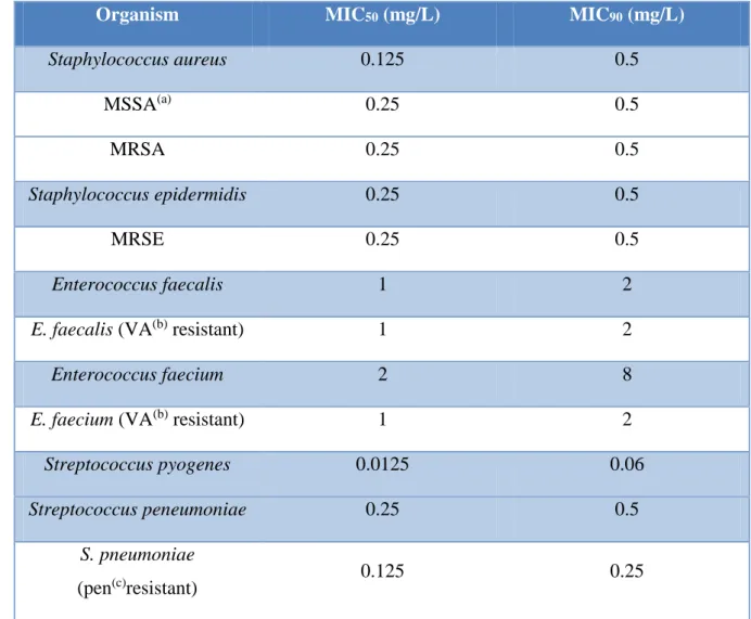
Daptomycin has activity against a broad range of Gram-positive aerobic and anaerobic bacteria, but no activity against Gram-negative bacteria (Enoch et al., 2007). Its major advantage is the bactericidal activity against species that are resistant to other antibiotics, including MRSA, penicillin-resistant Streptococcus pneumoniae (PRSP), vancomycin-resistant enterococci (VRE) and vancomycin-resistant Staphylococcus aureus (VRSA) (Nguyen et al., 2006). In vitro susceptibility data, i.e. minimal inhibitory concentrations, are presented in Table 1.1.
The European Committee on Antimicrobial Susceptibility Testing established the breakpoints for both staphylococci and streptococci strains (excluding S. pneumonia): ≤1 mg/L is susceptible and > 1 mg/ L is resistant. In the USA has been defined a breakpoint of 4 mg/L for enterococci (Enoch et al., 2007).
As a natural lipopetide antibiotic active against Gram-positive bacteria, particularly methicillin-resistant strains, daptomycin can be an interesting antibacterial tool to combat ocular infections caused by these microorganisms (Silva, 2012).
11
Table 1.1 – MIC50 and MIC90 of daptomycin among different species isolated. Adapted from
Enoch et al. (2007).
Organism MIC50 (mg/L) MIC90 (mg/L)
Staphylococcus aureus 0.125 0.5 MSSA(a) 0.25 0.5 MRSA 0.25 0.5 Staphylococcus epidermidis 0.25 0.5 MRSE 0.25 0.5 Enterococcus faecalis 1 2
E. faecalis (VA(b) resistant) 1 2
Enterococcus faecium 2 8
E. faecium (VA(b) resistant) 1 2
Streptococcus pyogenes 0.0125 0.06
Streptococcus peneumoniae 0.25 0.5
S. pneumoniae
(pen(c)resistant) 0.125 0.25
(a) MSSA: abbreviation for methicillin susceptible Staphylococcus aureus; (b) VA: abbreviation for vancomycin; (c) Pen: abbreviation for penincillin;
5.
Routes of drug delivery to the eye
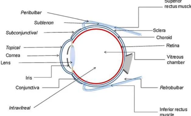
There are different possible routes of drug delivery into the ocular tissues, and the selection of the route of administration depends primarily on the target tissue (Urtti, 2006). The various routes of ocular drug delivery are represented in Figure 1.4.
Physicochemical properties of drugs, namely lipophilicity, solubility, molecular size, shape, charge and degree of ionization may affect the route and rate of permeation in cornea (Le Bourlais et al., 1998).
12
Figure 1.4 – Different routes of ocular drug delivery and their anatomical location (Gaudana et al., 2010).
5.1. Topical delivery
Topical administration, mostly in form of eye drops, is employed to treat anterior segment diseases and the site of action is usually different layers of the cornea, conjunctiva, sclera and the other tissues of the anterior segment such as the iris and ciliary body (Gaudana et al., 2010). However, the topical route is inefficient in delivering therapeutic concentrations of a drug to the posterior segment of the eye, owing to rapid drainage through the nasolacrimal ducts, low permeability of the corneal epithelium, systemic absorption and the blood-aqueous barrier (Thrimawithana et al., 2011). Typically, less than 5% of the applied drug penetrates the cornea and reaches intraocular tissues (Le Bourlais et al., 1998).
5.2. Systemic delivery
Following systemic administration, the blood aqueous barrier and the blood-retinal barrier are the major barriers for anterior and posterior segments ocular drug delivery, respectively, and restrict the entry of the therapeutic agents from blood (Gaudana et al., 2010). However, owing to the toxicity and delivery concerns, intravenous administration is not very common in treating ocular disorders (Gaudana et al., 2010).
13
5.3. Intravitreal delivery
Intravitreal injection of drugs into the eye involves direct injection of the formulation, in the form of solution, particles, suspension, depot or implants, into the posterior segment through the pars plana, a flat area in the ciliary body (Thrimawithana et al., 2011). Direct drug administration into the vitreous offers distinct advantage of more straightforward access to the vitreous and retina, providing increased drug concentrations at the neural retina and minimizing systemic side effects (Urtti, 2006; Thrimawithana et al., 2011). However, with intravitreal injections the drug distribution in the vitreous is non-uniform, as small molecules can rapidly distribute through the vitreous, whereas the diffusion of larger molecules is restricted, depending on the pathophysiological condition and the molecular weight of the administered drug (Gaudana et al., 2010).
5.4. Periocular delivery
The periocular route includes subconjunctival, subtenon, retrobulbar and peribulbar administration and is comparatively less invasive than intravitreal route, being considered the least painful and the most efficient route of drug delivery to the posterior eye (Gaudana et al., 2010; Thrimawithana et al., 2011). Drug solutions are placed in close proximity to the sclera which results in high retinal and vitreal concentrations as the periocular route enables the deposition of molecules against the external surface of the sclera because it is made up of fibrous tissue, which offers less resistance to permeability of drugs (Gaudana et al., 2010; Thrimawithana et al., 2011).
6. Ocular barriers
The delivery of drugs to the posterior segment of ocular tissue is prevented by ocular anatomical and physiological constraints, which include the relative impermeability of the corneal epithelial membrane, tear dynamics, nasolacrimal drainage and the high efficiency of the blood-ocular barrier (Ding, 1998).
14
6.1. Drug loss from the ocular surface
The exposed part of the eye is covered by a thin fluid layer, the so-called precorneal tear film that consists of a superficial lipid layer, a central aqueous layer and an inner mucus layer (Ludwig, 2005). After administration of eye drops, the most common dosage form, the flow of lacrimal fluid removes instilled compounds from the surface of the eye and the excess volume is flown to the nasolacrimal duct rapidly in a couple of minutes (Urtti, 2006).
In addition, most of small molecular weight drugs are absorbed into systemic circulation after few minutes, decreasing the drug concentration in lacrimal fluid extensively (Urtti, 2006). In this case, attempts to overcome the toxicity associated with the high initial concentration without a requirement for frequent dosing form is a challenging task, particularly in case of potent drugs (Ding, 1998).
6.2. Nasolacrimal drainage system
Corneal epithelium limits the drug absorption from the lacrimal fluid into the eye and is the major factor for drug loss that leads to poor ocular bioavailability. It is also the major route of entry into the circulatory system for drugs that are applied through topical administration (Urtti, 2006; Ding, 1998). The most apical corneal epithelial cells form tight junctions that limit the drug permeation, therefore lipophilic drugs have higher permeability in the cornea than the hydrophilic drugs (Urtti, 2006).
Furthermore, the systemic exposure through nasolacrimal drainage after topical administration can be sufficiently high to cause systemic toxicity (Ding, 1998).
6.3. Blood-ocular barrier
The eye is protected from xenobiotics in the blood stream by blood-ocular barriers and, consequently, the delivery of drugs to the retina from the peripheral circulation is also limited by this membrane impermeability (Urtti, 2006; Park et al., 2012). These barriers consist of two parts: blood-aqueous barrier and the blood-retina barrier, which are formed by complex tight junctions of retinal blood vessels and the RPE (Urtti, 2006; Park et al., 2012).
It is known that the anterior blood-eye barrier prevents the access of albumin into the aqueous humor and limits the access of hydrophilic drugs from plasma into the aqueous humor,
15
but these blood-eye barriers have not been extensively characterized in terms of drug transporter and metabolic enzyme expression (Urtti, 2006). The blood-ocular barrier can be overcome by intravitral injections of drugs, however this route of administration is associated with several problems, including risks of endophthalmithis (Bochot et al., 2000).
Cell culture models of ocular barriers constitute potent systems to explore the architecture, barrier function and regulation of ocular barriers in vitro. Monolayers of ARPE-19 cells have become a well-established in vitro model of the outer blood-retinal barrier (Hornof et al, 2005). This cell line was characterized by Dunn and colleagues (Dunn et al., 1996), who concluded that it has structural and functional properties characteristic of RPE cells in vivo and suggest that this cell line is valuable for in vitro studies of RPE physiology.
7.
Ophthalmic drug delivery systems
Ocular drug delivery remains among the most challenging approaches to the administration of therapeutic agents to human body, being circumvent of the ocular protective barriers in order to achieve therapeutically effective concentration of drugs in the intraocular tissues the ultimate task (Liu et al., 2012).
An idyllic ocular drug delivery system should hold the following characteristics:
- provide controlled and sustained release profile to keep therapeutic concentration of the medicine for a lengthy period of time, in order to shrink the regularity of administration;
- be precise on targeting and sustained holding in the sickly tissues, with the aim of increase therapeutic efficiency and moderate side effects;
- be patient friendly delivery routes that reduce or eradicate side effects resulting directly from these administration methods (Liu et al., 2012).
The ultimate goals are to improve relevant drug-related parameters, such as pharmacokinetics, pharmacodynamics, non-specific toxicity, immunogenicity, biorecognition and to improve therapeutic efficacy (Diebold and Calonge, 2010).
Controlled drug delivery systems aim to deliver drugs at predetermined rates and predefined periods of time, targeting drugs to a desirable group of cells. In this perspective, micro- and nano-scale intelligent systems can maximiza the efficacy of therapeutic treatments (Safari and Zarnegar, 2012)
16
Nanocarriers, such as nanoparticles, have the capacity to deliver ocular drugs to specific target sites, holding the promise to revolutionize the therapy of many eye diseases (Diebold and Calonge, 2010).
7.1. Nanoparticles for ocular drug delivery
Nanoparticles are particles of less than 1µm diameter, prepared from natural or synthetic materials, such as polymers (Hans and Lowman, 2002).
Nanoparticles can be prepared in different sizes, charge and other physicochemical features, conferring great versatility upon them; also, as a biomedical application, they should be biologically compatible with living tissues by not producing toxic, injurious or immunologic responses in them (Diebold and Calonge, 2010).
Nevertheless, the same properties that make nanoparticles attractive for biomedical applications may become reactive in biological systems and develop toxicity: for instance, smaller size nanoparticles are preferred for better interactions at the cellular level. However, smaller nanoparticles have larger surface area per unit mass, which may mean higher reactivity and consequently, cell or tissue toxicity (Diebold and Calonge, 2010).
Nanoparticles have the great advantage of higher drug loading capacity and higher stability in biological fluids and during storage compared to other similar carriers (Eljarrat-Binstock et
al., 2007).
Considering the fact that the cornea and conjunctiva have a negative charge, it was proposed that the use of mucoadhesive polymers, which may interact intimately with these extraocular structures, and increase the concentration and residence time of the associated drug (Motwani
et al., 2008).
Among the wide variety of natural polymers reported in literature, alginate and chitosan seem to have most of the desirable characteristics for the formulation of drug loaded nanoparticles for ocular delivery (Motwani et al., 2008).
17 7.1.1. Alginate
Alginate is a random, linear and anionic polysaccharide consisting of linear copolymers of α-L-guluronate and β-D-mannuronate residues (Motwani et al., 2008). Commercial alginate is primarily extracted from marine algae such as Laminaria hyperborean, Ascophyllum nodosum and Macrocystis pyrifera and its molecular variability depends on the source of algae, tissue from which alginate is extracted and also the season of crop harvesting (Liew et al., 2005). The composition, sequence of polymer blocks and molecular weight of alginate is also important, as these factors determine the physical properties of the gel formed (Liew et al., 2005).
Alginate is widely used in the pharmaceutical, cosmetic and food industry and their significance in the biomedical area is increasing, including the development of drug delivery systems and a variety of oral and topical pharmaceutical formulations, as they are biodegradable, non-toxic, biocompatible and mucoadhesive (Motwani et al., 2008). It has also been used in a variety of oral and topical pharmaceutical formulations and it has been specifically used for aqueous microencapsulation of drugs (Motwani et al., 2008). Alginate polymers are also hemocompatible, have not been found to accumulate in any major organs and show evidence of in vivo degradation (Motwani et al., 2008).
The use of alginate in the encapsulation of drugs for sustained release is already wide-ranging. For instance, Sangeetha and colleagues (Sangeetha et al., 2007) formulated sodium alginate nanospheres of amphotericin B by controlled gellification method and yielded particles with 419.6 nm approximately that were found to have better antifungal activity when compared to the free drug and yielded sustained release; Zahoor and colleagues (Zahoor et al., 2007) studied the chemotherapeutic potential of alginate nanoparticle-encapsulated econazole and other antitubercular drugs against murine tuberculosis and concluded that alginate nanoparticles are the ideal carriers of these drugs, and also reduce dosing frequencies.
Alginate has also been used in several ocular delivery systems, either alone or in combination with other materials (Zhu et al., 2012). Liu and co-workers (Liu et al., 2008) formulated an ophthalmic delivery system for gatifloxacin using alginate as gelling agent in combination with hydroxypropyl methylcellulose as a viscosity-enhancing agent and concluded that the mixture can be used as an in-situ gelling vehicle to enhance ocular bioavailability; the same authors (Liu
et al., 2012) developed a composite collagen hydrogel containing protein encapsulated alginate
18
mechanical strength, and excellent optical clarity for possible use as therapeutic lens for drug delivery or use as corneal substitute for transplantation.
An extensive literature review allows to conclude that, despite the many ocular applications of alginate already developed, none of them includes the use of alginate as a base for formulation of a potential topical delivery systems of antibiotics to the eye.
7.1.2. Chitosan
Chitosan is a cationic polysaccharide and a deacetylated form of chitin, which is the second-most abundant polymer in nature after cellulose (de la Fuente et al., 2010; Alonso and Sánchez, 2003). Many applications have been found in the food, pharmaceutical, textile, agriculture, water treatment and cosmetics industries (Kong et al., 2010).
Chitosan exhibits several favorable biological properties, such as biodegradability, non-toxicity, biocompatibility and mucoadhesiveness (de Campos et al., 2004). Furthermore, antimicrobial activity of chitosan has also been demonstrated against many bacteria, filamentous fungi and yeast (Kong et al., 2010).
Chitosan base is soluble in acidic solutions wherein it becomes protonated; this positive charge of the chitosan molecule enables its interaction with polyanions, a process that has been used to obtain complexes as well as micro and nanoparticulate drug delivery systems (Alonso and Sánchez, 2003).
Because of all these favorable properties and also the ability to increase mucosal epithelia permeability, chitosan is a very promising biomaterial in ophthalmology (Alonso and Sánchez, 2003). Moreover, it shows pseudoplastic and viscoelastic properties, desirable for ocular drug administration (de la Fuente et al., 2010).
Chitosan-alginate polyionic complexes are formed through the ionic gelation via interactions between the carboxyl groups of alginate and the amine groups of chitosan (Motwani et al., 2008). This complex revealed to protect the encapsulant and limit the release of encapsulated materials more effectively than either alginate or chitosan alone (Motwani et al., 2008).
19
7.1.3. Alginate-Chitosan polyelectrolyte complexes
Nanoparticles made of opposite surface charged polymers have potential application in ophthalmic delivery. Low stability and encapsulation efficiency have been observed with capsules formed by an alginate polymer (anionic), but these problems can be overcome using cationic polymers such as chitosan (Nagarwal et al., 2012). In fact, the combination of chitosan and sodium alginate has been widely investigated and is considered to be the most interesting chitosan-polyanion complex for colloidal carrier systems (Patil et al., 2011; Motwani et al., 2007). The formed nanoparticles are biocompatible, biodegradable, non-toxic, and capable to sustain the release of encapsulated materials more efficiently than either alginate or chitosan alone, which represents the major advantage for using chitosan coated alginate nanoparticles (Motwani et al., 2007; Nagarwal et al., 2012).
Motwani and colleagues (Motwani et al., 2007) designed chitosan-sodium alginate nanoparticles as a new vehicle for the prolonged topical ophthalmic delivery of gatifloxacin that revealed a fast release during the first hour followed by a more gradual release during a 24-hour period; Nagarwal and co-workers (Nagarwal et al., 2012) developed chitosan coated sodium alginate-chitosan nanoparticles loaded with 5-Fluorouracil for ophthalmic delivery and
in-vivo study in rabbit eye showed a great level of 5-FU in aqueous humor and high
bioavailability resulting from the mucoadhesiveness.
Despite the evident advantages of using alginate in combination with chitosan for ocular applications, it is possible to conclude, after a general literature review, that alginate-chitosan polymeric nanoparticles have never been used for encapsulation of antimicrobial peptides, such as daptomycin.
Topical administration of daptomycin into the eye by encapsulation into chitosan nanoparticles has been studied by Silva (Silva et al., 2013), concluding that chitosan nanoparticles are suitable for delivering daptomycin into the eye. However, considering the capacity of alginate-chitosan polymeric nanoparticles to sustain the release of encapsulated material more efficiently than chitosan nanoparticles, the development of chitosan coated sodium alginate nanoparticles for encapsulation of daptomycin and subsequent topical administration into the eye constitutes a promising system for treatment of bacterial endophthalmitis
20
8.
Objectives
The general objective of the present study is the improvement of an efficient topical delivery system of the novel antibiotic daptomycin to the posterior segment of the eye for the treatment of bacterial endophthalmitis. In order to achieve this objective, daptomycin will be encapsulated into chitosan coated alginate nanoparticles and its potential as ocular drug delivery system will be evaluated.
The specific objectives of this study include:
Preparation of feasible alginate-chitosan nanoparticles for encapsulation of daptomycin;
Physicochemical characterization of obtained nanoparticles and determination of drug loading capacity;
Determination of Minimal Inhibitory Concentrations (MICs) against some Gram-positive pathogens responsible for bacterial endophthalmitis for daptomycin solution and encapsulated daptomycin in alginate-chitosan nanoparticles, and establish the comparison between both;
Determination of the permeability of daptomycin solution and daptomycin-loaded nanoparticles in ARPE-19 and HCE cells monolayers;
21
II. Material and Methods
1.
Materials
Daptomycin (94.9% of purity) was offered by Cubist Pharmaceuticals, Inc. (Massachusetts, USA) and Novartis Pharma AG (Basel, Switzerland). Alginic acid sodium salt from brown algae (sodium alginate), low molecular chitosan (deacetylation degree of 85%) and acetic acid at 99.7% were acquired from Sigma-Aldrich® (Missouri, USA). Calcium chloride dehydrate (CaCl2.2H2O) was obtained from Merck (Darmstadt, Germany). Ultra-pure water was achieved
in our lab with a Millipore™ (Massachusetts, USA) water purification system.
Acetonitrile, trifluoroacetic acid and triethylamine were also purchased from Sigma-Aldrich® (Missouri, USA). Nanoparticles were characterized with ZetaPALS equipment from Brookhaven Instruments Corporation (New York, USA) and drug loading capacity was determined using a Beckman Coulter System Gold HPLC system (California, USA).
Nutrient Broth and Nutrient Agar mediums were obtained from Lab M™ (Lancashire, United Kingdom) and Mueller-Hinton medium was acquired from Biokar Diagnostics (Beauvais, France). 96-well microplates were purchased from Nunc (Roskilde, Denmark).
For cell culture assays, Dulbecco’s Modified Eagle Medium (DMEM) was purchased from PAA Laboratories (Pasching, Austria), Hanks Balanced Salt Solution (HBSS) was obtained from Biowhittaker (Verviers, Belgium), tissue culture test plates were acquired from Orange Scientific (Braine-l’ Alleud, Belgium) and 6-well- format cell culture transwell inserts were purchased from BD Falcon (New Jersey, USA).
2.
Development of chitosan coated sodium-alginate nanoparticles
2.1. Unloaded nanoparticles
The main purpose for previous formulation of unloaded nanoparticles is to achieve the best conditions of alginate-chitosan nanoparticles formulation to be used in daptomycin-loaded nanoparticles.
22
Unloaded chitosan coated alginate nanoparticles were prepared following the method of Zahoor et al. (2005) with slight modifications, using cation-induced controlled gellification of alginate. Nanoparticles were prepared from dilute alginate solution by inducing an ionotropic pre-gelation with calcium counter ions, followed by polyelectrolyte complex coating with chitosan.
For that purpose, 0.5 mL of CaCl2 and 2 mL of chitosan solutions were added dropwise to
9.5 mL of alginate solution, followed by stirring for 30 minutes. The pH of the final solution was adjusted to 3.5-3.6, in order to keep the conditions of formulation of unloaded and loaded nanoparticles similar, as at these pH values daptomycin is positively charged and alginate is negatively charged, allowing the polyelectrolyte interaction between them. Nanoparticles were stored at room temperature overnight.
Based on the concentrations of alginate, chitosan and CaCl2used by Zahoor (Zahoor et al.,
2005), it were first prepared the stock solutions. Alginate stock solutions were prepared by dissolving different amounts of alginate into ultrapure water, achieving concentration of 0.3, 0.6, 0.9 and 1.2 mg/mL. Chitosan was dissolved overnight in different acetic acid solutions (also prepared with ultrapure water), achieving stock solutions with concentrations of 0.2, 0.5 and 1.0 mg/ mL. At last, stock solutions of CaCl2 were set with ultrapure water, with
concentrations of 1.14 and 2.64 mg/ mL.
Table 2.1 - Different concentrations of alginate, chitosan and CaCl2used in the final solution
to prepare unloaded chitosan coated sodium alginate nanoparticles.
Alginate Concentration (mg/mL) Chitosan Concentration (mg/mL) CaCl2 Concentration (mg/mL) Alginate: Chitosan mass ratio 0.24 0.08 0.11 1: 0.07 0.48 0.04 0.11 1: 0.018 0.08 0.06 1: 0.035 0.11 1: 0.035 0.17 0.11 1: 0.07 0.71 0.08 0.11 1 : 0.024 0.95 0.08 0.11 1 : 0.018
After nanoparticle formation, alginate achieved final concentrations of 0.24, 0.48, 0.71 and 0.95 mg/ mL, chitosan achieved the concentrations of 0.04, 0.08 and 0.17 mg/mL and CaCl2
23
solutions the final concentrations of 0.06 and 0.11 mg/ mL, as suggested by Zahoor (Zahoor et al., 2005). In all cases, it was guaranteed that chitosan and CaCl2 concentrations were not
superior to alginate concentration. The different combinations of final concentrations used to prepare the nanoparticles are presented in table 2.1.
Considering the physicochemical characteristics of the nanoparticles obtained using these formulations, it was then prepared another set of unloaded nanoparticles using stock solutions of alginate with 0.1 mg/ mL, stock solutions of chitosan with 0.1, 0.3 and 0.5 mg/ mL and stock solutions of calcium chloride of 1.0, 1.5 and 2.0 mg/ mL. From these stock solutions, new nanoparticles were obtained with the concentrations in the final solution presented on table 2.2.
All alginate nanoparticles were characterized for their size, polidispersity index and zeta potencial.
Table 2.2 - Different concentrations of alginate, chitosan and CaCl2used in the final solution
to prepare unloaded chitosan coated alginate nanoparticles, after preliminary study.
Alginate Concentration (mg/mL) Chitosan Concentration (mg/mL) CaCl2 Concentration (mg/mL) Alginate: Chitosan mass ratio 0.08 0.02 0.04 1 : 0.05 0.06 0.08 0.05 0.04 1 : 0.14 0.06 0.08 0.08 0.04 1 : 0.22 0.06 0.08
2.2. Daptomycin-loaded nanoparticles
To prepare daptomycin-loaded nanoparticles, 0.5 mL of CaCl2 (0.04 mg/mL in the final
solution) was added to 9.5 mL of alginate (0.08 mg/mL in the final solution) containing different amounts of daptomycin, as described in table 2.3. The pH was adjusted to 3.5 - 3.6, because at this pH value, daptomycin molecules have an electric positive charge and the
24
alginate solution has a negative charge, enhancing electrical binding between alginate and daptomycin (Silva et al., 2013; Tønnesen and Karlsen, 2002).
After that, 2.0 mL of chitosan solution (0.02 mg/mL in the final solution) were then added, followed by stirring for 30 minutes and nanoparticles were stored at room temperature overnight. Alginate, chitosan and calcium chloride concentrations were chosen based in the optimal particle size, polydispersity index and zeta potential results obtained for unloaded nanoparticles.
Table 2.3 - Different concentrations of alginate, chitosan and CaCl2and daptomycin mass in
the final solution used to prepare chitosan coated nanoparticles loaded with daptomycin.
Alginate (mg/ mL) Chitosan (mg/ mL) CaCl2 (mg/mL) Daptomycin (mg/ mL) Alginate : Daptomycin mass ratio 0.08* 0.02 * 0.04 * 10 * 1 : 0.01 20 * 1: 0.03 40 * 1: 0.05 80 * 1 : 0.1 160 * 1 : 0.2 330 * 1 : 0.4
* Concentration in the final nanoparticle suspension
An aliquot of nanoparticle suspension was transferred into a cuvette for size, polydispersity index and zeta potential characterizing in the ZetaPALS equipment. Posterior, daptomycin loaded nanoparticles were recovered by centrifugation 5000 rpm for 120 minutes and the supernatants were collected for determination of daptomycin loading capacity through the reverse-phase high performance liquid chromatography (RP-HPLC).
25
3.
Physicochemical characterization of nanoparticles
Both loaded and unloaded alginate nanoparticles were submitted to deep physico-chemical characterization. Particle size, polydispersity index and Zeta potential were determined using a ZetaPALS equipment (Brookhaven Instruments Corporation, New York, USA).
Particle size and polydispersity index were determined by dynamic light scattering, using a 90º scattering angle and a temperature of 25ºC for the analysis. Zeta potential was measured by phase analysis light scattering technique with Smoluchowski model, using the same cell. Samples were submitted to 6 runs with 20 cycles each, at the temperature of 25ºC.
4.
Determination of encapsulation efficiency of daptomycin in
nanoparticles
The amount of daptomycin entrapped in alginate nanoparticles was calculated by estimating the amount of free drug in the supernatant after centrifugation of nanoparticles (Zahoor et al., 2005)
The daptomycin encapsulation efficiency was calculated as the percentage of drug entrapped in alginate nanoparticles compared with the initial amount of drug, using the following equation (1):
(1) Encapsulation Efficiency (%) = Final concentration of drug - drug concentration in supernatant x 100 Final concentration of drug
The daptomycin concentration in supernatant was determined by RP-HPLC, using the method of Martens-Lobenhoffer et al. (2008) with slightly modifications.
4.1. Instrumentation
The HPLC system consisted of a Beckman Coulter System Gold comprising a System Gold 508 Autosampler, a System Gold 126 Solvent Module, a System Gold 168 detector and a Gecko 2000 Column Oven. The analytical column used was a Zorbax Eclipse XDB-C8 150mm x 4.6mm 5 µm particle size (Agilent Technologies, Böblingen, Germany).
26
4.2. Chromatography method setup
The mobile phase for the chromatographic separation of daptomycin was mixed from pure acetonitrile and buffer solution, which consisted of 20mM trifluoroacetic acid and 15mM triethylamine, resulting in a pH of about 3.5 (Martens-Lobenhoffer et al., 2008). Both solutions were filtered under vacuum and sonicated for 15 minutes.
The applied gradient starts as 30% acetonitrile, rises to 40% in 5 minutes, then set constant until 11 minutes. After 11 minutes the column is flushed for 3 minutes with 100% acetonitrile. The flow rate was constant to 0.700 mL/min and the column temperature was adjusted to 30ºC. The volume injected was 50 µL. Detention took place at a wavelength of 224 nm (Martens-Lobenhoffer et al., 2008).
4.3. Preparation of calibration solutions and calibration function
First, it was prepared a daptomycin stock solution in ultra-pure water, achieving the final concentration of 5000 µg/mL. From the dilution of this stock solution, a working solution of 500 µg/mL was obtained and this one was also diluted, acquiring another 50 µg/mL working solution.
From the stock solution with final concentration of 500 µg/mL, the calibration solutions with concentrations of 500, 400, 350, 250, 140, 70 and 35 µg/mL were prepared. From the working solution with the final concentration of 50 µg/mL, the calibration solutions with concentrations of 25, 15, 12, 10, 8, 6, 4, 2, 1 and 0.5 µg/mL were set.
Calibration solutions were filtered with 0.22 µm filters and analyzed by RP-HPLC.
5.
Determination of Minimum Inhibitory Concentrations for Daptomycin
Determination of Minimum Inhibitory Concentration (MIC) was executed following standards for antimicrobial susceptibility testing, provided by Clinical and Laboratory Standards Institute (CSLI) in documents M7-A6 and M100-S15, and the procedure for susceptibility testing of daptomycin by broth microdilution method, provided by Novartis®. Both guidelines recommend the use of Broth Microdilution Method.
27
Assays were performed in 96-well microplates, allowing to compare the MIC of free daptomycin with the MIC of entrapped daptomycin, at concentrations of 0.03, 0.06, 0.12, 0.25, 0.5, 1.0, 2.0, 4.0, 8.0 and 16.0 µg/ mL, against the following organisms: methicillin-susceptible
Staphylococcus aureus ATCC 25913 (MSSA), methicillin-resistant Staphylococcus aureus
ATCC 43300 (MRSA), Staphylococcus epidermidis ATCC 14990, Staphylococcus capitis ATCC 27840, Staphylococcus hominis ATCC 13348, Staphylococcus lugdunensis ATCC 43809, Staphylococcus haemolyticus ATCC 29970 and Staphylococcus warmeri ATCC 13354.
First, it was performed a calcium adjustment of Mueller-Hinton broth to a final concentration of 50 mg/ L Ca2+, using a calcium stock solution. Inocula were prepared by suspending each bacteria colony into the calcium adjusted Mueller-Hinton broth, achieving a turbidity equivalent to 0.5 McFarland standard (1x10⁸ CFU/ mL), and then diluted in the calcium adjusted Mueller- Hinton, to reach the recommended concentration of bacteria in wells, 5 x 10⁵ CFU/ mL.
Fitfty microliters L of each inoculum were transferred to the microplate and every well was fulfilled with free daptomycin or daptomycin-loaded nanoparticles at concentrations cited above.
Further on, three controls were also done at the same time: the first one containing inoculum and Mueller-Hinton broth, the second one containing only daptomycin (free or entrapped) and the third one containing Mueller-Hinton broth.
6.
Determination of the permeability of daptomycin in ocular cells
The drug permeability prediction across the ocular tissues is important in the development of new drugs and drug delivery strategies. It is known that cornea is the major absorption route for topically applied drugs and corneal epithelium represents the rate-limiting barrier for transcorneal permeation (Hornof et al., 2005; Barar et al., 2009). In this experiment, permeability of daptomycin in corneal and blood-retinal epitheliums was tested using cell culture models.
Assays for determination of daptomycin permeability in ocular cells were realized for ARPE – 19 (human retinal pigment) and HCE (human corneal epithelium) epithelial cell lines, which are widely used in cell culture models, as they both have the structural and functional properties of retinal epithelium and corneal epithelium in vivo, respectively (Dunn et al., 1996; Barar et
28
al., 2009). This experiment was performed following the method used by Geiger and
co-workers (Geiger et al., 2005), with slight modifications.
6.1. Cell culturing
Human adult ARPE-19 and HCE cells were grown in DMEM supplemented with 10% fetal bovine serum (FBS), 1% penicillin-streptomycin and 1% amino acids mixture, on tissue culture-treated T-25 and T-75 flasks, at 37ºC. Medium was substituted every 2 – 3 days, until confluence was achieved.
6.2. Cell seeding
Cells were then plated on 6-well tissue culture test plates provided with PET membranes inserts: for each cell line, medium was aspired, cells were washed with PBS, incubated with trypsin for 5 minutes at 37ºC and DMEM was added again. For each well, it was passed 2.5 mL of DMEM, Transwell® inserts were then collocated and 1.5 mL of DMEM and cells were added, 120.000 for ARPE-19 cells and 100.000 for HCE cells. Figure 2.1 presents, schematically, the in vitro cell culture mode.
Figure 2.1 – Schematic representation of the in vitro cell culture model, using transwell inserts (Barar et al., 2009).
Medium was replaced once a week (2.5 and 1.5 mL from basolateral and apical compartments, respectively) and transepithelial resistance (TER) was measured with a
29
voltmeter, until 80-110 mΩ value was achieved. The final concentration of cells in each well was 200.000 cell/ cm2 for ARPE-19 cell line, and 90.000 cell/cm2 for HCE cell line.
6.3. Permeability Experiment
Thirty minutes before start of the experiment, cells were rinsed with complete HBSS (2.5 mL basolaterally and 1.5 mL apically). At the start of the experiment, the complete volume of HBSS was removed and replaced with new HBSS, with the respective volumes. Daptomycin in free and encapsulated forms was added to apical compartment.
Permeability measurements were made at 15, 30, 60, 120,180 and 240 minutes, removing 100 µL aliquots from basolateral compartment and adding 100 µL of HBSS to the same compartment. Removed aliquots were then measured for daptomycin concentration using RP-HPLC.
7.
Statistical Analysis
Obtained results were statistical analyzed with IBM SPSS Statistics 19, using Linear Regression, T-student for paired samples and One-Way Analysis of Variance (ANOVA) with Tukey HSD post-hoc test. The differences were considered significant at a level of 95% (p < 0.05).