INSTITUTO DE CIÊNCIAS BIOMÉDICAS ABEL SALAZAR
Sofia
Paulino
. Epigenetic Biomarkers in Diffuse Large B-Cell Lymphoma
Epigenetic B
iomarkers i
n D
iffuse L
arge B
-Cell L
ymphoma
Sofia M
argarida d
e C
astro P
aupério e
S
ilva P
aulino
2017M
.ICBAS
2017
MESTRADO ONCOLOGIA MOLECULAREpigenetic Biomarkers in Diffuse
Large B-Cell Lymphoma
Sofia Paupério Paulino
Sofia Margarida de Castro Paupério e Silva Paulino
EPIGENETIC BIOMARKERS IN DIFFUSE LARGE B-CELL LYMPHOMA
Dissertação de Mestrado em Oncologia apresentada ao Instituto de Ciências Biomédicas Abel Salazar da Universidade do Porto – Especialização Oncologia Molecular Orientador
Professor Doutor Rui Manuel Ferreira Henrique
Diretor, Serviço de Anatomia Patológica
Diretor, Escola Portuguesa de Oncologia do Porto (EPOP) Investigador sénior, Grupo de Epigenética e Biologia do Cancro - Centro de Investigação
Instituto Português de Oncologia do Porto Francisco Gentil, E.P.E.
&
Professor Catedrático Convidado
Departamento de Patologia e Imunologia Molecular
Instituto de Ciências Biomédicas Abel Salazar (ICBAS) da Universidade do Porto
Co-orientador
Professora Doutora Carmen de Lurdes Fonseca Jerónimo
Coordenadora do Grupo de Epigenética & Biologia do Cancro Centro de Investigação-LAB 3 (Edifício F, 1º piso)
Instituto Português de Oncologia do Porto Francisco Gentil, E.P.E.
&
Prof. Associada Convidada com Agregação Diretora do Mestrado em Oncologia
Instituto de Ciências Biomédicas Abel Salazar (ICBAS) da Universidade do Porto
Epigenetic biomarkers in Diffuse large B-cell lymphoma
V
A realização desta dissertação não seria possível sem o apoio, colaboração e compreensão de diversas pessoas, pertencentes à minha vida profissional e pessoal, a quem quero agradecer e passo a citar:
Ao meu orientador e diretor, Professor Doutor Rui Henrique, que com o seu “saber”, o seu “saber ser” e “saber estar” prontamente disponibilizou todo o seu apoio, orientando-me desde o início nesta caminhada. O orientando-meu muito obrigado pelo voto de confiança.
À minha co-orientadora, Professora Doutora Carmen Jerónimo, que prontamente me acolheu no seu grupo de trabalho. O seu cuidado e preocupação foram uma constante em cada fase de desenvolvimento do mesmo.
À minha coordenadora, Técnica Maria do Amparo e à minha supervisora na Histologia, Fernanda, que permitiram uma gestão e flexibilidade de horário de forma a que eu conseguisse conciliar o trabalho e a realização da tese.
A todos os meus colegas do Serviço de Anatomia Patológica do IPO-Porto, pelo apoio e compreensão durante este período, em especial ao Jorge e ao Diogo, que já fizeram esta caminhada e foram-me dando algumas “orientações” e à Paula e Renata pelas “dicas” relativas à imunocitoquímica.
À Paulinha, pelo incentivo constante, desde o primeiro dia em que falámos sobre a realização do mestrado.
À Isa, que de muito perto acompanhou toda esta fase, e sempre se demonstrou disponível para me ajudar. Um agradecimento especial pela paciência e pelos desabafos tão importantes para a minha tomada de consciência.
À Dra. Mariana Afonso, pelas palavras de carinho e incentivo constante.
Ao Dr. João Lobo, pela valiosa colaboração durante o desenvolvimento do trabalho. Ao Engenheiro Luís Antunes, pela preciosa colaboração no tratamento dos dados e análise estatística.
A todos os elementos do Grupo de Epigenética e Biologia do Cancro que conheci neste período, em especial à Sofia Salta, com quem trabalhei mais diretamente, mostrando-se disponível para ajudar sempre que foi necessário.
A todos os meus colegas do Mestrado em Oncologia, com quem partilhei ansiedades e que de diferentes formas contribuíram para a realização deste trabalho.
Epigenetic biomarkers in Diffuse large B-cell lymphoma
VI
Às minhas amigas de sempre, Andreína e Marta que estiveram sempre do meu lado, privando-se da minha companhia em diversos momentos, mas incentivando-me sempre que algo corria menos bem.
E por último, mas de forma alguma menos importante, à minha família:
Aos meus irmãos, que apesar das suas exigências profissionais, estiveram e estão sempre presentes.
Aos meus avós, em especial ao meu avô Paupério, que num ano extremamente difícil, nunca deixou de se preocupar com os netos, nem de os acompanhar nos percursos académicas, inclusive esta minha caminhada.
E aos meus pais, a quem devo tudo o que sou. A quem tenho de agradecer pela educação proporcionada e princípios incutidos. Têm sido o exemplo de que só conseguimos alcançar os nossos objetivos através do trabalho, espírito de sacrifício, nunca esquecendo valores como o respeito, simplicidade e humildade. Sempre procuraram adquirir novos conhecimentos e competências, conseguindo atingi-las mesmo em alturas mais complicadas. O vosso exemplo encorajou-me a fazer o mesmo e embarcar nesta caminhada. E não há palavras suficientes para expressar o meu mais sincero agradecimento por todo o apoio que me têm dado. Muito obrigada!
Epigenetic biomarkers in Diffuse large B-cell lymphoma
VII
RESUMOO Linfoma difuso de grandes células B (LDGCB) é o subtipo mais comum dos Linfomas Não-Hodgkin, correspondendo a cerca de 30% a 40% de novos casos e a mais de 80% dos linfomas agressivos. Devido à sua heterogeneidade clínica, morfológica, imunológica e genética, torna-se pertinente, aquando do diagnóstico, identificar os doentes que irão beneficiar da administração de um tratamento mais agressivo. O sistema de classificação segundo o estadio Ann Arbour, não é, por si só, suficiente, sendo necessário a utilização do Índice Internacional de Prognóstico como ferramenta mais completa. No entanto, têm surgido novos marcadores de prognóstico. Assim, torna-se essencial um melhor conhecimento das alterações epigenéticas, que constituem o alvo do presente estudo, para a aplicação de decisões terapêuticas ajustadas e personalizadas na tentativa de obter melhores resultados clínicos
Neste estudo, tivemos por objetivo avaliar a expressão das modificações epigenéticas pós-traducionais das histonas H3K4me3 (trimetilação da lisina 4 da histona H3) e H3K27me3 (trimetilação da lisina 27 da histona H3), correlacionando-as com os parâmetros clínicos e patológicos, determinando o seu potencial como biomarcadores de prognóstico em LDGCB.
Com este propósito, foi realizado um estudo transversal retrospetivo baseado em 155 casos de Linfoma difuso de grandes células B consecutivamente diagnosticados e tratados no Instituto Português de Oncologia do Porto, entre 2008 e 2013, do qual existia material biológico de arquivo disponível para avaliação da imunoexpressão das marcas epigenéticas H3K4me3 e H3K27me3, utilizando um sistema de quantificação baseado em análise de imagem digital para determinar os parâmetros “percentagem de células positivas” e o “Hscore”. Foi utilizada a base de dados do Serviço de Oncohematologia para obtenção dos parâmetros clínicos e patológicos relevantes.
Com base na análise da sobrevivência, o sistema de classificação Ann Arbour agrupado (p=0.015), o IPI agrupado (p=0.041) e a idade quando o diagnóstico (≤ ou > 60 anos e ≤ ou > 70 anos, p=0.006 e p=0.005, respetivamente) foram confirmados como fatores de prognóstico. Na avaliação das marcas epigenéticas H3K4me3 e H3K27me3, não se observou associação com a sobrevivência global nem com a sobrevivência livre de doença, embora se tenha identificado uma associação entre o IPI (p=0.41) e a marca epigenética H3K27me3.
Epigenetic biomarkers in Diffuse large B-cell lymphoma
VIII
A coorte de pacientes estudados é globalmente representativa dos casos de LDGBCB, conforme atestado pela verificação da associação entre diversos parâmetros clínicos e o prognóstico. Contudo, as marcas epigenéticas H3K4me3 e H3K27me3 não revelaram valor prognóstico nesta neoplasia. Fatores como a dimensão da amostra e a ausência de categorização de acordo com a célula de origem poderão contribuir para explicar este resultado negativo. Assim, será necessário incrementar a série e melhor caracterizá-la imunofenotipicamente para concluir mais definitivamente sobre o potencial valor clínico deste biomarcadores epigenéticos.
Palavras-Chave: Linfoma não Hodgkin, linfoma difuso de grandes células B, epigenética,
Epigenetic biomarkers in Diffuse large B-cell lymphoma
IX
SUMMARYDiffuse large B-cell lymphoma (DLBCL), a heterogenous category of aggressive lymphoid malignancies, is the most common type of B-cell non-Hodgkin lymphoma, accounting for approximately 30 to 40% of all newly diagnosed cases worldwide and 80% of aggressive lymphomas. The DLBCL lymphomagenesis remains complex and mostly unknown. Prognostic information for most NHL is not accurately provided by the Ann Arbour system and it is of limited value for treatment decisions. International Prognostic Index (IPI) and R-IPI remain the most reliable tools for predicting outcome in patients with DLBCL. Additional prognostic markers are under investigation. Epigenetic changes are now recognized as playing an important and early role in the lymphomagenesis process, contributing to the disease progression.
In this study, we aimed to quantitatively evaluate the expression of the post-translational epigenetic modification of H3K4me3 (trimethylation of lysine 4 on histone H3) and H3K27me3 (trimethylation of lysine 27 of histone 3) in a series of diffuse large B-cell lymphoma through immunoexpression and correlation with clinicopathological parameters, to determine its relevance as prognostic biomarker.
A retrospective transversal study was performed using archival biological material from 155 patients with diffuse large B-cell lymphoma, consecutively diagnosed and treated between 2008 and 2013 at the Portuguese Oncology Institute of Porto. The H3K4me3 and H3K27me3 marks were assessed by immunohistochemistry, using a digital imaging assisted system to determine the % of positively immunostained cells and the Hscore. Relevant clinical and pathological data was extracted from the database of the Department of Oncohematology.
In this study, grouped Ann Arbour stage (p=0.015), grouped IPI score (p=0.041) and age at the time of diagnosis (≤ or > 60 years and ≤ or > 70 years, p=0.006 and p=0.005, respectively) were all significantly associated with prognosis. Quantitative H3K4me3 and H3K27me3 imnunoexpression did not associate with overall or disease-specific survival, although a significant association between IPI score and H3K27me3 mark was disclosed (p = 0.041).
Our patient cohort is globally representative of DLBCL patients as depicted by the significant associations between established prognostic parameters and patient outcome. Nevertheless, H3K4me3 and H3K27me3 immunoexpression were not shown to display prognostic value in this dataset. This negative result might be partially explained by insufficient sample size and lack of discrimination of DLBCL subtypes according to cell of
Epigenetic biomarkers in Diffuse large B-cell lymphoma
X
origin. Thus larger studies, with increased sample size to provide a more robust statistical analysis and additional immunophenotyping for DLBCL subtyping are required to more conclusively determine whether H3K4me3 and H3K27me3 might be of prognostic value in DLBCL.
Keywords: Diffuse large B-cell lymphoma, epigenetics, molecular biomarkers,
Epigenetic biomarkers in Diffuse large B-cell lymphoma
XI
TABLE OF CONTENTSINTRODUCTION _____________________________________________________ 1
1 – DIFFUSE LARGE B-CELL LYMPHOMA _____________________________________ 7 1.1 - EPIDEMIOLOGY AND ETIOLOGY ______________________________________________ 7 1.2 - SITES OF INVOLVEMENT AND CLINICAL FEATURES _____________________________ 7 1.3 - MORPHOLOGY ____________________________________________________________ 8 1.3.1 – Morphological variants __________________________________________________ 8 1.3.2 – Molecular subgroups ____________________________________________________ 9 1.3.2 – Immunophenotipically subgroups __________________________________________ 9 1.4 - STAGING __________________________________________________________________ 11 1.5 - PROGNOSIS _______________________________________________________________ 11 1.6 - THERAPY _________________________________________________________________ 13 2 – EPIGENTICS __________________________________________________________ 14 2.1 - DNA METHYLATION _________________________________________________________ 14 2.2 - HISTONE MODIFICATION ____________________________________________________ 15 2.2.1 - Histone lysine methylation _______________________________________________ 16
2.3 – EPIGENETICS IN DIFFUSE LARGE B-CELL LYMPHOMA ________________________ 18
AIMS OF THE STUDY _________________________________________________ 19
MATERIAL AND METHODS ____________________________________________ 23
1 – PATIENTS AND SAMPLES ____________________________________________________ 25
2 – IMMUNOHISTOCHEMISTRY __________________________________________________ 25 2.1 – EPITOPE RETRIEVAL AND ENDOGENOUS PEROXIDASE NEUTRALIZING ____________ 25
3 – NUCLEAR IMMUNOSTAINING QUANTIFICATION ____________________________ 27
4 – STATISTICAL ANALYSIS ________________________________________________ 29
RESULTS ________________________________________________________________ 31
1 – CNILICAL AND PATHOLOGICAL CHARACTERIZATION OF THE PATIENTS ________ 33
2 – SURVIVAL ANALYSIS ________________________________________________________ 33
Epigenetic biomarkers in Diffuse large B-cell lymphoma
XII
DISCUSSION _______________________________________________________ 39
CONCLUSIONS AND FUTURES PERPECTIVES _________________________________ 45
REFERENCES ______________________________________________________ 49
ANNEX ____________________________________________________________ I
Epigenetic biomarkers in Diffuse large B-cell lymphoma
XIII
FIGURE INDEXFigure 1 - Estimated age-standardized incidence rates of NHL per 100,000, worldwide ____ 3 Figure 2 - Estimated incidence of distinct types of cancer in western Europe and _________ 4 Portugal in 2012.
Figure 3 - Diffuse large B-cell lymphoma typical cells _______________________________ 8 Figure 4 - Hans algorithm based on the immunohistochemical analysis of three markers ___ 10 (CD10, Mum-1 and Bcl-6).
Figure 5 - Example of immunohistochemical analysis for DLBCL ______________________ 10 Figure 6 - DNA methylation ___________________________________________________ 14 Figure 7 - Function of chromatin modifying and organizing genes _____________________ 15 Figure 8 - Deregulation of H3K27 methylation in cancer _____________________________ 17 Figure 9 - Equipment support for the digital image analysis system GenASIs™ __________ 27 Figure 10 - Example of capture digital images of GenASIs™ software __________________ 27 Figure 11 - Example of nuclear classification into 4 levels of intensity on GenASIs™ ______ 28 Figure 12 - Analysis of disease-specific survival according to individual or grouped _______ 34 Ann Arbour stage
Figure 13 - Analysis of disease-specific survival according to grouped IP score __________ 34 Figure 14 - Analysis of disease-specific survival according to age _____________________ 35 Figure 15 - Analysis of disease-specific survival according gender _____________________ 37 Figure 16 - Positive and negative nuclear immunostaining of H3K4me3 ________________ 36 Figure 17 - Positive and negative nuclear immunostaining of H3K27me3 _______________ 37 Figure 18 - Analysis of disease-specific survival by % Positivity, in the left panel _________ 38 and Hscore, on the right panel. The comparison is made using the median.
Figure 19 - Analysis of disease-specific survival by % Positivity, in the left panel __________ 39 and Hscore, on the right panel. The comparison is made using the 25th percentile.
Figure 20 - Analysis of disease-specific survival by % Positivity, in the left panel __________ 39 and Hscore, on the right panel. The comparison is made using the 75th percentile.
Epigenetic biomarkers in Diffuse large B-cell lymphoma
XV
TABLE INDEXTable 1 – 2016 Classification of mature B-cell, T and NK neoplasm ____________________ 4 Table 2 – Ann Arbour Classification _____________________________________________ 12 Table 3 – International Prognostic Index _________________________________________ 12 Table 4 – Eastern Cooperative Oncology Group Performance Status __________________ 13 Table 5 – Antibodies and conditions ____________________________________________ 26 Table 6 – Clinical and pathological characteristics of the patients ______________________ 33 Table 7 – Comparison between Hscore, clinical and pathological parameters (H3K4me3) __ 36 Table 8 - Comparison between Hscore, clinical and pathological parameters (H3K27me3) _ 37
Epigenetic biomarkers in Diffuse large B-cell lymphoma
XVII
LIST OF ABBREVIATIONSABC Activated B-cell like
BCR B-cell receptor
CHOP Cyclophosphamide, hydroxydaunorubicin, oncovin and prednisone
COO Cell of origin
CpG Cytosine-phosphatidyl-guanine
CpGs Cytosine-phosphatidyl-guanine islands
DLBCL Diffuse large B-cell lymphoma
DNMT DNA methytransferase
ECOG Eastern cooperative oncology group EUA United States of America
EZH2 Enhancer of zeste homolog 2
GCB Germinal center B-cell like
H3K27me3 Trimethylation of lysine 27 on histone H3
H3K4me3 Trimethylation of lysine 4 on histone H3
HHV8 Human herpes virus 8
HIV Human immunodeficiency virus
HP Helicobacter pylori
ICD-O International classification of diseases for Oncology IPI International prognostic index
LDH Serum lactate dehydrogenase
LH Hodgkin’s Lymphomas
MLL Mixed lineage leukemia
ncRNA Non-coding RNA
Epigenetic biomarkers in Diffuse large B-cell lymphoma
XVIII
NLH Non-Hodgkin’s Lymphomas
NOS Not otherwise specified
OS Overall survival
PMBCL Primary mediastinal large B-cell lymphoma
PRC2 Polycomb repressive complex 2
R-CHOP Rituximab, cyclophosphamide, hydroxydaunorubicin, oncovin and
prednisone
R-IPI Re-categorized international prognostic index
WHO World Health Organization
Epigenetic biomarkers in Diffuse large B-cell lymphoma
3
Lymphoma is the malignant neoplasm from lymphoid system cells. It is predominantly located in the lymph nodes or other extra lymph node location, such as spleen, bone marrow, skin, gastrointestinal tract and other organs(1,2). The World Health
Organization (WHO) classifies lymphomas into two major groups, comprising Hodgkin's Lymphomas (LH) and Hodgkin's Lymphomas (NHL). Hodgkin lymphoma and Non-Hodgkin lymphoma can be differentiated pathologically, by its pattern of disease, treatment and prognosis (2).
Non-Hodgkin’s Lymphomas are a heterogeneous group of diseases that originate from mature and immature B, T or Natural Killer (NK) cells (2), representing 4% of all cancers,
which are more frequent in developed countries, such as United States of America (EUA), Australia and Western Europe (2,3) (Figure 1).
In Western Europe, the estimated incidence in 2012 was 34,188 new cases, accounting for 3.1% of all diagnosed cancers (Figure 2) (3). The same trend was reported
for Portugal, in which the estimated incidence rate, for the same year, was 1,842 new cases, accounting for 3.7% of all cancers (Figure 2) (3).
o Data .6 .6 . . . . 7.0 7.0 on Hodgkin lymphoma Source GL A 0 IAR Incidence ASR oth se es
Epigenetic biomarkers in Diffuse large B-cell lymphoma
4
In 2016, the WHO classification of hematopoietic and lymphoid tumours was revised
(4). Although alterations are limited compared with the 2008 version, it will include a large
set of information published over the last 8 years with significant diagnostic, prognostic and therapeutic implications (4). The updated classification of the mature B-cell neoplasms is
depicted in Table 1 and constitutes the worldwide gold standard.
Table 1 – 2016 WHO classification of Mature B-Cell, T and NK neoplasms. Adapted from (4).
Mature B-cell neoplasm
Chronic lymphocytic leukemia/small lymphocytic lymphoma Monoclonal B-cell lymphocytosis*
B-cell prolymphocytic leukemia Splenic marginal zone lymphoma Hairy cell leukemia
Splenic B-cell lymphoma/leukemia, unclassifiable
• Splenic diffuse red pulp small B-cell lymphoma
• Hairy cell leukemia-variant
Lymphoplasmacytic lymphoma
• Waldenström macroglobulemia
Monoclonal gammopathy of undetermined significance(MGUS), IgM* µ heavy-chain disease
γ heavy-chain disease α heavy-chain disease
Monoclonal gammopathy of undetermined significance(MGUS), IgG/A*
Figure 2 - Estimated incidence of distinct types of cancer in Western Europe and Portugal in 2012, number of newly diagnosed cases and proportion of each cancer comparing to all types of cancer (in both genders, excluding non-melanoma skin cancers). Adapted from (3).
Epigenetic biomarkers in Diffuse large B-cell lymphoma
5
Table 1- (continued)
Plasma cell myeloma
Solitary plasmacytoma of bone Extraosseous plasmacytoma
Monoclonal immunoglobulin deposition diseases*
Extranodal marginal zone lymphoma of mucosa-associated lymphoid tissue (MALT lymphoma) Nodal marginal zone lymphoma
• Pediatric nodal marginal zone lymphoma
Follicular lymphoma
• In situ follicular lymphoma*
• Duodenal-type follicular lymphoma*
Pediatric-type follicular lymphoma*
Large B-cell lymphoma with IRF4 rearrangement*
Primary cutaneous follicle center lymphoma Mantle cell lymphoma
• In situ mantle cell neoplasia*
Diffuse large B-cell lymphoma (DLBCL), NOS
• Germinal center B-cell Type*
• Activated B-cell type*
T-cell/Histiocyte-rich large B-cell lymphoma
Primary DLBCL of the central nervous system (CNS) Primary cutaneous DLBCL, leg type
EBV+ DLBCL, NOS*
EBV+ mucocutaneous ulcer*
DLBCL associated with chronic inflammation Lymphomatoid granulomatosis
Primary mediastinal (thymic) large B-cell lymphoma Intravascular large B-cell lymphoma
ALK+ large B-cell lymphoma
Plasmablastic lymphoma Primary effusion lymphoma
HHV8+ DLBCL, NOS*
Burkitt lymphoma
Burkitt-like lymphoma with 11q aberration*
High-grade B-cell lymphoma, with MYC and BCL2 and/or BCL6 rearrangements* High-grade B-cell lymphoma, NOS*
Epigenetic biomarkers in Diffuse large B-cell lymphoma
6
Table 1- (continued)
Mature T and NK neoplasms T-cell prolymphocytic leukemia
T-cell large granular lymphocytic leukemia
Chronic lymphoproliferative disorder of NK cells
Aggressive NK-cell leukemia
Systemic EBV+ T-cell lymphoma of childhood*
Hydroa vacciniforme-like lymphoproliferative disorder* Adult T-cell leukemia/lymphoma
Extranodal NK/T-cell lymphoma, nasal type Enteropathy-associated T-cell lymphoma
Monomorphic apitheliotropic intestinal T-cell lymphoma*
Indolent T-cell lymphoproliferative disorder of the GI tract*
Hepatosplenic T-cell lymphoma
Subcutaneous panniculitis-like T-cell lymphoma Mycosis fungoides
Sézary syndrome
Primary cutaneous CD30+ T-cell lymphoproliferative disorders
• Lymphomatois papulosis
• Primary cutaneous anaplastic large cell lymphoma
Primary cutaneous γδ T-cell lymphoma
Primary cutaneous CD8+ aggressive epidermotropic cytotoxic T-cell lymphoma
Primary cutaneous acral CD8+ T-cell lymphoma
Primary cutaneous acral CD4+ small/medium T-cell lymphoproliferative disorder* Peripheral T-cell lymphoma*
Angioimmunoblastic T-cell lymphoma
Follicular T-cell lymphoma*
Nodal peripheral T-cell lymphoma with TFH phenotype*
Anaplastic large-cell lymphoma, ALK+
Anaplastic large-cell lymphoma, ALK-*
Breast implant-associated anaplastic large-cell lymphoma* Italics - Provisional entities/ *Changes from the 2008 classification.
In this Dissertation, the focus will be on a specific type of the NHL, the Diffuse Large B-Cell Lymphomas (DLBCL), which will be characterized in the next sections.
Epigenetic biomarkers in Diffuse large B-cell lymphoma
7
1 - DIFFUSE LARGE B-CELL LYMPHOMA1.1 - Epidemiology and Etiology
Diffuse large B-cell lymphoma (DLBCL) is a heterogeneous category of aggressive lymphoid malignancies and represents the most common type of B-cell NHL, accounting for approximately 30% to 40% of all newly diagnosed cases worldwide (5,6,7,8). Given the
aggressiveness of this disease, more than 50% of patients may be cured by current immune-chemotherapy protocols (5,9,10). The International Classification of Diseases for
Oncology (ICD-O) is used to code malignant tumours using a coherent system and ICD-O code for DLBCL is 9680/3 (1). DLBCL lymphoma may affect patients of all ages but are
mostly diagnosed in the elderly. The median age is at 60-70 years but it may occur in children and young adults as well (11,12,13). DLBCL is slightly more common in men than in
women (1,2) with a M/F ratio of 1.7:1(14).
The lymphomagenesis of DLBCL remains complex and mostly unknown. It can arise
de novo, i.e., as a primary lymphoma but it may also derive from progression or
transformation of a less aggressive (clinically indolent) lymphoma. There is a diversity of factors that may be involved on lymphomagenesis such as immunosuppression (significant risk factor), infectious agents (including human immunodeficiency virus - HIV, human herpes virus 8 - HHV8 and Helicobacter pylori - HP), toxicity from cytotoxic drugs and tumour biology (1, 2, 15).
1.2 - Sites of involvement and clinical features.
Patients may present nodal and/or extranodal disease. The most common extranodal sites are gastrointestinal tract, bone, head and neck, liver, kidney and thyroid (1, 16). Nearly 10% to 25% of patients with DLBCL disease disclose bone marrow involvement
(17, 18, 19).
The clinical manifestations of DLBCL are diverse and depend on the site of disease involvement. Patients usually present with rapidly growing masses, causing some symptoms, but usually patients are asymptomatic (1).
Epigenetic biomarkers in Diffuse large B-cell lymphoma
8
1.3 - Morphology
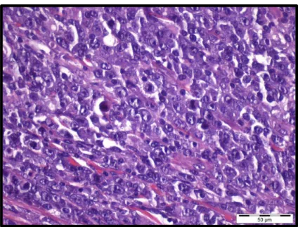
DL L is characterize by lymphoid cells with “nuclear size equal to or exceeding
normal macrophage nuclei or more than twice the size of a normal lymphocyte, that has a diffuse growth pattern” (1) (Figure 3).
It is divided into morphological variants, molecular and immunophenotypically subgroups and different disease entities (1). The heterogeneity of some cases make it
impossible sometimes to include them on the previously defined subgroups, and these are classified as DLBCL, not otherwise specified (NOS). Thus, DLBCL, NOS includes al DLBCL cases that not fill in the specific subgroups mentioned above (1).
1.3.1 - Morphological variants
There are three morphological variants recognized (1):
• Centroblastic variant – predominantly composed of centroblasts which
“are medium-sized to large lymphoid cells with oval to round, vesicular nuclei containing fine chromatin”. It may contain two to four nuclear
membrane-bound nucleoli. The cytoplasm is frequently scanty and amphophilic to basophilic. Frequently the tumour is polymorphic with an admixture of centroblasts and immunoblasts (<90%). This is the most common variant.
Figure 3 – Diffuse large B-cell lymphoma typical cells (Hematoxylin and Eosin stain-H&E, case 286. Source: Department of Pathology, IPO PORTO.
Epigenetic biomarkers in Diffuse large B-cell lymphoma
9
• Immunoblastic variant – in which “greater than 90% of the cells are
immunoblasts with a single centrally located nucleolus and an appreciable amount of basophilic cytoplasm”.
• Anaplastic variant - characterized by “large to very large round, oval or
polygonal cells with bizarre pleomorphic nuclei”.
1.3.2 - Molecular subgroups
Based on similarities to the cell of origin (COO), DLBCL can be subdivided into three subtypes: the germinal center B-cell like (GCB) DLBCL, activated B-cell-like (ABC) DLBCL and primary mediastinal large B-cell lymphoma (PMBCL) (1,5,6,9). This classification includes
gene alterations and drug sensitivities.
GCB cells are produced from mature B cells in lymphoid organs that have been activated by interactions with an antigen and T-helper cells. The differentiation on GCB cells activates somatic hypermutation, leading to diversity in the immunoglobulin gene regions
(5,20). ABC DLBCL has a gene expression feature identical to plasma cells, arise from post
germinal center B cells that are arrested during plasmocytic differentiation (5,20). Primary
mediastinal large B-cell lymphoma presents with mediastinal lymphadenopathy and has some molecular genetic similarities with Hodgkin lymphoma (5, 20).
By next-generation sequencing techniques it was found that alterations of chromatin remodelling genes are predominant in GCB subtype, whereas gene mutations of B-cell
receptor (BCR) signalling and NF-kB pathway are frequent in ABC subtype (6). The ABC
DLBCL subgroup has a poorer overall survival compared to GCG DLBCL subgroup (5,6).
1.3.3 - Immunophenotypical subgroups
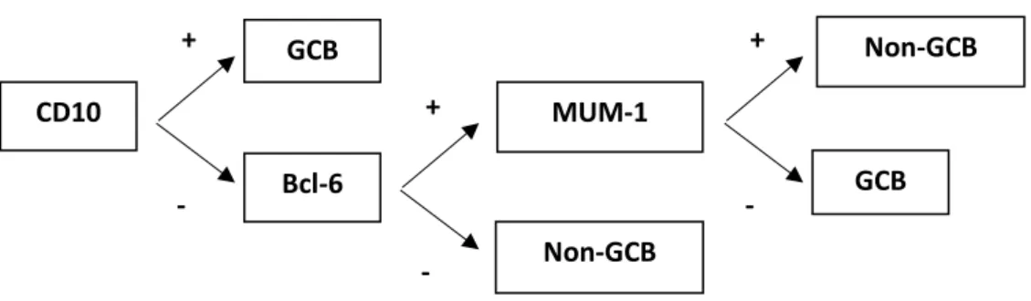
Immunohistochemical staining is used to make an approximation to the cell-of-origin subtype of DLBCL. It allows for the discrimination of GCB from non-GCB subgroups. The Hans algorithm (9) which uses the antibodies CD10, BCL6, and IRF4, also called MUM1
Epigenetic biomarkers in Diffuse large B-cell lymphoma
10
Figure 4 – Hans algorithm based on the immunohistochemical analysis of three markers (CD10, Mum1 and bcl6). Adapted from (9).
CD10 Bcl-6 GCB MUM-1 Non-GCB Non-GCB GCB + - + - + -
Figure 5 - Example of immunohistochemical staining for DLBCL. Case 286: CD10 -, BCL6-, MUM1 +, a Non-GCB subtype. Source: Department of Pathology, IPO Porto.
BCL-2 BCL-6
MUM-1 CD10
Epigenetic biomarkers in Diffuse large B-cell lymphoma
11
1.4 - StagingChoosing the most appropriate therapy involves accurate diagnosis and careful staging evaluation. The first staging concept was developed during the 20th century in the
United States. Disease heterogeneity was a challenge for development of a uniform staging system. The first staging system was named Ann Arbour Staging System and it was used for Hodgkin lymphomas, but it was later modified and adapted for NHL, providing an anatomic staging (24).
The Ann Arbour Staging System divides patients into four stages, depending on whether it is localized disease, multiples sites of disease on one or the other side of the diaphragm, lymphatic disease on both sides of diaphragm and disseminated extranodal disease (1, 24) (Table 2).
Table 2 – Ann Arbour Classification. Adapted from (24).
Stage Features
I Involvement of a single lymph node region or lymphoid structure (e.g. spleen, thymus, Waldeyer’s
ring)
II Involvement of two or more lymph node regions on the same side of diaphragm
III Involvement of lymph node regions on both side of diaphragm
IV Involvement of extranodal site(s) beyond that designated E
For all stages
A No symptoms
B Fever (>38ºC), drenching sweats, weight loss (10% body weight over 6 months)
For Stages I to III
E Involvement of a single, extranodal site contiguous or proximal to known nodal site
1.5 - Prognosis
Prognostic information for most NHL is not accurately provided by the Ann Arbour system and it is of limited value for treatment decisions (24).
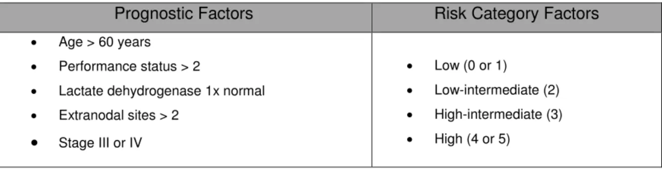
In 1993, the International Prognostic Index (IPI) (25) was developed as a primary
clinical tool to predict outcome in patients with aggressive NHL (26). This classification is
based on some negative prognostic factors at the time of diagnosis (Table 3). It includes age over 60 years, Eastern Cooperative Oncology Group (ECOG) poor performance status
Epigenetic biomarkers in Diffuse large B-cell lymphoma
12
(Table 4), Ann Arbour Stage I and II versus Stage III or IV, elevated serum lactate dehydrogenase (LDH) and involvement of two or more extranodal sites as high-risk factors
(24, 25, 26).
Table 3 – International Prognostic Index. Adapted from (1,24).
Prognostic Factors Risk Category Factors
• Age > 60 years
• Performance status > 2
• Lactate dehydrogenase 1x normal
• Extranodal sites > 2 • Stage III or IV • Low (0 or 1) • Low-intermediate (2) • High-intermediate (3) • High (4 or 5)
Table 4 – Eastern Cooperative Oncology Group Performance Status. Adapted from (24, 28).
Grade Description
0 Fully active, able to carry on all predisease performance without restriction
1 Restricted in physically strenuous activity but ambulatory and able to carry out work of a light
or sedentary nature (eg, office work)
2 Ambulatory and capable of self-care but unable to carry out any work activities. Up and about
more than 50% of waking hours
3 Capable of only limited self-care, confined to bed or chair more than 50% of waking hours
4 Completely disabled. Cannot carry on any self-care. Totally confined to bed or chair.
5 Dead
The IPI stratified DLBCL into four discrete outcome groups with a 5-year overall survival (OS) ranging from 26 to 73% (25,26, 27). Further studies demonstrated that a better
risk discrimination was achieved with a re-categorized IPI (R-IPI) for rituximab-based immunochemotherapy (26). Thus, Sehn et al stratified IPI into three categories with
significantly different outcome:
• “Very good” score 0 – patients with zero risk factors, with more than 90% chance of long-term progression-free survival
• “Good” or – patients with one or two risk factors, with approximately 80% chance of long-term progression-free survival
• “Poor” 3-5) – patients with three, four or five risk factors, with a 50% chance of long-term progression-free survival.
Epigenetic biomarkers in Diffuse large B-cell lymphoma
13
Risk group with less than 50% likelihood of long-term progression-free survival is not identified by neither IPI or R-IPI (26).
Many studies have subsequently focused on recognizing novel molecular and genetic markers to define more precise prognostic factors. Although several molecular prognostic markers have been identified, they have still not been validated (29,30,31). Thus,
IPI and R-IPI remain the most reliable tools for predicting outcome in patients with DLBCL
(26), and it is recommended that evaluation of a new patient should involve both systems:
Ann Arbour Staging and International Prognostic Index.
1.6 – Therapy
Although DLBCL are very aggressive, they are potentially curable with multimodal chemotherapy (1). The cyclophosphamide, hydroxydaunorubicin, oncovin and prednisone
(CHOP) chemotherapy regimen has been considered the standard treatment for several decades (32). In 1994, the results from Czuczman et al encouraged the use of rituximab with
CHOP (rituximab, cyclophosphamide, hydroxydaunorubicin, oncovin, prednisone - R-CHOP)for indolent lymphoma (34). Subsequently, improved failure-free survival (FFS) and
OS with R-CHOP (administered as rituximab on day 1 of each of eight CHOP cycles) compared with CHOP, especially in older patients, was disclosed (32, 35, 36). Thus, R-CHOP
is currently the standard of care in treatment of older patients with DLBCL. Toxicity data and efficacy associated with the number of chemotherapy cycles and the number of rituximab infusions must, however, be considered (7,32,33). Nevertheless, about 35% to 50%
of patients with advanced-stage disease are not cured using R-CHOP (8). Therefore, it is
important to understand and make efforts to identify biologic subgroups among DLBCL and develop more appropriate treatments to address DLBCL heterogeneity (32).
Epigenetic biomarkers in Diffuse large B-cell lymphoma
14
2 – EPIGENETICS
“Epigenetics”, a term firstly used by Conrad Waddington (37,87), consist on the study
of heritable changes in gene expression, involving active and inactive genes, without change in the DNA sequence. There is a change in phenotype without a change in genotype
(39, 40, 41). Epigenetic change is a regular and natural occurrence but can also be influenced
by several factors including age and the environment/lifestyle (40). Epigenetic change can
induce damaging effects, helping the cell to adapt or promoting pathological behaviour, that can result in cancer formation. It may occur at distinct stages of tumorigenesis and contribute to the development or progression of cancer. There is variable heterogeneity in epigenetic alterations that may give similar phenotypes. Thus, the varied clinical behaviour of cancers can range from indolent, slow-growing to aggressive, fast-growing tumors (41).
The epigenetic mechanism embrace three main systems: DNA methylation, histone modification and non-coding RNA (ncRNA)-associated gene silencing, that are currently considered to initiate and sustain epigenetic change (40).
2.1 – DNA Methylation
DNA methylation is one of the most well studied and characterized epigenetic modifications, which consists on the addition of a methyl group to carbon 5 of the cytosine residue within the nucleotide cytosine-phosphatidyl-guanine (CpG) (43, 44) (Figure 6). CpG
methylation of DNA is found at sites with a high percentage of CpGs (so-called CpG islands), leading to transcriptional repression (40).
De novo methylation marks are established by DNA methyltransferase (DNMT) 3a
and 3b, through embryonic development. The formation of heterochromatin and gene Figure 6 – DNA methylation. DNA gets methylated at the pyrimidine ring of the cytosine. The methyl mark leads to changes in the chromatin structure and recruits effector molecules. (A-adenosine; C-cytosine; G-guanine; T-thymine). Adapted from (40).
Epigenetic biomarkers in Diffuse large B-cell lymphoma
15
silencing depends on DNA methylation (40, 44). In almost all forms of cancer aberrant
hypermethylation and silencing of tumor suppressor genes has been found. Both hypometylation and hypomethylation of CpG islands located at gene promoters may affect the expression of protein coding genes and of non-coding R A’s, eventually resulting in tumorigenesis (44-47).
2.2 – Histone modification
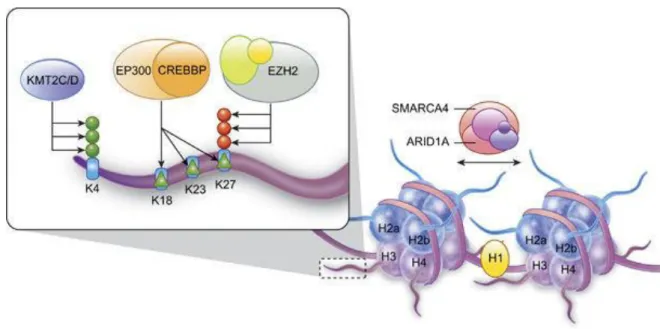
The second major mechanism in epigenetic gene regulation is modification of histone tails by chromatin modifying enzymes, that has significant impact on intra and inter-nucleossomal interactions (44,48,49,50). DNA is packaged around an octamer comprising
homodimers of four different histone proteins: H2A, H2B, H3 and H4. The nucleosome is stabilized in place by H1. Every histone protein has an N-terminal tail, which is the main target of a diversity of histone modifications (40) (Figure 7).
Modifications of histones can make changes into two ways: modifications can be recognized by “readers” that can recruit additional factors like other chromatin-modifying enzymes, and acetylation and phosphorylation may act on chromatin structure by reducing Figure 7 -Function of chromatin modifying and organizing genes. DNA is wrapped around histone octamers to form a nucleosome. Adapted from (50).
Epigenetic biomarkers in Diffuse large B-cell lymphoma
16
the positive charge of histones and alter their association with negatively charged DNA
(51-55).
2.2.1 – Histone lysine methylation
Histone lysine methylation may occur at residues 4, 9, 27, 36 and 79 of Histone 3 and residue 20 of Histone 4 (56). Three degrees of methylation are known: mono-methylation
(me1), di-methylation (me2) or trimethylation (me3) that may be associated with active euchromatin or inactive heterochromatin states (40,57)
.
H3K4 methylation is frequently associated with active transcription and, thus, H3K4me3 is usually located around promotor regions whereas H3K4me1 is located around enhancer regions. In contrast to H3K4me3, trimethylation of lysine 27 on histone H3 (H3K27me3) is associated with transcriptional repression. Histone methylation does not have a direct effect on chromatin structure (57-59).Histone lysine methylation is also a dynamic process that encodes a diversity of chromatin states (40).
➢ H3K4 methylation
One of four members in the mixed lineage leukemia (MLL) family proteins - KMT2D (MLL2) – is involved in H3K4 methylation. KMT2D somatic mutations are found in most follicular lymphomas (FL) but is less common in GCB-like DLBCL. The polycomb repressor complex 2 is unable to methylate H3K27me3 if H3K4 is trimethylated in the same histone tail. The H3K4me3 is recognized by reader proteins such as the product of ING1 gene, which is deleted in 1/3 of GCB-like DLBCL. Until now, there is no evidence of the effect of KMT2D mutation on H3K4 methylation or a suggestion of the mechanism by which these mutations promote lymphomagenesis (40,51,60).
➢ H3K27 methylation
Enhancer of zeste homolog 2 (EZH2) is the enzymatic subunit of an epigenetic gene-silencing complex named polycomb repressive complex 2 (PRC2). It is a SET domain histone methyltransferase that catalyses histone 3 lysine 27(H3K27) methylation, leading to a transcriptional repression of the target gene
Epigenetic biomarkers in Diffuse large B-cell lymphoma
17
Components of PRC2 are highly expressed in germinal centers. The first chromatin-modifying gene alteration described in DLBCL was EZH2 mutation. An increase or decrease of H3K27 methylation activity may lead to malignancy, depending on the cell of origin of the tumor (57, 67-69). The H3K27me3 mark is
self-sustaining, as DNA replicates, H3K27 methylated histones recruits PRC2 and PRC1 complexes to nucleosomes of the nascent DNA strand to continue gene silencing (70). Heterozygous EZH2 mutations suggest that cancer cells were
haploinsufficent for the enzymatic activity. It can result in deficient H3K27 methylation and widespread derepression of gene expression. Overexpression of EZH2 may cause silencing of growth-suppressive genes (71). Some studies
propose that increased or decreased H3K27methylation activity may lead to malignancy, depending on the cell of origin (72,73, 74).
Figure 8 – Deregulation of H3H27 methylation in cancer. A – EZH2 trimetylates H3K27 to inhibit gene expression; loss of EZH2 in cancer may lead to derepression of genes that promote cell growth. B – EZH2 overexpression silences additional targets. C-UTX removes H3K27me3 marks, loss of UTX increases H3K27me3 and silences tumor suppressors. Adapted from (57).
Epigenetic biomarkers in Diffuse large B-cell lymphoma
18
2.3 – Epigenetics of diffuse large B-cell lymphoma
Complex diseases require clarifications about genetic and environmental influences in the causative factors and prediction parameters. Epigenetic code alterations have been found in diverse pathological conditions (55,75), including cancer (76). In DLBCL, it was
demonstrated that EZH2 is important for G2/S transition and represses cell cycle-related tumor suppressor genes, through trimethylation of H3K27, contributing disease progression
(77). Mutations of EZH3 were identified in 22% of GCB DLBCL (55, 58, 64, 68). The most frequent
EZH2 mutations are found in the SET domain of EZH2 affecting tyrosine 641 (Y641), alanine 677 (A677) and alanine 687 (A687). EZH2 gain-of-function mutations favours increased H3K27me3 levels and loss-of-function mutations were identified in H3K4 trimethylase MLL2 in 32% of DLBCL (80,81). Whereas H3K27 trimethylation due to EZH2
activity has been associated with gene repression, methylation of H3K4 was associated with gene expression, with MLL and EZH2 displaying opposite functions (82). Indeed,
elevated levels of H3K27me3 in approximately one third of the DLBCL cases has been reported, suggesting that it may be involved in a subgroup of DLBCL. Moreover, an elevated level of H3K27me3 was associated with poor overall survival within non-GCB DLBCL patients (83).
Epigenetic biomarkers in Diffuse large B-cell lymphoma
21
As previously stated, altered activity of MLL and EZH2 has been reported in DLBC lymphoma, affecting the levels of the respective marks, H3K4me3 and H3K27me3, eventually causing altered expression of several critical genes. Thus, we hypothesized that quantitative evaluation of H3K4me3 and H3K27me3 might constitute biomarkers of DLBC progression, providing novel prognostic parameters for risk stratification of patients with DLBCL.
Hence, the specific aims of this Dissertation are:
- To evaluate quantitatively the immunohistochemical expression of histone-marks H3K4me3 and H3K27me3 in diffuse large B-cell lymphoma;
- To correlate the immunohistochemical expression with standard histopathological and clinical parameters, as well as disease-specific and overall survival;
- To verify their potential value as prognostic biomarkers for patients with diffuse large B-cell lymphoma.
Epigenetic biomarkers in Diffuse large B-cell lymphoma
25
1 - PATIENTS AND SAMPLESFor the purposes of this retrospective study, patients diagnosed (Department of Pathology) and treated (Department of Onco-Hematology) for diffuse large B-cell lymphoma at Portuguese Oncology Institute of Porto, Portugal between 2008 and 2013 were identified. Then, all cases with available representative archived tissue were enrolled, corresponding to 155 patients.
Pertinent clinical data were collected from the clinical charts by an experienced Hematologist (Dr. Sérgio Chacim) from the Department of Onco-Hematology.
This study, the use of samples and access to clinical data were approved by the institutional review board (Comissão de Ética para a Saúde) of Portuguese Oncology Institute of Porto, Portugal.
2 – IMMUNOHISTOCHEMISTRY
Three µm thick paraffin sections were obtained from representative formalin-fixed paraffin embedded tissue from each case, and placed in positively charged slides (Histobond®, Marienfeld, Germany) for immunohistochemistry assays.
For deparaffinization, slides were placed in an oven at 60ºC for 1 hour and 30 minutes, and then immersed in xylene (Sigma-Aldrich®, St. Louis, MO, USA). Subsequently, sections were hydrated in decreasing series of ethanol solutions (Merck, Darmstadt, Germany).
2.1 - Epitope retrieval and endogenous peroxidase neutralizing
For antigens retrieval, slides were placed in sodium citrate buffer (1:100; pH6; Vector® Antigen Unmasking Solution; H-3300; Burlingame; CA 94010 U.S.A) in a Bosch microwave oven at 700W (2 cycles of 10 minutes each).
Endogenous peroxidase activity was neutralized with 0.6% hydrogen peroxide for 20 minutes (Merck).
Protein detection was performed using the NovolinkTM Max Polymer Detection System (Leica Biosystems, Nussloch, Germany), according to a standard protocol (Annex 1). Depending of the antibody used, the incubation period varied, as depicted in Table 5.
Epigenetic biomarkers in Diffuse large B-cell lymphoma
26
Table 5 – Antibodies and Conditions
Antibodies Company Clone Positive Control Dilution Incubation H3H4me3 Abcam Rabbit polyclonal-ChIP Grade ab8580 Hepatocellular carcinoma 1:1000 1h (Humid chamber) H3K27me3 Cell Signaling Technology Rabbit monoclonal antibody (mAb) -C36B11 Colorectal cancer 1:1500 Overnight (4ºC – Humid chamber)
Successive washing steps were performed with tris buffered saline with Tween® 20 (TBS-T) (Sigma-Aldrich®).
Antigen-antibody binding reaction was revealed by means of incubation with a 0.0 % m/v 3,3’- diaminobenzidine (DAB) solution (Sigma-Aldrich®) in phosphate-buffered saline (PBS) (Biochrom Ltd., Cambridge, United Kingdom) previously activated with a 0,1% hydrogen peroxide solution, for 7 minutes, in the dark.
Counterstaining of the slides was performed with hematoxylin (Merck) for 30 seconds and then slides were washed for 1 minute in a tap water.
Finally, slides were dehydrated in an increasing series of ethanol solutions, diaphanized in xylene and mounted.
Negative control consisted on the omission of the primary antibody. Appropriate positive controls (see Table 5) were used in each batch.
Positive control slides were evaluated in an optical microscope by an experienced Pathologist.
Epigenetic biomarkers in Diffuse large B-cell lymphoma
27
3 - NUCLEAR IMMUNOSTAINING QUANTIFICATIONThe nuclear immunostaining quantification was done by a digital image analysis system (GenASIs, Version 7. .7.3 0, ASI, “Applied SpectralImaging”, 0 3 (Figure 9).
The slides were scanned at 200x magnification and high-resolution digital images were obtained. Manual selection of 5 to 8 areas of tumour cells were captured to reach at least 5000 cells fit for evaluation (Figure 10).
Figure 9 - Equipment support for the digital image analysis system GenASIs™.
Epigenetic biomarkers in Diffuse large B-cell lymphoma
28
Subsequently, positive nuclei were automatically detected and a nuclear classification into 4 levels of intensity (0-3) was generated, each one identified with a different color (Figure 11):
• 0 (negatively stained nuclei) – blue; • 1 (low intensity stained nuclei) – yellow;
• 2 (intermediate intensity stained nuclei)– orange; • 3 (higher intensity stained nuclei) – red.
Epigenetic biomarkers in Diffuse large B-cell lymphoma
29
The percentage of immunostained nuclei and the H-score were determined for each case and used for subsequent analyses.
4 - STATISTICAL ANALYSIS
For statistical analysis, patients were divided into two Ann Arbour Stage groups (stage I/II, stage III/IV) and into three IPI scores (0/1-2/3-5). Cutoffs for H3K4me3 and H3K27me3 nuclear immunostaining (high vs. low) were set at 25th and 75th percentile (P25
and P75). Comparison between % Positivity, Hscore, clinical and pathological parameters was performed using non-parametric tests (Mann-Whitney or Kruskall-Wallis, for IPI). Survival curves were created using Kaplan-Meier non-parametric estimator. Survival between groups was compared using Log-rank test. Statistical significance was considered when p-value was inferior to 0.05. All statistical analyses were performed using R version 3.1.2 (R Foundation for Statistical Computing, Vienna, Austria).
Epigenetic biomarkers in Diffuse large B-cell lymphoma
33
1 – CLINICAL AND PATHOLOGICAL CHARACTERIZATION OF THE PATIENTSFor the purposes of this study, 155 patients diagnosed with DLBCL and treated at the Portuguese Oncology Institute of Porto, from 2008 to 2013 were included. The clinical and pathological characteristics of this cohort are shown in Table 6. In this cohort, 43.6% of patients were male, and the median age at diagnosis was 66 years. Recurrent/transformed disease was experienced by 40 % of the patients.
Table 6 - Clinical and pathological characteristics of the patients
N % Gender Female 87 56,1% Male 68 43,9% Age at Diagnosis ≤ 70 years 99 63,9% > 70 years 56 36,1%
Ann Arbour Stage
I/II 79 50,6%* III/IV 72 46,2%* IPI Low risk (0) 21 13,5%** Intermediate risk (1,2) 66 42,6%** High risk (3-5) 51 32,9%**
*3,2% of patients has unknown Ann Arbour Stage information. ** 11% of patients has unknown IPI information.
2 –SURVIVAL ANALYSIS
The follow-up time was determined at date of the last consultation and the median was 55.4 months. The overall survival was estimated as 76.7% at 2 years and 68.1% at 5 years. The disease-specific survival was estimated as 79.1% at 2 years and 74.9% at 5 years.
For survival analysis, we considered the association between the disease-specific survival and the Ann Arbour stage, IPI score, and age at the time of diagnosis (≤ or > 60 years and ≤ or > 70 years . In our dataset, individualized Ann Arbour stage was not associated with prognosis (p=0.088) (Figure 12), whereas grouped Ann Arbour stage (stages I/II vs. III/IV) was associated with prognosis (p=0.015).
Epigenetic biomarkers in Diffuse large B-cell lymphoma
34
IPI score (p=0.005) and age at the time of diagnosis (≤ or > 60 years and ≤ or > 70 years) were significantly associated with prognosis (p=0.006 and p=0.005, respectively) (Figures 13 and 14). Indeed, lower IPI score and age at diagnosis were associated with a better outcome. 0 20 40 60 80 100 0. 0 0. 2 0. 4 0. 6 0. 8 1. 0 Ann Harbour
Time since diagnosis (months)
D S S I II III IV p = 0.088 0 20 40 60 80 100 0. 0 0. 2 0. 4 0. 6 0. 8 1. 0 Ann Harbour
Time since diagnosis (months)
D S S I/II III/IV p = 0.015
Figure 12 - Analysis of disease-specific survival according to individualized or grouped Ann Arbour stage.
0 20 40 60 80 100 0. 0 0. 2 0. 4 0. 6 0. 8 1. 0 IPI
Time since diagnosis (months)
D S S 0 1/2 3/4/5 p = 0.005
Figure 13 - Analysis of disease-specific survival according to grouped IPI score.
Epigenetic biomarkers in Diffuse large B-cell lymphoma
35
No difference in prognosis was observed concerning gender (p=0.154) (Figure 15).
0 20 40 60 80 100 0. 0 0. 2 0. 4 0. 6 0. 8 1. 0 Age group
Time since diagnosis (months)
D S S <=60 >60 p = 0.006 0 20 40 60 80 100 0. 0 0. 2 0. 4 0. 6 0. 8 1. 0 Age group
Time since diagnosis (months)
D S S <=70 >70 p = 0.005
Figure 14 - Analysis of disease-specific survival according to age (≤ or > 60 years and ≤ or > 70 years).
0 20 40 60 80 100 0. 0 0. 2 0. 4 0. 6 0. 8 1. 0 Sexo
Time since diagnosis (months)
D S S M F p = 0.154
Figure 15 - Analysis of disease-specific survival according to gender.
Epigenetic biomarkers in Diffuse large B-cell lymphoma
36
3 – IMMUNOHISTOCHEMICAL PROFILE
The percentage of immunostained nuclei and the H-score were determined for each case and used to evaluate the immunoexpression profile.
• H3K4me3
For H3K4me3 epigenetic mark, Hscore varied between 4.1 and 276.2, with a median value of 157.1 (Figure 16). In statistical analysis of the distribution of Hscore of H3K4me3 according to gender, age, Ann Arbour stage, transformation and IPI there were no statistically significant associations. We used Mann Whitney’s test for all the variables except for IPI score, in which Kruskal Wallis’ test was used (Table 7).
Table 7 - Comparison between Hscore, clinical and pathological parameters. P
Gender 0.216
Age (≤ or > 70 years) 0.118
Ann Arbour Stage 0.148
Transformation 0.536
IPI 0.472
The immunoexpression of H3K4me3 was not associated with disease-specific survival using either % Positivity and Hscore median (p=0.643 and p=0.917, respectively) or the 25th and 75th percentile for Hscore (p=0.978 and p=0.518, respectively) (Figures 18,
19 and 20).
Epigenetic biomarkers in Diffuse large B-cell lymphoma
37
• H3K27me3
Concerning H3K27me3 epigenetic mark, Hscore varied between 38.6 and 247, with a median of 117.2 (Figure 17). In statistical analysis of the distribution of Hscore of H3K4me3 according to gender, age, Ann Arbour stage and transformation there were no statistically significant results except for IPI score (p = 0.041). We used Mann Whitney’s test for all the variables except for IPI score, in which Kruskal Wallis’ test was used (Table 8).
Table 8 - Comparison between Hscore, clinical and pathological parameters. P
Gender 0.634
Age (≤ or > 70 years) 0.530
Ann Arbour Stage 0.094
Transformation 0.422
IPI 0.041
The immunoexpression of H3K27me3 was not associated with disease-specific survival using either the % Positivity and Hscore median (p=0.979 and p=0.959, respectively) or the 25th and 75th percentile (p=0.291 and p=0.683), respectively) (Figures
18, 19 and 20).
Epigenetic biomarkers in Diffuse large B-cell lymphoma
38
Figure 18 - Analysis of disease-specific survival by % Positivity, in the left panel and Hscore, on the right panel. The comparison is made using the median.
Epigenetic biomarkers in Diffuse large B-cell lymphoma
39
Figure 19 - Analysis of disease-specific survival by % Positivity, in the left panel and Hscore, on the right panel. The comparison is made using the 25th percentile.
Figure 20 - Analysis of disease-specific survival by % Positivity, in the left panel and Hscore, on the right panel. The comparison is made using the 75th percentile.
Epigenetic biomarkers in Diffuse large B-cell lymphoma
43
Diffuse large B-cell lymphomas are a heterogeneous group of lymphoid neoplasms that have a wide range of clinical presentations, immunologic characteristics, genetical features and clinical outcomes in patients that experience standard therapy (8). It is important
to emphasize the diversity of DLBCL as it must be taken into consideration when investigating new therapies (9, 27, 33). Currently, prognosis of patients with DLBCL is
estimated using the clinical parameters of the International Prognostic Index (27). The
predictive ability of IPI is, however, limited and estimating the risk that a DLBCL poses to an individual patient remains a challenging task. Thus, improved means of prognostication are critical to allow for individualized risk-adapted therapy. Likewise, development of predictive biomarkers will be essential to select appropriate patients for novel target approaches with the goal of achieving a personalized cancer care approach (33,47). Taking
in consideration these principles and considering that altered patterns of histone modifications associated with EZH2 and MLL2 mutations occur in DLBCL (55, 58, 64, 68), we
hypothesized that a quantitative evaluation of histone marks, specifically H3K4me3 and H3K27m3, might provide candidate prognostic biomarkers which might be used as ancillary tools for risk-stratification of DLBCL patients.
This study was based in a consecutive series of 155 patients diagnosed with DLBCL and treated in IPO Porto between 2008 and 2013. The median age at diagnosis, 66 years, and the percentage of patients more than 70 years old (36,1%) are in accordance with published literature (11). However, the observed M/F ratio was 1/1.3, which is an inverse
proportion compared to most published studies (1,2,14). There is no clear explanation for this
finding, which might bias subsequent analyses. Nevertheless, gender is not considered a prognostic factor in DLBCL and, thus, we are confident that the bias, if it exists, it is likely to be negligible. Indeed, more relevant parameters including the distribution of IPI score (13.5%, 42.6% and 32.9%) and the proportion of patients with transformation/recurrence (40%) are similar to those of previous studies (27,1,5). Sample size, however, might account
for some results, such as the lack of association of individualized Ann Arbour stage (i.e., stages I vs. II vs. III vs. IV), which was, however, disclosed when stages I/II and III/IV were lumped together. Moreover, and as expected, IPI score and age at the time of diagnosis (both ≤ or > 60 years and ≤ or > 70 years were significantly associated with prognosis, providing indirect validation of this patient cohort.
Epigenetic biomarkers in Diffuse large B-cell lymphoma
44
The main goal of this study was to investigate the potential of quantitative H3K4me3 and H3K27me3 immunoexpression as prognostic biomarker in DLBCL, which, to the best of our knowledge, has not been attempted before. The rationale for this approach was that the enzymes that catalyse H3K4me3 and H3K27me3, specifically MLL2 and EZH2, display altered activity in a proportion of DLCBL cases. Whereas the EZH2 activating mutations, which were found in up to 22% of DLCBL (55, 58, 64, 68), are deemed to increase H3K27me3,
MLL2 inactivating mutations, occurring in 32% of DLBCL (80,81), are expected to decrease
H3K4me3. Di- and trimethylation of H3K4 is linked to transcriptional activation and high levels of H3K4me3 trimethylation are associated with the promoters of actively transcribed genes (57-59). H3K27me3, on the other hand, is an epigenetic mark associated with
transcriptional silencing by promoting a compact chromatin structure (57-59). Thus, both
alterations might lead to inappropriate silencing of genes that are critical for the control of cell growth and proliferation. We expected that more aggressive DLCBL would associate with lower and/or higher H3K4me3 and H3K27me3 Hscore/percentage of immunostained cells, respectively. However, only a significant association between IPI and H3K27me3 Hscore was found. Moreover, survival analysis did not disclose any association between quantitative H3K4me3 and H3K27me3 immunoexpression and patients’ outcome, although several cutoff values were tested (P25, median, P75). A previous study has shown an association between increased H3K27me3 and poor overall survival in DLBCL (83) and in
other tumor models increased EZH2 expression has been associated with worse prognosis and an increase of invasion and tumour progression capacities (41). Nevertheless, it is
noteworthy that the association observed between increased H3K27me3 expression and poor overall survival was restricted to non-GCB DLBCL patients (83). Our cohort includes
both GCB and non-GCB DLBCL, which we did not discriminate owing to the lack of sufficient immunophenotypical information. Thus, a direct comparison between the two studies cannot be made. Moreover, our negative results might also derive from an insufficient sample size, not having statistical power to disclose the previously reported associations. It should also be recalled that the immunohistochemical assay used in our study provides a global estimate of H3K4me3 and H3K27me3 expression, which might not reflect important gene-specific silencing events, which we are not able to identify with this methodology. However, the selection of an immunohistochemical assay was based on the widespread availability of this technique in most Pathology labs, eventually facilitating translation into routine clinical practice.
Several limitations of this study should be acknowledged. Firstly, and as previously stated, sample size may not be sufficient to ascertain a significant association between