IT U T O D E C IÊ N C IA S B IO M ÉD IC A S A B EL S A LA Z A R U LD A D E D E C IÊ N C IA S U LD A D E D E M ED IC IN A
dr
o M
. M
ira
nd
a D
ie
t-M
icr
ob
io
ta
In
te
ra
ctio
n i
n H
ea
lth
a
nd
et
-m
icr
ob
io
ta i
nt
er
ac
tio
n i
n h
ea
lth a
nd d
is
ea
se
:
as
e
dr
o M
igu
el M
ira
nd
a
Diet-microbiota interaction in health
and disease: role of high salt diet in
Pedro Miguel Miranda
D
2018D
PEDRO MIGUEL GOMES DA SILVA MIRANDA
Diet-microbiota interaction in health and disease: a role
of high salt diet in inflammatory bowel disease
Tese de Candidatura ao grau de Doutor em
Biologia Básica e Aplicada submetida ao
Instituto de Ciências Biomédicas Abel
Salazar da Universidade do Porto.
Orientador – Premysl Bercik
Categoria – Médico Gastroenterologista e
Investigador Principal
Afiliação – Farncombe Institute, McMaster
University, Hamilton, Canada
Co-orientador – Didier Cabanes
Categoria – Investigador Principal
Afiliação – Instituto de Biologia Molecular e
Celular, Porto
This work was supported by Fundação para a Ciências e Tecnologia (FCT) by means of a Ph.D. fellowship SFRH/BD/51278/2010 awarded to Pedro Miguel Gomes da Silva Miranda through the Graduate Program in Areas of Basic and Applied Biology (GABBA), Universidade do Porto, Portugal. The research was also supported by the Canadian Institutes of Health Research (CIHR).
Acknowledgments
Looking back at this PhD journey, I could not feel more grateful and humbled for what I have been given by so many people. The PhD is not a lonely ride and I could not have asked for a better team. I would like to express my gratitude and admiration for everyone who was part of this incredible experience. This work is for you.
First, I would like to thank the GABBA program for giving me the opportunity to embark on this incredible experience that the PhD has provided. Thank you Professor Maria de Sousa, Professor António Amorim, Professor Alexandre do Carmo, and everyone from the GABBA family who opened me the door to enter the inspiring world of science and research. Thank you for being always present and curious listeners. I thank Didier, my co-supervisor, for always being present and making sure I was on track.
Thank you to my supervisors, Premek and Steve, who believed in me and adopted this fresh-off-the-boat naïve Portuguese into their lab family. Thank you for your guidance, your support and for inspiring me with your passion for what you do. Premek, I could not have asked for a better mentor. Thank you for always find the time to receive me at your office, for the late nights, early mornings and weekends working on my case. Your contagious joy and optimism always kept me going! We both know how my life in Canada, at times, seemed to be ruled by Murphy’s laws but you have never let me fall. You have trusted in my decisions, guided me, supported me and were always available, even when we were apart. Thank you!
To my dearest lab and Farncombe “team mates”. I am very proud of being part of such a wonderful and selfless group of people, who were always available for avid discussions about our projects and to help in any possible way, in big and smaller experiments. Jun, Marc, Sacha, Vivek, Viktoria, Justin, Alberto, Heather, Jane, Jen, Michelle, Giada, you have all set the bar very high
for how a work team should be. A special thanks to Giada, for being the rock of the lab. Especially for your incredible multitasking ability, which at times gives the impression that you multiply in many Giadas. Thank you for always finding the time to listen, to laugh and to help. I could not have made it without your patience and hard work. Thank you to Elena and Mike Surette for leaving your office doors always open for our scientific discussions. Justin and Alberto, thank you for keeping us scientifically inspired with those Phoenix beer afternoons.
À Mariana, Ferreira, Inês, Neto, Milene, Catarina, Ricky, Bruno, Daniel,
Lindo e Martins, os inigualáveis GABBA 14th, obrigado por todos os momentos
memoráveis que passámos no Porto e por serem uma verdadeira família. Este doutoramento é de todos vós tambem! Mariana, obrigado pela força e motivação contagiante e por seres a minha crónica parceira das últimas horas.
To the Cline Avenue gang: Chris, David, Arnaud, Gabby, Justine, Caitlyn, Sean, Stevie and Alison. Thank you for being my family in Canada! For the BBQs, games night, cocktail parties, Mario Kart, Art crawls, and most of all, for making me feel like home.
Esta aventura começou muito antes de ter sido selecionado pelo programa GABBA. Gostaria de agradecer à minha mãe e pai por fazerem com que o meu caminho está livre para que eu o possa tomar. Obrigado por apoiarem as minhas decisões, mesmo que isso signifique que eu irei viver a 5000 Km de distância. Mãe, Pai, Avó Teresa, Avó Fernanda, Bruno e todos na família, o vosso apoio e amor incondicional tornam tudo o resto possível. Muito obrigado! Ao meu irmão Bruno, obrigado por me inspirares a procurar a diversão e felicidade em tudo o que fazemos.
Finally, this is for you and because of you, Alison. Thank you for being my rock through this journey that we shared together. You were always there, keenly listening to my greatest exciting discoveries and to my failures and concerns. Thank you for your unlimited patience and for your therapeutic smile and giggles when I most needed. For covering for me when I was overwhelmed,
for those meals you would bring to the lab at late hours. Thank you for your unconditional love and care, and for being my number one fan.
Table of Contents
Abstract ... 2
Resumo ... 3
List of abbreviations ... 5
I – General Introduction ... 8
1. Chronic inflammatory disorders: incidence in the “Western world” ... 82. “Western diet” ... 9
2.1. General features and implications in chronic inflammatory disorders ... 9
2.2 Salt: a new player in chronic inflammatory disorders ... 10
3. Gut Microbiota ... 11
3.1. Overview ... 11
3.2. Development of the human gut microbiota ... 13
3.3 The gut microbiota functions in health ... 14
3.3.1. Microbiota role in host metabolism ... 15
3.3.2. Microbiota role in host immune system ... 17
3.3.3. Microbiota role in promoting epithelial barrier ... 18
3.3.4. Microbiota role in protection against pathogens ... 19
3.3.5. Microbiota role in central nervous system ... 19
3.4. Factors influencing gut microbiota ... 20
3.4.1. Genetics ... 21
3.4.2. Lifestyle, geography and diet ... 21
3.4.3. Microbe-microbe interaction ... 22
3.5. Diseases associated with microbiota ... 22
4. Diet-microbiota interaction in health and disease ... 24
5. Inflammatory Bowel Disease ... 27
5.1. Epidemiology and etiologies ... 27
5.2. Colitis models in mice ... 29
5.2.1. DSS-colitis ... 29
5.2.2. DNBS-Colitis ... 30
5. AIMS ... 30
II – Research Work ... 33
1. Abstract ... 362. Background ... 37
3. Materials and Methods ... 39
4. Results ... 45
5. Discussion ... 62
6. Conclusions ... 66
7. Supplementary Figures ... 67
8. Supplementary Tables ... 71
9. Supplementary methods ... 78
10. Declarations ... 80
III – General Discussion ... 83
IV - Publications ... 90
V – References ... 92
Abstract / Resumo
Abstract
Changes in hygiene and dietary habits, including increased consumption of foods high in fat, simple sugars, and salt that are known to impact the composition and function of the intestinal microbiota, may explain the increase in prevalence of chronic inflammatory diseases. High salt consumption has been shown to worsen autoimmune encephalomyelitis and colitis in mouse models through p38/MAPK signaling pathway. However, the effect of high salt diet (HSD) on gut microbiota and on intestinal immune homeostasis, and their roles in determining vulnerability to intestinal inflammatory stimuli are unknown. Here, we investigate the role of gut microbiota alterations induced by HSD on the severity of murine experimental colitis.
Compared to control diet, HSD altered fecal microbiota composition and function, reducing Lactobacillus sp. relative abundance and butyrate production. Moreover, HSD affected the colonic, and to a lesser extent small intestinal mucosal immunity by enhancing the expression of pro-inflammatory genes such as Rac1, Map2k1, Map2k6, Atf2, while suppressing many cytokine and chemokine genes, such as Ccl3, Ccl4, Cxcl2, Cxcr4, Ccr7. Conventionally raised mice fed with HSD developed more severe DSS- (dextran sodium sulfate) and DNBS- (dinitrobenzene sulfonic acid) induced colitis compared to mice on control diet, and this effect was absent in germ-free mice. Transfer experiments into germ-free mice indicated that the HSD-associated microbiota profile is critically dependent on continued exposure to dietary salt.
Our results indicate that the exacerbation of colitis induced by HSD is associated with reduction in Lactobacillus sp. and protective short-chain fatty acid production, as well as changes in host immune status. We hypothesize that these changes alter gut immune homeostasis and lead to increased vulnerability to inflammatory insults. This study provides an example of how the gut microbiota plays an essential intermediary role between environmental factors, such as diet, and host physiology. Furthermore, this thesis indicates the high salt consumption as a potential risk factor for inflammatory bowel diseases.
Resumo
Mudanças nos hábitos higiénicos e alimentares, incluindo o consumo de alimentação com alto teor de gordura, açucares simples e sal - que se sabe exercerem um impacto na composição e função da microbiota intestinal – podem explicar o aumento da prevalência de doenças imunes crónicas. Foi recentemente demonstrado que o consumo de alto teor de sal tem um efeito adverso no desenvolvimento de encefalomielite autoimune e colite em ratinhos através da cascata de sinalização p38/MAPK. No entanto, ainda é desconhecido o efeito da dieta com alto teor de sal (DS) na estrutura da microbiota intestinal (ou flora intestinal) e na homeostasia do sistema imune; bem como o seu papel na vulnerabilidade para responder a estímulos imunológicos no intestine. Neste estudo, investigámos o papel que as alterações na microbiota intestinal induzidas por DS, exerce na severidade do desenvolvimento de colite em ratinhos.
Comparado com a dieta controlo, a DS induz a alteração da composição e função da microbiota fecal, nomeadamente a redução da abundancia relativa de Lactobacillus sp. e produção de butirato. Além disso, DS afectou a imunidade da mucosa intestinal no cólon e, de uma maneira menos proeminente, do intestino delgado. DS induziu a expressão de genes pro-inflamatórios como por exemplo Rac1, Map2k1, Map2k6, Atf2, e suprimiu genes de várias citocinas e quimiocinas, dos quais Ccl3, Ccl4, Cxcl2, Cxcr4, Ccr7. Ratinhos alimentados com DS desenvolveram uma colite mais severa usando os modelos de colite induzida por DSS e DNBS, quando comparados com ratinhos em dieta controle. Este efeito esteve ausente quando usados ratinhos desprovidos de bactérias (Germ Free mice). Experiências de transferência de microbiota para ratinhos desprovidos de bactérias indicaram que a microbiota associada a DS precisa duma exposição continuada a DS para manter o seu perfil.
Os nossos resultados indicam que o agravamento da colite induzido pela DS está associada à redução de Lactobacillus sp e à produção de butirato, bem como a mudanças no status imunológico do hospedeiro. Nós propomos que estas mudanças alteram a homeostasia da imunidade nos intestinos, o que leva
ao aumento da vulnerabilidade aos insultos inflamatórios. Este estudo apresenta-se como um exemplo do papel essencial que a macrobiota intestinal desempenha na intermediação entre factores ambientais, como a dieta, e a fisiologia do hospedeiro. Além disso, esta tese indica o consumo elevado de sal como um potencial factor de risco em doenças intestinais inflamatórias.
List of abbreviations
5-HT
Serotonin
ASD
Autism spectrum disorder
BDNF
Brain-derived neurotropic factor
BMI
Body mass index
B. fragilis
Bacteroides fragilis
C. difficile
Clostridium difficile
CD
Crohn’s disease
CDI
Clostridium difficile infection
CID
Chronic immune disorders
CVD
Cardiovascular disease
DA
Dopamine
DNBS
Dinitrobenzene sulfonic acid
DSS
Dextran sulfate sodium
EAE
Experimental autoimmune encephalomyelitis
FMT
Fecal microbiota transplant
GALT
Gut-associated lymphoid tissues
GF
Germ-free
GWAS
Genome-wide association studies
HFD
High fat diet
HSD
High salt diet
IBD
Inflammatory bowel disease
IBS
Irritable bowel syndrome
IL
Interleukin
LPS
Lipopolysaccharide
MAMP
Microbial-associated molecular pattern
MLN
Mesenteric lymph node
NA
Noradrenaline
NaCl
Sodium chloride (salt)
NFAT5
Nuclear factor of activated T-cells 5
NLR
Nod-like receptors
NMDA
N-methyl-D-aspartate
P38/MAPK
p38 mitogen-activated protein kinase
PP
Peyer’s patches
PRR
Pattern recognition receptors
RA
Rheumatoid arthritis
SCFA
Short-chain fatty acid
SFB
Segmented filamentous bacteria
SGK1
Serum/glucocorticoid-regulated kinase 1
SNP
Single nucleotide polymorphism
TLR
Toll-like receptor
TMAO
Trimethyl amine N-oxide
TNF
Tumor necrosis factor
UC
Ulcerative colitis
I – General Introduction
1. Chronic inflammatory disorders: incidence in the
“Western world”
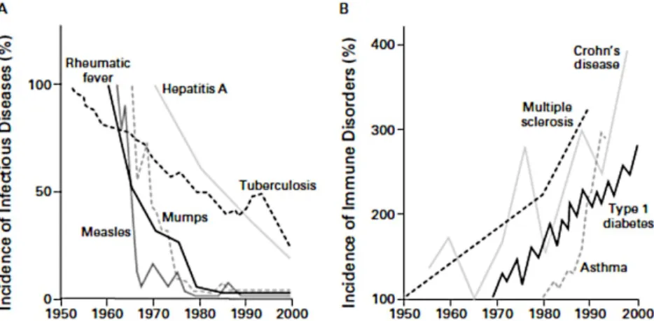
The incidence of chronic diseases, namely related to some form of immune disorder, such as inflammatory bowel disease (IBD), multiple sclerosis (MS), diabetes, asthma, rheumatoid arthritis (RA), and others has been increasing over the past 50 years, namely in the “Western” (industrialized) regions of the planet [1,2]. Although many of these chronic immune disorders (CID) have been linked to genetic susceptibility factors, genetic drift alone can not explain the population incidence rising of CID over the last half-century [3]. Moreover, a relatively low concordance rate for most of autoimmune diseases between monozygotic twins suggests non-genetic factors as important triggers of disease [4]. Several of these non-genetic factors have been proposed to explain the rise of chronic immune related disorders, including decreased incidence of in infectious diseases (the so called “hygiene hypothesis”) [1,5] (Fig. 1), as well as environmental factors related to changes in life-style observed in the western world, in particular dietary and hygiene factors [6].
Figure 1. Hygiene hypothesis. Inverse relationship between the incidence of prototypical infectious diseases (Panel A) and the incidence of chronic immune disorders (Panel B) from 1950 to 2000. (Figure from Bach JF, 2002[1]).
2. “Western diet”
2.1. General features and implications in chronic inflammatory
disorders
Diet have undergone significant changes over the last few decades, that have been considered too quick on an evolutionary time scale, for the human genome to adjust [7]. The Western Diet (WD) is generally characterized by high content of saturated fats, red meats, refined sugars and salt, while low in fresh fruits and vegetables with consequent low fiber content [7,8]. There is growing evidence from human and animal studies implicating WD factors in the development of CID. Epidemiological studies revealed a strong correlation of dietary factors such as low fiber [9] and high fat [10] consumption with CID, in particular IBD, diabetes and asthma [8]. However, careful approach must be taken when interpreting diet-disease epidemiological studies because of the multifactorial etiology of chronic diseases, and the complexity and often subjective nature of self-reported dietary patterns [11]. Interventional studies have uncovered a link between high fat and low fruit and vegetables consumption and worse asthma outcomes [12,13]. Although population studies have demonstrated a correlation between diet and CID, the mechanisms involved in diet-mediated inflammatory processes are largely unknown. Feeding mice with high fat diet (HFD) was shown to spontaneously increase the circulating proinflammatory cytokines TNFα, IL-1β and IL-6 in the plasma, and to induce macrophage infiltration and inflammation in the adipose tissue, in a TLR4 signaling pathway dependent way [14]. Moreover, it was shown in rodent models that a high-fat diet exacerbates models of IBD [15], experimental autoimmune encephalomyelitis (EAE, a rodent model mimicking aspects of MS) [16] and collagen-induced arthritis (CIA) [17].
2.2 Salt: a new player in chronic inflammatory disorders
One dietary feature of WD that has been overlooked for its potential role in contributing for CID is its high salt (sodium chloride, NaCl) content. The consumptions of salt has increased in the Western world societies, mainly driven by the increased intake of processed foods and “fast food” in the industrialized societies, which can contain more than 100 times the salt found in similar home made meals [7,18,19]. In addition to well-documented effects of excess salt intake in increased blood pressure, cardiovascular disease and stroke [20], salt was recently shown to modulate aspects of the immune system and was linked to EAE severity in mice [21,22]. Increased NaCl concentration was able to induce the differentiation of a pathogenic form of Th-17 T cells from human and rodent
naïve CD4+ T cells in vitro. This process was dependent on the activation of p38
mitogen-activated protein kinase (P38/MAPK), nuclear factor of activated T-cells 5 (NFAT5) and serum/glucocorticoid-regulated kinase 1 (SGK1) [21,22]. Th17
cells are a group of CD4+RORγt+ T cells that secrete interleukin-17 (17A and
IL-17F) and IL-22, and play an important role in protecting against bacterial and fungal pathogenic infections, particularly at mucosal surfaces [23]. Pathogenic IL-23-dependent Th-17 cells have been shown to be critical for the development of EAE in rodents and autoimmune diseases [24]. Indeed, mice receiving a salt-enriched diet (high salt diet, HSD) developed an exacerbated form of EAE, thus showing for the first time a link between salt consumption and a CID in an animal model [21,22]. Moreover, an observational clinical study using regression analysis, found a positive correlation between sodium intake and increased disease activity in a cohort of relapsing-remitting patients with MS followed for two years [25]. Interestingly, in a longitudinal experiment performed under highly controlled spaceflight simulation program, human subjects were exposed to fixed salt intake changes and their immune system was assessed. Subjects, when exposed to HSD, displayed increased numbers of circulating monocytes and pro-inflammatory cytokines IL-6 and IL-23, along with decrease of anti-inflammatory cytokine IL-10 [26].
A possible mechanism by which dietary factors modulate physiological processes and in particular the immune system, is through interaction with gut microbiota. In fact, it is well established that diet determines the composition and function of the gut microbiota, with repercussions for health and disease [27].
3. Gut Microbiota
3.1. Overview
Accumulating data suggest that gut microbiota plays an essential role in host health and disease. It consists of a living consortium of different microorganisms such as Bacteria and Archaea, Eukarya and Viruses inhabiting the gastro-intestinal tract (GI tract), that have co-evolved with the host over thousands of years into a complex symbiotic mutualistic relationship. While the host provides a protected habitat and the necessary substrate, the gut microbiota contributes by controlling a vast enzymatic repertoire, essential for gut physiology, metabolic, immune and neuronal functions.
Bacteria numbers in the human body were estimated to account for ≈
1014, thus exceeding human body cells by 10:1. These numbers were however
recently revisited, after the original estimation was traced back to an old “back-of-the-envelope calculations” published in the 1970’s [28,29]. In the new
proposed estimation, the number of total bacteria was estimated to ≈ 3.813,
resulting in a ≈ 1:1 ratio of bacteria to human cells [29,30]. Even more relevant, the largest human gut microbiome (the sum of all genes in the microbiota) metagenomics analysis to date [31], has identified around 10 million non-redundant genes from a multi-continental human consortium including Europeans, Chinese and American subjects, comprising of an average of ≈ 750,000 genes per sample. These numbers completely dwarf the 19,000-20,000 protein-coding genes that comprise the human genome [32], indicating that the gut microbiome gene pool is 500x larger than human genome, and that an
average human gut microbiome contains 40x more genes than the human host genome itself.
Microbiota populates the entire GI tract, from mouth to rectum, but the density and composition of bacteria varies greatly along the gut. While the upper GI tract contains low density of predominantly aerobic bacteria, the lower GI tract harbors higher density numbers of anaerobic and facultative anaerobic
bacteria, ranging from 103-104 bacteria/ml in the stomach and duodenum, to
1011 bacteria/ml in the colon, which harbors the vast majority of the body’s
bacterial load [33]. This heterogeneous pattern is a reflection of several determinant factors such as pH and oxygen level, as well as the content flow speed through the tract. Low pH and faster content flow, typical of the stomach and the upper part of small intestine impose a harsh environment that limits bacteria colonization, and vice-versa. This diversity makes the GI tract a continuous collection of different micro ecosystems, with specialized functions and microbiota-host interactions.
The development of culture-independent technologies based on the sequencing of gut microbiota genetic information, started a new era in our understanding of the role of microbiota in host’s health and disease, by allowing a deeper monitoring and quantification of microbiota composition, function and dynamics, in a relatively fast and low cost manner. Perturbations in gut microbiota composition and function, known as dysbiosis, have been extensively associated to multiple diseases, ranging from intestinal disorders to metabolic, hepatic, neuronal, respiratory and immune-related diseases. In fact, gut microbiota has gained tremendous attention as a potential key mediator between the rapid changes occurring in environmental factors and life-style and the rise of the incidence of several diseases, as this cannot be explained by the human population genetic drift alone.
This thesis will be focus on the bacteria that inhabits the GI tract. So for simplicity purposes, I will be referring to bacteria when mentioning microbiota.
3.2. Development of the human gut microbiota
Historically, the newborn infant was considered to be sterile until the mother has given the initial microbiota inoculum at birth, from the vaginal or skin microbiota, at natural birth or C-section respectively. However, this dogma was challenged by DNA-based studies that shown undeniable evidence of the microbial presence in the healthy placenta, amniotic fluid [34,35], umbilical cord [36] and meconium [37,38] (the first stool of a newborn). It is still unclear how bacteria penetrate the intrauterine environment but in one experiment, specific labeled bacteria fed to pregnant mice has been detected in the meconium of the offspring, suggesting that intrauterine bacteria can be transmitted from the mother’s GI tract [37].
After birth, the GI tract becomes rapidly colonized, with the neonatal gut microbiota composition being determined by several factors such as the mode of delivery (vaginal or C-section), gestational age, diet (breast milk or milk formula) and early antibiotic treatment [39–42]. Early assembly of gut microbiota is a non-random process with different species appearing at different time points, beginning with the establishment of oxygen tolerant bacterial species mainly from Firmicutes and Proteobacteria phylum, which reduce the GI tract environment allowing the further establishment of strict anaerobic species such as Bifidobacterium and Bacteroides [40,41,43]. Over time, the microbiota diversity rapidly increases until an adult-like microbiota community dominated by the bacteria phylum Bacteroidetes and Firmicutes becomes established at around 2-4 years old [40,41,43]. In the elderly, the gut microbiota suffers a general loss of diversity, a reduction of beneficial bacteria such as
Bifidobacterium spp and changes in metabolic pathways such as the decrease of
Short Chain Fatty Acid (SCFA) production, a class of bacteria metabolites essential for intestinal function, that will be discussed bellow [44–46].
Several studies have attempted to define healthy gut microbiota enterotypes (defined types of microbial communities) [47,48] based on diet habits or geographical distribution of the population. However, and despite the efforts this concept remains controversial due to the disparities found across multiple studies and the sensitivity of the clustering methods applied in
defining microbiota enterotypes [49,50]. In the past decade, two major population-scale studies, The Human Microbiome Project (HMP, [51,52]) and the Metagenomics of the Human Intestinal Tract (MetaHIT, [31]), helped shedding light on the structure and diversity of healthy human microbiota. These large population studies confirmed that the human gut microbiota is clearly dominated by two bacteria phyla, Bacteroidetes and Firmicutes; accompanied by bacteria from Proteobacteria, Actinobacteria, Fusobacteria, Verrucomicrobia phyla and others. Overall, mice share the same gut core microbiota as humans, with a strong dominance of Bacteroidetes and Firmicutes phyla, although the proportions of some phyla and genera can be distinct [53,54].
3.3 The gut microbiota functions in health
The beneficial properties that the gut microbiota provides to the host are diverse and include breaking down diet-derived molecules and generating new essential metabolites with beneficial functions to the host, contributing to intestinal barrier function, development and maturation of the host’s immune system, protecting against pathogens and even playing a role in the central nervous system function.
Studies using animals deprived of any microbes (germ-free) and studies using antibiotics to induce dysbiotic gut microbiota, provide an important proof-of-principal to understand the functions and benefits of the microbiota. GF animals are reared in sterile isolators in order to control for the exposure of any microorganism. These animals can be studied in their sterile condition (germ-free animals) or be colonized with a defined microbiota mixture (gnotobiotic animals). When compared to conventional mice harboring normal microbiota, GF mice suffer from a generalized metabolic inefficiency, as demonstrated by the need to consume more food to maintain the same body weight [55] or the resistance of these mice to diet-induced obesity [56]. GF mice are also deficient in some vitamins and completely lack secondary bile acids [57,58]. Moreover, GF mice have an immature IS, with extensive deficits in the development of the gut-associated lymphoid tissues (GALT), and in the production of antibodies,
defensins and other anti-microbial proteins. GF mice contain abnormal immune cell levels and have deficiencies in immune responses [59]. Defects in the gut barrier were also observed of GF mice, including thinner mucus layer, lower expression of proteins important for gut epithelial integrity and a general increased permeability [59]. Finally, GF mice were shown to have altered responses to stress, behavioral baseline and brain chemistry when compared to conventional mice [60].
Similarly to studies in GF animals, the action of antibiotics in disrupting the gut microbiota has helped shedding light on the beneficial role of gut microbiota. Most notably, antibiotics have been shown to be associated with intestinal pathogenic infections such as Clostridium difficile, reinforcing the function of commensal microbiota in the protection against pathogens [61]. Likewise, the use of antibiotics during development is associated with detrimental health consequences such as increased risk of infections, asthma, allergies, diabetes and obesity [62], supporting the role of gut microbiota in the development of the IS and metabolic function of the host.
3.3.1. Microbiota role in host metabolism
The gut microbiota controls a vast enzymatic repertoire that exceeds by a large scale the host enzymatic capacity. This bacterial enzymatic machinery play an essential role complementing the digestion of the host’s diet, as well as by providing the host with a vast diversity of metabolites that are important for host’s health function. The large quantities of complex carbohydrates (fiber) that escape digestion and absorption by the small intestine are assimilated by colonic bacteria and fermented into different SCFA, representing the major flow of carbon from the diet, through the microbiome to the host [63]. SCFA are fatty acids with two to six carbon atoms that are small enough to be absorbed by the epithelial cells in the GI tract, and play an important function in host physiology as sources of energy and signaling molecules that act as regulators of cellular processes such as gene expression, chemotaxis, differentiation, proliferation and apoptosis [64]. The three major SCFA are acetate, propionate and butyrate
and they differ in their potential function in host physiology. While most anaerobic bacteria can produce acetate, which invariably achieves the highest concentrations among the SCFA, propionate and butyrate are produced by specific groups of gut bacteria that use distinct metabolic pathways [64]. Propionate is produced by members of Bacteroidetes phylum, such as certain
Bacteroides and Prevotella bacteria, whereas butyrate is mostly produced by
members of Firmicutes phylum, mainly by bacteria of Lachnospiraceae and
Ruminococcaceae families, but also by Actinobacteria species [64]. Butyrate has
received much attention for its many roles in health and disease and potential therapeutic value [65]. Butyrate serves as the preferred metabolic substrate of
colonic epithelial cells (colonocytes), where it is oxidized to CO2 in the presence
of circulatory O2, resulting in the formation of ATP [66]. Butyrate has been
shown to have anti-inflammatory properties, mainly via the inhibition of nuclear factor kB (NF-kB) in colonocytes [67]; and anti-cancer activity via the ability to induce apoptosis in colonic cancer cells [65]. Butyrate has also been shown to contribute to intestinal barrier integrity and to regulate the intestinal pH [68]. A decrease in butyrate-producing bacteria has been found in IBD patients [69] and is associated with expansion of pathogenic bacteria such as Salmonella [70], supporting the view that butyrate plays a critical role in disease resistance.
Vitamins are another class of beneficial metabolites that can be synthesized by gut microbiota. These include vitamin K and several B group vitamins such as biotin (B8), cobalamin (B12), folate (B9), pyridoxine (B6), riboflavin (B2) and others [57]. In fact, a study analyzing the biosynthesis pathways of eight different B-vitamins in the human gut microbiome, estimated that the gut microbiota contributed with over a quarter of the suggested dietary intake of four B vitamins [71]. Colonic bacteria can also metabolize bile acids that escape the enterohepatic circulation, into secondary bile acids. These metabolites play an important role in dietary digestion and as a microbial community selective pressure due to its anti-microbial properties [72].
3.3.2. Microbiota role in host immune system
The co-evolution of the immune system and the gut microbiota created a symbiotic relationship, where the immune system maintains a tolerant stand towards the microbial communities inhabiting the intestine while providing microbial population control that ultimately benefits the community. In turn, the microbiota promotes and calibrates multiple aspects of the immune system [73]. The communication between the gut microbiota and the immune system is mainly mediated by the recognition of conserved microbial-associated molecular patterns (MAMPs) such as lipopolysaccharide (LPS) and peptidoglycans, by the host cells. These molecular effectors can be recognized by pattern recognition receptors (PRRs) in epithelial and immune cells, such as toll-like receptors (TLRs) and Nod-like receptors (NLR).
Studies in GF mice have revealed that these mice have fewer and smaller Peyer’s patches (PP) and mesenteric lymph nodes (MLNs), reduced numbers of
CD4+ T cells, IgA-producing plasma cells and reduced expression of
antimicrobial molecules and TLRs; thus illustrating the key role that microbiota plays in the development of host’s immune system [59]. The role of microbiota in the immune system starts at birth, when the first members of the microbial community start to colonize the GI tract and interact with the naïve IS. This early interaction is vital to promote the healthy colonization of the microbial community and benign relationship between microbiota and the immune system, while tuning a healthy responsive mucosal and systemic immune system for the long-term. In fact, early antibiotic interventions are associated with increased risk for diseases related to hyperreactivity of the IS such as asthma, allergies, Crohn’s disease, Type 1 and Type 2 Diabetes [62,74].
One of the first described direct effects of a specific commensal bacteria on the host immune system was the role of polysaccharide A (PSA) produced by
the commensal Bacteroides fragilis in stimulating the expansion of CD4+ T cells
and development of lymphocyte follicles in the spleen [75]. Observations in GF mice and antibiotic studies have shown that the differentiation and expansion of Th17 populations is highly dependent on the gut microbiota [76]. Indeed, Ivanov et al observed that mice from a specific animal commercial vendor
facilities presented abnormally low numbers of Th17 cells, which could not be attributed to genetic differences but instead to the gut microbial composition [76]. Moreover, the authors showed that a particular bacteria, the segmented filamentous bacteria (SFB), induced differentiation and expansion of Th17 cells in intestinal lamina propria [77].
3.3.3. Microbiota role in promoting epithelial barrier
An example of the mutualistic dialogue between the gut microbiota and the immune system is the team effort to limit bacteria contact with the epithelial cell surface. This is an essential strategy for maintaining the homeostasis between the two parts, and is accomplished by the combination of several factors including the epithelial cell monolayer, production of mucus, IgA, antimicrobial peptides and immune cells, a phenomenon that has been referred as the “mucosal firewall” [78]. Microbiota derived molecules such as LPS and SCFA stimulate different types of host cells to produce this panoply of immune factors that will limit microbial translocation to lamina propria and tissue inflammation, which could result in damaging consequences for both the microbial community and the host health [79]. Deficiencies in this balance are suggested to play a role in the development of IBD. Indeed, patients with IBD have increased numbers of epithelial cell surface-associated bacteria [80] and patients with PRR NOD2 defects, one of the first identified IBD risk allele, have lower antimicrobial α-defensin production [81].
Several bacteria species have been linked to promoting epithelial integrity. Akkermansia muciniphila and Lactobacillus plantarum were shown to upregulate tight junction proteins and reduce gut permeability [82,83]. In addition, Faecalibacterium prausnitzii and Bacteroides thetaiotaomicron species have been shown to modulate the production of mucus glycans and the development of goblet cells [84].
3.3.4. Microbiota role in protection against pathogens
Gut microbiota plays an essential role in protecting the host against potential pathogens by limiting pathogen colonization through competing for metabolites and ecological niches within the intestine habitat, in a process referred to as colonization resistance [85]. External factors such as diet and antibiotics can disrupt the microbiota ecological balance and release competitive pressure that will benefic pathogens [85]. This microbiota function is well illustrated by the antibiotic-induced Clostridium difficile infection (CDI), which prevalence and severity have been increasing in the past two decades [86]. CDI can be resolved by fecal microbiota transplantation (FMT) with remarkable efficacy, likely by restoring microbial diversity and altering the metabolic environment in the intestine of recipient [87]. In fact, specific commensal bacteria belonging to XIVa cluster of Clostridia were shown to be sufficient to confer resistance to CDI by synthesizing C. difficile-inhibiting metabolites from host-derived bile acids [88].
3.3.5. Microbiota role in central nervous system
Gut microbiota is in constant communication with the enteric and central nervous system, through bidirectional neural, immune and endocrine pathways, the so-called microbiota-gut-brain axis [89]. Over the last decades, a tremendous body of evidence has demonstrated the essential role of gut microbiota in the development and function of the host brain, and host behavior [89]. The earliest descriptions that the gut microbiota could impact brain function and behavior arise from the observation that administration of oral antibiotics can reverse hepatic encephalopathy [90]. GF mice exhibit altered brain chemistry, including the expression of neurotransmitters such as noradrenaline (NA), dopamine (DA) and serotonin (5-HT); NMDA and 5-HT receptors, as well as brain-derived neurotropic factor (BDNF) [91,92]. Additionally, GF mice exhibit an increased response to stress and exploratory behavior, as well as learning, memory and sociability deficits [91–94]. Treating
mice with antibiotics was also sufficient to trigger changes in brain chemistry and behavior, and these changes were reversible upon normalization of microbiota [95], or treatment with probiotics and physical exercise [96]. Moreover, in an important proof-of-principal experiment demonstrating the influence of gut microbiota on host’s behavior, Bercik et al were able to modulate the behavior of different mice strains, the timid BALB/c mice and the exploratory NIH Swiss mice, by transplanting the gut microbiota from one strain to the other. When the microbiota of NIH Swiss was transplanted to the GF BALB/c mice, the recipient mice acquired a more exploratory behavior characteristic of the NIH Swiss donor. However, GF NIH Swiss mice become less exploratory when received microbiota from BALB/C mice [95]. Studies with specific probiotics have also showed the dynamics of the microbiota-gut-brain axis. Feeding mice with specific beneficial bacteria was shown to modulate brain chemistry and ameliorate stress, anxiety and depressive-like behaviors, promote social behavior and improve cognitive function [97]. Gut microbiota has been also implicated in neuropsychiatry disorders including depression and anxiety, autism spectrum disorder (ASD), schizophrenia and Alzheimer’s disease [98].
In addition to CNS, gut microbiota was shown to be important for the normal post-natal development of the enteric nervous as demonstrated by a decreased in nerve density and abnormal neurochemical composition and function of enteric neurons in the small intestine of GF mice [99].
3.4. Factors influencing gut microbiota
A multitude of endogenous host factors and external factors contribute to shaping the gut microbiota composition. Host factors such as genetic background [100], gender, body mass index (BMI) [101], age [102], the immune response [103], gut function (e.g. gut motility and pH) [33], central factors (e.g. stress, mood, psychiatry disorders) [104] and even diurnal rhythm [105] have been described to exert influence on the composition of gut microbiota. Likewise, environmental factors such as diet [106], xenobiotics (e.g. antibiotics) [107], probiotics/prebiotics [108], FMT [109] and life style factors (e.g. smoking)
[110], and mode of delivery of a neonate [102] were reported to have an impact in shaping microbiota composition and function.
3.4.1. Genetics
In the last few years, several studies have attempted to analyze the role of host genetics shaping gut microbiota. In particular, three independent reports using different large population cohorts published in the same issue in Nature Genetics (Nov 2016) have found association between host single nucleotide polymorphisms (SNPs) and genomic loci and individual microbiota taxonomies and functional pathways [111–113]. However, the fact that even highly heritable microbiome traits identified associations have small effect size, and recently described concerns about the statistical associations of these studies, have raised the question of the true weight of host genetic in modulating the microbiota [114–116]. Indeed, a recent study has found limited evidence of microbiome-genetic association after analyzing the previous published cohorts, and using a new independent cohort [116]. The study showed that sharing a common household, not host genetic relationship, determines microbiome similarity, and concludes that environmental factors dominate over host genetics in shaping human gut microbiota [116].
3.4.2. Lifestyle, geography and diet
To determine how lifestyle, including geographic, ethnic factors and long-term diet, impacts the gut microbiota, scientists have employed comparative population studies between rural communities and industrialized/westernized societies. These studies have revealed that industrialization had a profound impact on gut microbiota including a decrease in the diversity within the individual (α-diversity) and an increased diversity between the individuals (β-diversity) [117,118], the acquisition and loss of specific gut microbes [119], and major shifts in gut metabolic patterns [120]. These differences were mainly
attributed to major changes in long-term dietary habits between rural and industrial societies. In particular, the high fiber plant-based diet of rural communities was shown to favor Prevotella bacteria while low-fiber animal based diet typical of industrialized societies favors Bacteroides [117,120–122].
Short-term diet-intervention studies in humans and animals have helped corroborating the impact of diet and specific dietary ingredients on gut microbiota composition and function. The content of fiber, protein, fat, artificial sweeteners, and emulsifying agents have all been reported to modulate the diversity and specific microbes of the GI tract [27,123–126].
3.4.3. Microbe-microbe interaction
Gut microbes live in close interaction with each other, either in co-operative or competitive relationships. For example, certain carbohydrate fermentation products such as lactate, succinate and 1,2-propanediol, are not usually found in high levels in the human colon of healthy adults, because they can serve as substrates for other bacteria, including propionate and butyrate producers, a mechanism called microbial cross-feeding [64]. On other hand, microbes compete for specific substrates and niches in the GI tract. Indeed, specific perturbation of gut microbiota by the depletion (e.g. antibiotics, dietary factors) and addition of specific microbial species (e.g. probiotic and FMT interventions) will impact the composition of microbiota [127].
3.5. Diseases associated with microbiota
Considering the important role of the gut microbiota in the host’s health, it is not surprising that association studies in humans and animals have revealed a wide range of dysbiosis-related chronic diseases. These include IBD, IBS, celiac disease, colorectal cancer, metabolic disorders (such as diabetes and obesity), atherosclerosis, multiple sclerosis, asthma, chronic kidney disease, and autism spectrum disorder [128]. Although there has been certain demonstrated
causality in some of these associations, many are still based on epidemiological and observational studies, with the mechanisms linking the microbiota and the host disorders still largely unknown. Adoptive transfer experiments, where the gut microbiota is transferred from a human or animal to germ free animals, have provided a major contribution to understand causality of the effect of microbiota in chronic disorders. Different studies were able to demonstrate that transplanting patient microbiota in mice was sufficient to transfer the human phenotypes related to obesity [129], Crohn’s disease, Ulcerative colitis [130], IBS [131] and MS [132].
4. Diet-microbiota interaction in health and disease
As described above, diet exerts a major influence on gut microbiota composition and function. The specific dietary components that arrive as substrate to gut microbes, will determine the metabolic pathways that the microbes will favor among its enzymatic repertoire. The cocktail of metabolites generated will, in turn, have an impact on the dynamics of that specific microbe and its “neighbors”, as well as on the host physiology (Fig. 2).
Figure 2. Diet-microbiota-host interaction. Diet provides substrate to gut microbiota, determining microbial metabolic pathways and composition. Host health and disease is affected by dietary nutrients and microbial metabolites and MAMPs. SCFA (short-chain fatty acids), 2nd BA
(Secondary bile acids), Vit. (vitamins), TMAO (Trimethyl amine N-oxide), LPS (Lipopolysaccharide), PGN (Peptidoglycan).
Gut microbiota can be described an intermediate catalyzer between diet and the host, with the ability to generate de novo molecules such as SCFA, vitamins and secondary bile acids, which directly benefit or exert negative
Microbiota
Diet
Host
Dietary substrate
(e.g. fiber, fat, salt, L-carnitine)
Microbial metabolites (e.g. SCFA, 2nd BA, Vit., TMAO) MAMPs (e.g. LPS, PG N) Dietary nutri ents
influences on the host. The beneficial diet-microbiota-host interaction is well illustrated by the induction of SCFA-producing bacteria with high fiber diet, and consequent production of SCFA, such as butyrate and acetate, with major implications for gut homeostasis [6]. Diet-microbiota-host interaction can also have a detrimental impact on the host. Trimethyl amine N-oxide (TMAO) is a microbial metabolite generated from dietary lecithin and L-carnitine, molecules that are enriched in foods such as red meats, eggs, milk and some fish. TMAO was found to accelerate atherosclerosis in mice [133,134], and associated with increased risk of cardiovascular disease (CVD) in humans [134]. Moreover, vegans and vegetarians do not only have lower levels of red-meat derived TMAO compared to omnivores, but their microbiota also has a lower capacity to synthesize it from ingested carnitine [134]. In a study analyzing the glycemic response to semi-standardized diet, Zeevi et al, observed a high inter-personal variability for the same foods [135]. Using an algorithm that integrated the glycemic response data with clinical and microbial features, the authors were able to demonstrate that gut microbiota is an important predictor of the glycemic outcome to specific dietary factors. Moreover, the authors were able to integrate the microbiota information with other clinical features and design personalized diets with beneficial glycemic outcomes [135]. These and other examples of diet-microbiota-host interaction (Table 1) provide a strong evidence of how gut microbiota plays a key role in mediating the outcomes of diet in host health and disease.
Ar ticle Year Model Di et /P re b io ti c In te rv en ti o n Mi crobi ot a/ met abol ic s hi ft Ph en o typ e o n t h e H o st Oth er s/Co m m en ts K oe th R A e t al , N at. M ed . [134] 2013 Human and Mice L-ca rn iti ne s up pl em en te d di et In
creased TMA/TMAO lev
els A cc el er ate d ath er os cl er os is a nd in hi bi ti on o f re ve rs ed ch ol es tr ol tr an sp or t in a m ic ro bi ota d ep en de nt fa shio n Re ve al s a m ic ro bi ota -d ep en de nt pa th w ay li nk in g di eta ry r ed m ea t in ge sti on w ith C V D Ze ev i D et al , Cell [135] 2015 Humans Pe rs on al iz ed Di et M ic ro bi ota m od ul ati on is v er y co ns is te nt an d hi gh ly pr ed ic ti ve o f th e PP G Rs o utc om e Pr ed ic te d "G oo d" d ie t im pr ov ed P os tp ra nd ia l G ly ce m ic Res pon se De y N e t al , Cell [152] 2015 Mice Di ff er en t w or ld d ie ts w er e te ste d se qu en ce lly to m im eti ze a " tr av el " sc en ar io M ic ro bi ota s hi fts a cc or di ng ly to d ie t w ith co ns eq ue nc es o n bi le a ci d m eta bo lite c on ce ntr ati on s Di et-in du ce d m ic ro bi ota a nd b ile a ci d sh if ts de te rm in e gu t m oti lity p he no ty pe s De vk ota S e t al , Di g. Di s. [148] 2015 Mice H ig h sa tu ra te d (m ilk d er iv ed )-fa t di et (M F) a nd n -6 po ly un sa tu ra te d (s af fl ow er o il) -f at di et (S F) M F: P ro m ote s Biloph ila wads wor th ia expan sion an d in cr ea se in ta ur oc ho lic ( TC ) bi le a ci d B. wads wor th ia b lo om in M F pr om ote s a Th 1-m ed ia te d im m un e re sp on se a nd d ev el op m en t of co liti s in IL -1 0- m ic e. Po ss ib le e xp la na ti on f or th e lin k be tw ee n IB D an d di eta ry -m ed ia te d se le cti on o f gu t m ic ro bi al pa th ob io nts in g en eti ca lly s us ce pti bl e ho sts Pe rr y RJ et al , N atu re [ 21 3] 2016 Ra ts H ig h Fa t Di et In cr ea se d ac eta te tu rn ov er Dr iv es G SI S se cr eti on in a m ic ro bi ota d ep en de nt fa sh io n. A ce ta te s up pl em en ta ti on w ith n or m al d ie t w as a bl e to in du ce O be si ty A ce ta de w as f ou nd to in du ce in su lin v ia pa ra sy m pa th eti c ac ti va ti on in vo lv in g th e va gu s ne rv e. A uth or s de sc ri be d an a ce ta te m ed ia te d m ic ro bi om e-brain -β c el ls a xi s pr om oti ng m eta bo lic s yn dr om e De vl in A S et
al, Cell H&M
[153] 2016 Mice Fr uc to -o lig os ac ch ar id es ( FO S) d ie t Fa vo ur ed g ro w th o f in do le -n on -p ro du ci ng B ac te ro id es sp ec ie s, le ad in g to r ed uc ti on o f in do le Re du cti on in in do xy l s ul fa te , w hi ch c an b e of c ri ti ca l im po rta nc e in p ati en ts w ith c hr on ic k id ne y di se as e ba cte ri a m eta bo lite s w er e m od ul ate d us in g a co m bi ne d ap pr oa ch ed o f di et in te rv en ti on a nd an ti bi oti cs Th ai ss C A e t al , N atu re [204] 2016 Mice H ig h Fa t Di et (H FD) In cr ea se le ve ls o f fl av on oi d-co nv er ti ng b ac te ri a, le ad in g to a c hr on ic d ec re as e le ve ls o f th e ce rta in fla vo no id s im pa ir m en t of e ne rg y ex pe nd itu re in b ro w n ad ip os e ti ss ue ( BA T) , l ea di ng to s us ce pti bi lity to a cc er el ate d w ei gh t re ga in u po n H FD By u se o f po st-bi oti cs f la vo no id es , a uth or s r ev er te d th e H FD-in du ce d m ic ro bi ota -d ep en de nt ph en oty pe G ei rn ae rt A e t al, S ci. Rep. [205] 2017 Human m ic ro bi ota / in v itr o Bu ty ra te -p ro du ci ng b ac te ri a su pp le m en te d in v it ro to C ro hn 's d is ea se ( C D) p ati en t m ic ro bi ota In cr ea se d bu ty ra te le ve ls En ha nc ed in te sti na l e pi th el ia l b ar ri er in te gr ity Pi lo t stu dy u si ng m ic ro bi ota o f 10 C D pa ti en ts th at pr ov id e a pr oo f-of -c on ce pt da ta w ith th er ap eu ti ca l po te nti al Ab b reviatio n s: T M A ( Tr im eth yl am in e) ; T M A O ( Tr im eth yl am in e-N -O xi de ); C V D (C ar di o V as cu la r Di se as e) ; G SI S (G lu co se S ti m ul ate d In su lin e Se cr eti on ); IB D (In fl am m ato ry B ow el Di se as es )
(See table on previous above)
Table 1. Evidence of diet-microbiota-host interaction. This table provides examples of studies demonstrating microbiota-mediated host phenotypes using diet or pre/probiotic interventions.
5. Inflammatory Bowel Disease
5.1. Epidemiology and etiologies
The inflammatory bowel diseases, Crohn’s disease (CD) and ulcerative colitis (UC), are chronic idiopathic disorders causing inflammation of the gastrointestinal tract. CD can involve any part of the GI tract in a non-continuous fashion and is commonly associated with complications such as strictures, abscesses and fistulas. UC is characterized by inflammation that is limited to the colon and which usually starts in the rectum. The microscopic features of CD include transmural inflammation with thickened submucosa, while in UC inflammation is limited to the mucosa and submucosa tissue, with cryptitis and crypt abscesses [136].
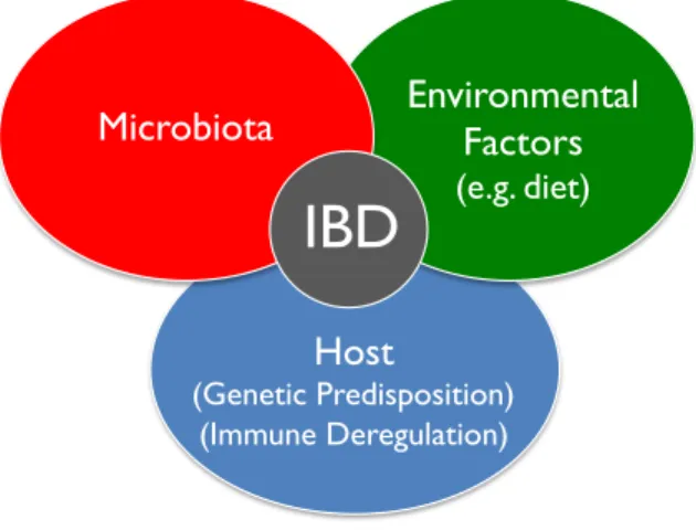
The prevalence of IBD rapidly increased in the Western world in the second half of the twentieth century and is becoming more common in different parts of the world that are adopting a Western lifestyle [137]. Epidemiological observations indicate that there are strong environmental influences on IBD, which is corroborated by the relatively low concordance rate in identical twins (50% for CD, and 10% for UC) [138]. Nevertheless, genome-wide association studies (GWAS) have found associations between specific genes and genetic loci with IBD, many of them involved in intestinal barrier function, epithelial restitution, innate and adaptive immune regulation and microbial defense [139,140]. The etiology of IBD remains an enigma but it is hypothesized that IBD results from an adverse interaction between environmental, host factors and genetic susceptibility with gut microbiota playing a key role mediating the pathology [136] (Fig. 3).
Host
(Genetic Predisposition) (Immune Deregulation)
Microbiota
Environmental
Factors
(e.g. diet)
IBD
Figure 3. Interaction of various factors contributing to chronic intestinal inflammation in a genetically susceptible host.
Indeed, despite some disparate results, studies assessing gut microbial features of IBD patients support the concept that there is a generalized reduction in biodiversity (α-diversity) and altered representation of several specific bacterial taxa [141]. Moreover, some of the genes with the strongest association to IBD, such as NOD2 and genes involved in the differentiation of Th17 cells (such as IL-23 and IL-23R), play a key role in gut microbial surveillance [142,143]. Recently, it has been shown that transplanting gut microbiota from patients with CD to GF mice induced a pro-inflammatory gene expression profile in the mice gut that resembles the immunological signatures found in CD patients. Furthermore, microbiota of CD patients exacerbated experimental colitis in IL-10 deficient mice, a murine model of genetic susceptibility to colitis [130].
Diet has also been considered as an additional risk factor contributing to the etiology of IDB. While no effect was found for the intake of fiber, sugar, macronutrients, total energy, vitamin A, C, D, E and carotene on the risk of ulcerative colitis, n-6 polyunsaturated fatty acid was identified as a risk factor for UC, whilst n-3 polyunsaturated fatty acid may protect against UC. Furthermore, high animal protein diet was associated with increased risk of IBD and relapses [144].
5.2. Colitis models in mice
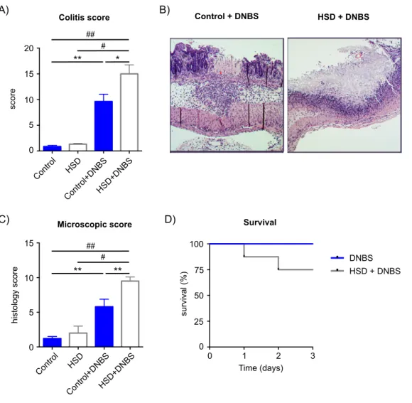
Experimental colitis models have widely served as useful tools for understanding the etiology of IBD and the preclinical evaluation of the efficacy of new drugs. Several models of acute and chronic intestinal inflammation have been developed to simulate various aspects of IBD in humans. Different models use very distinct strategies to induce GI inflammation including 1) disruption of epithelial barrier, 2) disruption of innate immunity, 3) excessive effector immune cell responses, 4) inadequate regulatory response, and others [145]. The methodology is also highly variable, including chemical-induced, genetic manipulation, pathogen inoculation, immune cells transfer and whole microbiota transfer [145]. Each experimental colitis model results in different phenotypic outcomes, including immunological and histological features, with various degrees of modeling human CD and UC (For a review in colitis models in mice, see [145]).
In this study, we used two different chemical-induced models, the dextran sulfate sodium (DSS) and the dinitrobenzene sulfonic acid (DNBS) models. Both models trigger a disruption of the GI tract epithelial layer although with different mechanisms of action and phonotypical outcomes.
5.2.1. DSS-colitis
DSS is delivered in drinking water of rodents and results in an inflammatory response in the GI tract with the recruitment of M1 macrophages and neutrophils to the mucosa. Histologically, DSS colitis induces submucosal erosions, inflammatory cell infiltration, erosion of the epithelium and mucosal hyperplasia. Mice with DSS colitis will show signs of acute colitis including body weight loss, bloody stools and diarrhea, accompanied with the increase in mucosal production of pro-inflammatory cytokines IL-6, TNFα and IL1β [146]. The damage induced by DSS affects preferentially the colon and the cecum. The mechanisms of action are not fully understood but it has been proposed that DSS establishes linkages with medium-chain-fatty acids (MCFA) to form vesicles
that can fuse with colonocyte membranes, leading to a loss of barrier function, increased translocation of luminal antigens and activation of intestinal inflammatory signaling pathways [147]. The concentration and the number of cycles of DSS administration can be customized in order to induce different severity degrees of acute colitis and to trigger chronic colitis, respectively [146].
5.2.2. DNBS-Colitis
DNBS is a hapten reagent delivered rectally in the rodent colon which induces a localized colonic inflammation. DNBS induces significant body weight loss and bloody diarrhea. The colon damage is characterized by hyperplasia, excess of fibrotic tissue and lymphocytic pus, as well as areas of mucosal necrosis and typical visible ulcerations. Histological features include a transmural necrosis, loss of epithelium and diffuse leukocyte cellular infiltrate in the submucosa, namely neutrophils. DNBS is dissolved in a 50% ethanol solution to help break the mucosal barrier. The hapten properties will trigger a T cell response, including the production of Th1 profile cytokines (IFNγ, TNFα and IL12) [146].
5. AIMS
High salt consumption was described to have an impact in modulating the immune system in a pro-inflammatory manner, with a direct effect on the differentiation and expansion of Th17 cells, and to exacerbate the development of MS, both in rodents and humans.
Considering that gut microbiota and Th17 T cells play a pivotal role in the development of the pathogenesis of IBD, we hypothesized that a diet-enriched in salt could affect the development of colitis in mice, through the modulation of gut microbiota.
First, we characterized the effect of HSD on the composition and metabolic pathways of mouse gut microbiota. We proceeded by characterizing the impact of HSD on the mucosal immunity of intestinal lamina propria. After that, we assessed the effect of HSD on the development of gut inflammation using two different models of experimental colitis, DSS and DNBS colitis. Finally, using GF mice and gut microbiota transplant experiment, we sought to determine whether gut microbiota plays a role in the HSD-induced colitis severity.
These data open new insights on the potential mechanisms for the development of IBD, as well as reveal a new pathway of diet-microbiota-host interaction.
II – Research Work
High salt diet exacerbates colitis in mice by decreasing
Lactobacillus levels and butyrate production
The research work “High salt diet exacerbates colitis in mice by decreasing Lactobacillus levels and butyrate production” consists of a first-authorship publication. Here is clarified the contribution of each of the authors:
Publication:
Pedro M. Miranda 1,2, Giada De Palma1, Viktoria Serkis1, Jun Lu1, Marc P.
Louis-Auguste1, Justin McCarville1, Elena F. Verdu1, Stephen M. Collins1,
Premysl Bercik1*, 2018. High salt diet exacerbates colitis in mice by
decreasing Lactobacillus levels and butyrate production. Microbiome
Journal (published).
1Farncombe Family Digestive Health Research Institute, Department of Medicine,
McMaster University, Hamilton, Ontario, Canada. 2Graduate Program in Areas of Basic
and Applied Biology, Instituto de Ciências Biomédicas Abel Salazar, Universidade do Porto, Porto, Portugal
* Corresponding author E-mail: [email protected]
Pedro Miguel Miranda, the candidate for the PhD degree at the University of Porto, contributed to the paper by initially single-handed establishing the HSD-colitis mice model in the laboratory. Pedro also developed the study concept and design, with the supervision of the authors GDP and PB. Moreover, Pedro prepared and carried out all experiments that led to this publication, including DNA/RNA isolation, 16S rRNA gene library preparation, sequencing analysis, interpretation of microbiome/immune/colitis data, SPF mice experiments. Finally, Pedro wrote this manuscript with the supervision of the authors GDP and PB.
The contribution of each author to the publication in clarified as the following:
Authors contribution: GDP and PB contributed to develop the study concept and design, interpretation and discussion of the results. GDP performed all experiments with the GF mice. VS and JM assisted with all immune cell isolation experiments, cell-staining and flow cytometry procedures. JL assisted with all animal work, including blind colitis and histological assessments. MPLA carried out Q-PCR experiments. EFV helped coordinating all GF mice experiments. GDP, PB and EFV were major contributors to the critical review of the manuscript. All authors read and approved the final manuscript.
High salt diet exacerbates colitis in mice by decreasing
Lactobacillus levels and butyrate production
Keywords: Salt, NaCl, Western Diet, Colitis, Microbiota, Lactobacillus, Butyrate, MAPK-pathway
1. Abstract
Background: Changes in hygiene and dietary habits, including increased consumption of foods high in fat, simple sugars, and salt that are known to impact the composition and function of the intestinal microbiota, may explain the increase in prevalence of chronic inflammatory diseases. High salt consumption has been shown to worsen autoimmune encephalomyelitis and colitis in mouse models through p38/MAPK signaling pathway. However, the effect of high salt diet (HSD) on gut microbiota and on intestinal immune homeostasis, and their roles in determining vulnerability to intestinal inflammatory stimuli are unknown. Here, we investigate the role of gut microbiota alterations induced by HSD on the severity of murine experimental colitis.
Results: Compared to control diet, HSD altered fecal microbiota composition and function, reducing Lactobacillus sp. relative abundance and butyrate production. Moreover, HSD affected the colonic, and to a lesser extent small intestine mucosal immunity by enhancing the expression of pro-inflammatory genes such as Rac1, Map2k1, Map2k6, Atf2, while suppressing many cytokine and chemokine genes, such as Ccl3, Ccl4, Cxcl2, Cxcr4, Ccr7. Conventionally raised mice fed with HSD developed more severe DSS- (dextran sodium sulfate) and DNBS- (dinitrobenzene sulfonic acid) induced colitis compared to mice on control diet, and this effect was absent in germ-free mice. Transfer experiments


![Figure 1. Self reported survey on mean sodium intake among (A) males and (B) females in the USA National Health and Nutrition Examination Surveys (NHANES) (Adapted from Loria CM et al, 2001[209])](https://thumb-eu.123doks.com/thumbv2/123dok_br/15751404.1073711/96.918.140.782.134.320/figure-reported-females-national-nutrition-examination-surveys-adapted.webp)