Prevalence and determinants of oral microflora among portuguese
adolescents
Carlos Pereira1, Nélio Veiga2,3, Carlos Resende4, Odete Amaral1, Joana Pereira5
2Research Centre for Education, Technology and Health Studies – Polytechnic Institute of Viseu, Viseu, Portugal.
2Health Sciences Institute – Universidade Católica Portuguesa, Viseu, Portugal.
3Center for Interdisciplinary Research in Health (CIIS) – Universidade Católica Portuguesa, Portugal.
4Institute of Molecular Pathology and Immunology of the University of Porto, Porto, Portugal. 5Riga Stradins University, Latvia.
Abstract
Objectives: Determine the prevalence and determinants of salivary S. mutans, Lactobacillus and A. actinomycetemcomitans and the associated risk of development of dental pathologies on a sample of portuguese adolescents.
Design: A cross-sectional study. Setting: Public school in Sátão, Portugal
Participants: Final sample of 447 adolescents aged between 12 and 18 years old
Measurements: A self-administered questionnaire was filled out by the adolescents. Clinical examination of oral health status was carried out and saliva collection was accomplished by the passive drool method. The identification of the different types of bacterial strains was accomplished using the Polymerase Chain Reaction technique.
Results: The prevalence of S. mutans in the sample studied was 80.8%, Lactobacillus 99.5%, and A. actinomycetemcomitans only 15.2%. The presence of S. mutans was associated with gender (male=76.1% vs female=83.6%, p=0.04) and dental pain in the presence of severe dental caries (77.3% vs 87.8%, p=0.006). The infection with A. actinomycetemcomitans was associated with age (<15yrs=12.3% vs ≥15yrs=20.3%, p=0.03) and residence area (rural=18.2% vs urban=11.0%, p=0.04), and may be related with a higher risk of periodontal disease development in adulthood.
Conclusions: A. actinomycetemcomitans infection was found to be associated with socio-demographic variables, suggesting that, if not clinically well identified and treated, may cause serious oral diseases during adulthood. It has been described that the oral microflora is one of the main etiological factors for dental caries and periodontal diseases development, but cannot be considered in an isolated manner.
Introduction
The oral cavity is inhabited by different bacterial species that play vital roles in maintaining oral health or in shifting to a diseased state such as dental caries and periodontal disease1,2. Dental caries consists in a post-eruptive bacterial infectious disease characterized by a progressive demineralization process that affects the mineralized dental tissues. It is considered the most prevalent oral disease and the main responsible for tooth loss among the population3,4. A carious lesion initiates with the production of organic acids by the microorganisms of the oral cavity that metabolize the extracellular carbohydrates of the individual´s diet5,6. The presence of the organic acids produced will decrease the pH in the interface between the tooth surface and the bacterial plaque, which permits the development of the demineralization process on the tooth enamel4. When the oral cavity has a pH below 5,5 (considered the critical pH), the saturation of the dental tissues initiates causing an initial lesion that will be the precursor of the dental carie7. Prevention methods have the main goal of decreasing the time of exposure of the tooth tissues to the low values of pH. Therefore, it is strictly necessary the frequent removal of bacterial plaque to avoid the increased contact with tooth surfaces8.
The researcher Paul Keyes developed a diagram that describes the multifactorial aetiology of dental caries. In this diagram, we can observe that there are three main etiological factors that are essential for the initiation and development of the disease: susceptible host; cariogenic oral microflora; substratum that depends on the host´s diet, which is then metabolized by the microorganisms that constitutes that bacterial plaque5.
Dental caries, as an infectious disease, correlates directly with bacterial strains that co-exist in the oral cavity, like S. mutans and Lactobacillus9. The cariogenic properties of S. mutans and Lactobacillus are widely recognised and, as significant odontopathogens, the former group is linked to enamel lesion formation while the latter is associated with cavity progression10. A. actinomycetemcomitans is one of the most well studied periodontal bacterial strains. It stays in the periodontal pocket of the oral cavity and damages tooth supporting tissues, being considered as the major cause of periodontitis11. Kaplan and colleagues found that all A. actinomycetemcomitans strains possess strong virulence potential12. A study developed by Hart et al., indicated the main bacterial species or groups that could be implicated in caries onset and progression. Both S. mutans and Lactobacillus were reported in that study has been linked with caries onset and progression13.
Aas et al., reported a distinctive predominant bacterial flora of the healthy oral cavity that is highly diverse and subject specific. It is important to fully define the human microflora of the healthy oral cavity before we can understand the role of bacteria in oral disease14. Furthermore, it has concurrently
been clearly established that social, economic, cultural, ethnic, and environmental factors also play an important role in the formation of dental caries and also influences the individual oral microflora highly related with oral health behaviours15.
The aim of the present study was, therefore, to investigate the prevalence of salivary S. mutans, Lactobacillus and A. actinomycetemcomitans and the influence on the risk of development of dental caries and the association with socio-demographic aspects among a sample of Portuguese adolescents. Materials and Methods
Material collection
A sample of 447 adolescents aged between 12 and 18 years old, attending a public school in Sátão, Portugal, was enrolled in this study. All samples were obtained from September to December of 2012. Questionnaires without information about gender and age were excluded of the study as well as the adolescents whose parents did not sign the informed consent before data collection.
All participants in this study filled out a self-administered questionnaire focusing socio-demographic variables, social and daily habits and oral health behaviours. Questions about socio-demographic variables such as gender (male/female), age, school grade at the moment of the study, residence area (urban/rural), parents´ educational level (choosing the higher educational level between father and mother), parents´ professional situation (employed/unemployed) and the number of rooms and people living in the house were used to determine the crowding index.
This research has been performed in accordance with the Declaration of Helsinki and was submitted and approved by the Ethics Committee of the Health School and Research Centre for Education, Technology and Health Studies of the Polytechnic Institute of Viseu, Portugal (CI&DETS). The information collected by the questionnaires was provided voluntarily and confidentially, guaranteeing, anonymity of the information collected by telling the adolescents not to sign their names or write down any other form of identification in any part of the questionnaire. Data collection was made only for adolescents whose parents signed an informed consent that explained the objectives of the study. After
collection, the questionnaires were numbered, stored and processed by computer. The results do not refer to nominal adolescents or contain any information that may identify any of the participants.
Clinical sample characterization
Clinical examination of oral health status was carried out according to the World Health Organization (WHO) criteria16. The teeth were clinically examined with dental instruments using visual-tactile method with the use of a dental mirror and a probe (approved by the WHO for caries diagnosis) and took place in the classroom under standardized conditions recommended by the WHO. Cotton rolls and gauze were available to remove moisture and plaque when necessary. There was only one observer that registered the results of each clinical observation during the study. The recorded variables of the clinical examination were caries experience, using the decayed, missing and filled permanent tooth index (DMFT) as oral health indicator, which consists in the sum of teeth decayed, teeth missing due to dental caries and teeth filled for each analysed adolescent. Each tooth would be classified with only one of the following codes:0: sound crown or root, showing no evidence of either treated or untreated caries; 1: indicates a tooth with caries; 2: filled teeth, with additional decay; 3: filled tooth with no decay; 4: tooth that is missing as a result of caries; 5: a permanent tooth missing for any other reason than decay; 6: teeth on which sealants have been placed; 7: indicate that the tooth is part of a fixed bridge; 8: this code is used for a space with an unerupted permanent tooth where no primary tooth is present; 9: erupted teeth that cannot be examined; T: indicates trauma in the presence of a fractured crown.
Bacterial strain identification
Saliva collection was accomplished by the passive drool method into a polypropylene tube until reaching 2 mililiters of saliva in each tube per adolescent. Next, DNA was extracted using the MagNA Pure LC DNA Isolation Kit (Bacteria, Fungi) (Roche), quantified with Nanodrop (Thermo Lifesciences), and bacterial DNA was amplified using the Multiplex Polymerase Chain Reaction (PCR) kit (Qiagen) with primers specific for the bacterial strains analysed. To validate primers specificity, DNA bands with the expected molecular from 3 positive-cases were excised from the agarose gel, purified with ULTRAPrep® Agarose Gel Extraction kits (AHN), and sequenced with BigDye Terminator Sequencing Kit (Applied Biosystems). The obtained DNA sequences were compared with a control and with NCBI database (http://www.ncbi.nlm.nih.gov/). Primers employed in this study are as described: Lactobacillus (GGAATCTTCCACAATGGACG and CGCTTTACGCCCAATAAATCCGG); S. mutans (TCGCGAAAAAGATAAACAAACA and GCCCCTTCACAGTTGGTTAG); A. actinomycetemcomitans (AAACCCATCTCTGAGTTCTTCTTC and ATGCCAACTTGACGTTAAAT) (17-19).
Statistical analysis
Data analysis was carried out using SPSS for Windows (version 18.0). Prevalence was expressed in proportions and were compared by the Chi-square test. The significance level established the inferential statistics was 5% (p<0.05).
Results
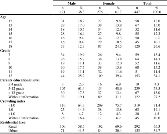
The sample used in this research was composed by 447 adolescents (38.3% were males and 61.7% females), all between the age of 12 and 18 years old, from a public school of Sátão, Portugal. When analysing the parents’ educational level, we observed that 4.3% have parents that only frequented school to or less than 4th grade, 53.5% stayed in school from the 5th to the 12th grade and 15.0% went to a superior degree after finishing the 12th grade. Crowding index < 1.0 is presented among 71.4% of adolescents, while 14.1% are equal to 1 and only 4.5% > 1.0, which indicates possible overcrowding at home. The analysis of the distribution of the sample by residence area revealed that the majority of adolescents live in rural areas (65.3% vs 34.7%) (Table 1).
Table 1: Socio-demographic characterization of the studied sample of portuguese adolescents.
The prevalence of S. mutans in the sample studied was 80.8%, Lactobacillus 99.5% and A. actinomycetemcomitans 15.2%. When analysing the combinatorial presence of two or more bacterial strains, we observed the following: S. mutans and Lactobacillus: 80.5%; Lactobacillus and A. actinomycetemcomitans: 15.0%; S. mutans and A. actinomycetemcomitans: 12.1%; S. mutans, Lactobacillus and A. actinomycetemcomitans: 12.1%.
Looking for the association between the bacterial species and gender, we observed a significant difference among gender (higher in females) for S. mutans (male=76.1% vs female=83.6%, p=0.04) and the co-infection with both S. mutans and Lactobacillus. Focusing on socio-demographic variables, we found that infection with A. actinomycetemcomitans was significantly associated with both age (<15 years=12.3% vs ≥15 years=20.3, p=0.03) and residence area (rural=18.2% vs urban=11.0%, p=0.04). The same situation was observed in adolescents infected with A. actinomycetemcomitans and any of the other bacterial species mentioned above. Taken together, these results demonstrate that A. actinomycetemcomitans may be the main bacterial strain associated with socio-demographic variables independently of the presence of the other bacterial strains analysed in the present study. These results may also be associated with a higher prevalence of periodontal disease among older adolescents and those that present worse socioeconomic status that live in rural areas as verified in table 2.
Male Female Total
n % n % n % 171 38.3 276 61.7 447 100.0 Age 12 31 18.2 27 9.8 58 13.0 13 29 17.0 38 13.8 67 15.0 14 18 10.5 34 12.3 52 11.6 15 28 16.4 27 9.8 55 12.3 16 16 9.4 34 12.3 50 112 17 16 9.4 29 10.5 45 10.1 18 33 12.3 87 24.3 120 26.6 Grade 7 34 19.9 26 9.4 59 13.4 8 26 15.2 38 13.8 64 14.3 9 19 11.1 33 12.0 52 11.6 10 30 17.5 38 13.8 68 15.2 11 19 11.1 32 11.6 51 11.4 12 44 25.2 109 39.4 153 34.1 Parents´educational level ≤ 4 grade 3 2.0 16 6.9 19 4.3 5-12 grade 105 61.4 134 48.6 239 53.5 > 12 grade 30 17.5 37 13.4 67 15.0 Without information 33 19.1 89 31.1 122 27.2 Crowding índex <1.0 110 64.3 209 75.7 319 71.4 1,0 25 14.6 38 13.8 63 14.1 >1.0 8 4.7 12 4.3 20 4.5 Without information 28 16.4 17 6.2 45 10.0 Residential área Rural 100 58.5 192 69.6 292 65.3 Urban 71 41.5 84 30.4 155 34.7
Table 2: Association between bacterial strains S. mutans (SM), Lactobacillus (LA) and A. actinomycetemcomitans (AA) and socio-demographic variables.
Our study revealed the lack of a significant correlation between worse oral health behaviours and the presence of the various bacterial species analysed. However, some bacterial strains were associated with dental pain, which occurs in the presence of severe dental caries. We observed that adolescents who suffered one or more episodes of dental pain due to dental caries had a higher rate of S. mutans infection (episode of dental pain= 87.8% vs no dental pain=77.3%, p=0.006) and both S. mutans and Lactobacillus as seen in table 3. The results demonstrate that dental pain might be associated mainly with S. mutans infection than with Lactobacillus infection, considering that 99.5% of the sample used in this study showed infection with this bacterial strain.
Table 3: Association between bacterial strains S. mutans, Lactobacillus and A. actinomycetemcomitans and oral health behaviours and dental pain.
Despite the observed association between dental pain and the presence of S. mutans alone or in combination with Lactobacillus, no significant association was found between the presence of the bacterial species studied and the risk of dental caries development. In fact, we detected the presence of the three species studied not only adolescents that have active caries during the time of intra-oral observation but also in caries-free individuals.
Gender Age Residential area
Bacterial strain Male Female p <15 yrs ≥15 yrs p Rural Urban p
N % N % N % N % N % N % SM 121 76.1 219 83.6 0.04 161 79.3 107 77.5 0.4 169 79.0 78 99.0 0.5 LA 158 99.4 261 99.6 0.6 203 100.0 136 98.6 0.2 213 99.5 126 99.2 0.6 AA 19 11.9 45 17.2 0.09 25 12.3 28 20.3 0.03 39 18.2 14 11.0 0.04 SM+LA 121 76.1 218 83.2 0.05 161 79.3 107 77.5 0.4 169 79.0 99 78.0 0.5 LA+AA 18 11.3 45 17.2 0.07 25 12.3 27 19.6 0.05 39 18.2 13 10.2 0.03 SM+AA 14 8.8 37 14.1 0.07 21 10.3 20 14.5 0.2 32 15.0 9 7.1 0.02 SM+LA+AA 14 8.8 37 14.1 0.07 21 10.3 20 14.5 0.2 32 15.0 9 7.1 0.02
Oral hygiene Dental appointment Dental pain
Bacterial strain ≤1/day ≥2/day p No Yes p No Yes p
N % N % N % N % N % N % SM 56 80.0 205 78.2 0.5 117 81.8 219 79.9 0.4 218 77.3 122 87.8 0.006 LA 70 100.0 260 99.2 0.6 143 100.0 272 99.3 0.4 282 100.0 137 98.6 0.1 AA 9 12.9 44 16.8 0.3 22 15.4 42 15.3 0.5 46 16.3 18 12.9 0.2 SM+LA 56 80.0 205 78.2 0.5 117 81.8 218 79.6 0.3 217 77.0 122 87.8 0.005 LA+AA 9 12.9 43 16.4 0.3 22 15.4 41 15.0 0.5 46 16.3 17 12.2 0.2 SM+AA 9 12.9 32 12.2 0.5 18 12.6 33 12.0 0.5 36 12.8 15 10.8 0.3 SM+LA+AA 9 12.9 32 12.2 0.5 18 12.6 33 12.0 0.5 36 12.8 15 10.8 0.3
Discussion
The results obtained in this research demonstrate that the salivary presence of S. mutans, Lactobacillus, and A. actinomycetemcomitans is not associated with the development of dental pathologies in our community sample of adolescents. Dividing the sample in caries-free adolescents and caries-active adolescents, we observed that both groups have similar levels of infection by S. mutans and
Lactobacillus. Nevertheless, our findings go against some studies that correlate the presence of S. mutans and Lactobacillus. with a higher risk of caries formation20,21. Also Fysal et al., observed that the number of S. mutans and A. actinomycetemcomitans determined by microscopic counts was twice as high in caries patients22. On the other hand, and strengthening our results, Reyes et al. demonstrated that Streptococcus species were present in caries-active and also in caries-free individuals. In this study, the authors also observed that dental caries can occur in the apparent absence of the bacterium Streptococcus and can even be associated with healthy states1. Parisotto et al. explored the association between caries development, colonization with caries-associated microflora, and immunity as children begin the transition to mixed dentition. In baseline level there was no significant differences in S. mutans and Lactobacillus between caries-free and caries-active groups23. Beighton confirmed that dental caries may develop in the absence of these species and their presence does not necessarily indicate dental caries activity24. Additionally, Wolff et al. demonstrated that there is no significant differences of oral bacterial strains between caries-free and caries subjects25.
A question may arise from the present study: can an association of other bacterial strains with S. mutans, Lactobacillus and A. actinomycetemcomitans, potentiate and increase the risk of oral disease development? Looking at the oral health behaviours, no differences were observed between oral hygiene habits and the presence or absence of the three bacterial strains in study. Our results are different from those obtained by Plonka et al. that determined that the presence of S. mutans and Lactobacillus would increase with children´s age and was also associated with worse oral health behaviours26. Levels of S. mutans and Lactobacillus were found to be strongly associated with
socioeconomic status among Palestinian children in East Jerusalem. The relatively high prevalence of cariogenic bacteria suggests that oral care prevention and treatment demands special attention from the health care institutions and authorities15.
In the present study, A. actinomycetemcomitans was associated with age and residence area. Adolescents from rural areas showed a higher prevalence of this bacteria in comparison with the ones who live in urban areas. Paolantonio et al. demonstrated that A. actinomycetemcomitans colonization in children and adolescents from central Italy is affected by socioeconomic and cultural factors, namely residence area (urban vs rural areas), and that these factors may also affect the periodontal condition of the subjects27. The same situation is verified in the presence of Lactobacillus and A. actinomycetemcomitans, S. mutans and A. actinomycetemcomitans and in the presence of all three bacterial strains. This fact can be considered important, because the adolescents that present A. actinomycetemcomitans can have, in the near future, a higher risk of periodontal disease development28,29. Therefore, special attention should be given to adolescents living in rural areas, which present worse oral health behaviours and more difficulties in attending frequent dental appointments biannually.
We verified that adolescents who suffered one or more episodes of dental pain due to dental caries had a higher incidence of S. mutans alone or in combination with Lactobacillus. This may be justified by the fact of adolescents that suffer dental pain have worse dental caries lesions and more retentive sites on the tooth surface that may increase the levels of anchored S. mutans, as previously suggested by Thaweboon et al.30. However, it is important to understand that this association between the prevalence of certain bacterial strains and the occurrence of dental pain does not occur in an isolated manner. Even knowing that S. mutans is one of the main bacterial strains responsible for dental caries development, we must have into account other aetiological factors. The consumption of sweet foods and oral hygiene habits are clearly described as being significantly associated with the severity of dental caries development4,5.
Conclusions
The presence of oral microflora is clearly one of the main etiological factors for dental caries and periodontal diseases development, but cannot be considered in an isolated manner. For the development of oral disease, various other factors need to be present such as high and daily sugar intake, inadequate oral hygiene habits and even genetic susceptibility. A. actinomycetemcomitans, even among adolescents, can be considered associated with socio-demographic variables, and, if not clinically well identified and treated, may cause serious oral diseases during adulthood. Adolescents
who suffered one or more episodes of dental pain due to dental caries had a higher incidence of S. mutans.
Probably the imbalances in the resident microflora may be the ultimate mechanism of oral disease development. Oral diseases can appear in the presence of changes of the oral bacterial communities’ structure and that may be related with the shift from health to disease. The enormous diversity of oral
microbiota allowed for a better understanding of oral micro ecosystem, and these pathogenic populations present in the oral cavity provide new insights into the aetiology of oral diseases and suggest new targets for interventions of the disease. The present study indicates that epidemiological surveys with the assessment of etiologic risk factors are crucial tools for oral health planners and clinicians in order to implement a risk-based preventive strategy.
Acknowledgments
The authors are deeply indebted to the researchers that participated in the development of this study and data collection phase: Dr. Marco Baptista and Dr. Inês Coelho. We also thank the teachers and students of the School Group of Sátão, Portugal, for the participation and important contribution to this study.
Funding
This study was funded by the Fundação para a Ciência e Tecnologia and the Research Centre for Education, Technology and Health Studies of the Polytechnic Institute of Viseu, Portugal (CI&DETS).
Competing interests
The authors have declared that no competing interests exist.
What we know about the theme?
1. The oral cavity is inhabited by different bacterial species that play vital roles in maintaining oral health or in shifting to a diseased state such as dental caries and periodontal disease.
2. Aggregatibacter actinomycetemcomitans is one of the most well studied periodontal bacterial strains.
3. Social, economic, cultural, ethnic, and environmental factors play an important role in the formation of dental caries and also influences the individual oral microflora.
What we get out the study?
1. Aggregatibacter actinomycetemcomitans, among adolescents, can be considered associated with socio-demographic variables, and, if not clinically well identified and treated, may cause serious oral diseases during adulthood.
2. Adolescents who suffered one or more episodes of dental pain due to dental caries had a higher incidence of S. mutans.
3. It has been described that the oral microflora is one of the main etiological factors for dental caries and periodontal diseases development, but cannot be considered in an isolated manner.
References
1. Reyes C, Dalmacio L. Bacterial Diversity in the Saliva and Plaque of free and Caries-active Filipino Adults. Philippine J Sci. 2012; 141(2):217-27.
2. Kleinberg I. A Mixed-bacteria Ecological Approach to Understanding the Role of the Oral Bacteria in Dental Caries Causation: an Alternative to Streptococcus mutans and the Specific-plaque Hypothesis. Crit Rev Oral Biol Med. 2002; 13(2):108-25. doi: 10.1177/154411130201300202.
3. Lamont R, Jenkinson H. Caries as an infectious disease. In Oral Microbiology at a Glance. 1st ed. Singapore: Wiley-Blackwell; 2010. ISSN: 9780813828923.
4. Pereira A. Odontologia em Saúde Colectiva - Planejando acções e promovendo saúde. 1st ed. Porto Alegre: Artmed Editora; 2003. ISSN: 853630166X.
5. Lima J. Cárie Dentária: um novo conceito. Rev Dental Press Ortodon Ortop Facial. 2007; 12(6):119-30. doi: 10.1590/S1415-54192007000600012.
6. Cortelli S, Cortelli J, Prado J, Aquino D, Jorge A. DMFT in school children relate to caries risk factors. Cienc Odontol Bras 2004; 7(2):75-82.
7. Axelsson P. Diagnosis and Risk Prediction of Dental Caries. 1st ed. Germany: Quintessence Publishing; 2000. ISSN: 978-0867153620.
8. Daniel S, Harfst S, Wilder R, Francis B, Mitchell S. Mosby´s Dental Hygiene: Concepts, Cases and Competencies. 2nd ed. St Louis, USA: Mosby Elvesier; 2008. ISSN: 9780323043526.
9. Fejerskov O, Kidd E. Dental Caries: The Disease and its Clinical Management. 2nd ed. Oxford: Blackwell Munksgaard; 2003. ISSN: 978-1405138895.
10. Brambilla E, Twetman S, Felloni A, Cagetti M, Canegallo L, Garcia-Godoy F, Strohmenger L. Salivary mutans streptococci and lactobacilli in 9- and 13-year-old Italian schoolchildren and the relation to oral health. Clin Oral Investig. 1999; 3(1):7-10.
11. Gasparetto A, Arana-Chavez V, Avila-Campos M. Actinobacillus actinomycetemcomitans attached to oral epithelial cells: stability and ultrastructural aspects. Pesqui Odontol Bras. 2000; 14(4):311-8. doi: 10.1590/S1517-74912000000400002.
12. Kaplan J, Schreiner H, Furgan D, Fine D. Population structure and genetic diversity of Actinobacillus actinomycetemcomitans strains isolated from localized juvenile periodontitis patients. J Clin Microbiol. 2002; 40:1181-7. doi: 10.1128/JCM.40.4.1181-1187.2002.
13. Hart T, Corby P, Hauskrecht M, Ryu O, Pelikan R, Valko M, et al. Identification of Microbial and Proteomic Biomarkers in Early Childhood Caries. Int J Dent. 2011; Article ID 196721:1-13. doi: 10.1155/2011/196721.
14. Aas J, Paster B, Stokes L, Olsen I, Dewhirst F. Defining the normal bacterial flora of the oral cavity. J Clin Microbiol. 2005; 43(11):5721-32. doi: 10.1128/JCM.43.11.5721-5732.2005.
15. Steinberg D, Eskander L, Zini A, Sgan-Cohen H, Bajali M. Salivary levels of mutans streptococci and Lactobacilli among Palestinian school children in East Jerusalem. Clin Oral Investig. 2014; 18(3):979-83. doi: 10.1007/s00784-013-1028-x.
16. World Health Organization. Oral Health Survey - Basic Methods. Geneva: WHO; 2003.
17. Abu Bakar F, Abdulamir A, Nordin N, Yoke T. Methods for precise molecular detection of probiotic microflora: using adjusted molecular biology protocols, primer sets and PCR assays. Biotechnol. 2010; 9(1):25-32. doi: 10.3923/biotech.2010.25.32.
18. Zhou C, Saxena D, Caufield P, Ge Y, Wang M, Li Y. Development of species-specific primers for detection of Streptococcus mutans in mixed bacterial samples. FEMS Microbiol Lett. 2007; 272(2):154-62. doi: 10.1111/j.1574-6968.2007.00756.x.
19. Ashimoto A, Chen C, Bakker I, Slots J. Polymerase chain reaction detection of 8 putative periodontal pathogens in subgengival plaque of gingivitis and advanced periodontitis lesions. Oral Microbiol Immunol. 1996; 11:266-73. doi: 10.1111/j.1399-302X.1996.tb00180.x.
20. Bhayat A, Ahmad M, Hifnawy T, Mahrous M, Al-Shorman H, Abu-Naba´a L, et al. Correlating dental caries with oral bacteria and the buffering capacity of saliva in children in Madinah, Saudi Arabia. J Int Soc Prev Community Dent. 2013; 3(1):38-43. doi: 10.4103/2231-0762.115712.
21. Luo A, Yang D, Xin B, Paster B, Qin J. Microbial profiles in saliva from children with and without caries in mixed dentition. Oral Dis. 2012; 18:595-601. doi: 10.1111/j.1601-0825.2012.01915.x. 22. Fysal N, Jose S, Kulshrestha R, Arora D, Hafiz K, Vasudevan S. Antibiogram pattern of oral microflora in periodontic children of age 6 to 12 years: a clinicomicrobiological study. J Contemp Dent Pract. 2013; 14(4):595-600. doi: 10.5005/jp-journals-10024-1370.
23. Parisotto T, King W, Duque C, Mattos-Graner R, Steiner-Oliveira C, Nobre-dos-Santos M, et al. Immunological and Microbiologic Changes during Caries Development in Young Children. Caries Res. 2011; 45:377-85. doi: 10.1159/000330230.
24. Beighton D. The complex oral microflora of high-risk individuals and groups and its role in the caries process. Community Dent Oral Epidemiol. 2005; 33(4):248-55. doi: 10.1111/j.1600-0528.2005.00232.x.
25. Wolff D, Frese C, Maier-Kraus T, Krueger T, Wolff B. Bacterial biofilm composition in caries and caries-free subjects. Caries Res. 2013; 47(1):69-77. doi: 10.1159/000344022.
26. Plonka K, Pukallus M, Barnett A, Walsh L, Holcombe T, Seow W. A longitudinal study comparing mutans streptococci and lactobacilli colonisation in dentate children aged 6 to 24 months. Caries Res. 2012; 46(4):385-93. doi: 10.1159/000339089.
27. Paolantonio M, di Bonaventura G, di Placido G, Tumini V, Catamo G, di Donato A, et al. Prevalence of Actinobacillus actinomycetemcomitans and clinical conditions in children and adolescents from rural and urban areas of central Italy. J Clin Periodontol. 2000; 27(8):549-57. doi: 10.1034/j.1600-051x.2000.027008549.x.
28. Haubek D. The highly leukotoxic JP2 clone of Aggregatibacter actinomycetemcomitans: evolutionary aspects, epidemiology and etiological role in aggressive periodontitis. APMIS Suppl. 2010; 130:1-53. doi: 10.1111/j.1600-0463.2010.02665.x.
29. Haubek D, Ennibi O, Poulsen K, Vaeth M, Poulsen S, Kilian M. Risk of aggressive periodontitis in adolescents carriers of the JP2 clone of Aggregatibacter (Actinobacillus) actinomycetemcomitans in Morocco: a prospective longitudinal cohort study. Lancet. 2008; 371(9608):237-42. doi: 10.1016/S0140-6736(08)60135-x.
30. Thaweboon S, Thaweboon B, Soo-Ampon S, Soo-Ampon M. Salivary mutans streptococci and lactobacilli after self arresting caries treatment. Southeast Asian J Trop Med Public Health. 2005; 36(3):765-8.