UNIVERSIDADE DE LISBOA
FACULDADE DE CIÊNCIAS
DEPARTAMENTO DE BIOLOGIA VEGETAL
Study and Characterization of CFTR Mutations in Vitro and in
Native Tissues from non CF Patients with Chronic Airways
Diseases
Arsénia Joana Massinga
Mestrado em Biologia Molecular e Genética
Dissertação orientada por:
Professora Doutora Margarida D. Amaral
III Table of Contents Acknowledgments/Agradecimentos ... VII Summary ... VIII Resumo ... IX List of abbreviations ... XI Section I: Introduction ... 1
1.1. Cystic Fibrosis (CF) and CF Diagnosis ... 1
1.2. CFTR: from Gene to Protein and Function ... 3
1.3. CFTR mutations: classification according to their functional defect and class-specific therapies ... 4
1.4. Class V mutations: splicing mutations and correction by RNA-based therapies ... 6
1.5. Objectives of the present work ... 8
Section II: Materials and Methods ... 9
Part I: Methods to analyse patients’ materials ... 9
2.1. The screen of mutations in CF and non-CF patients ... 9
Part II: Methods to study splicing mutations in cellular systems ... 9
2.2. Generation of models to study splicing mutations in IVS5 ... 9
2.2.1. Plasmid vectors ... 9
2.2.2. Site-directed mutagenesis ... 10
2.2.3. Cloning into pLVX-Puro ... 10
2.2.4. Production of lentiviral particles ... 11
2.2.5. Generation of stably transduced cells: lentiviral infection ... 12
2.3. Cell culture ... 12
2.3.1. Cell lines and culture conditions ... 12
2.3.2. Transient transfections ... 13
2.3.3. Stable transfections in HEK Flp In cells ... 13
2.3.4. Treatment with an AON ... 13
2.4. Biochemical ... 13
2.4.1. Western Blot ... 13
2.4.2. Immunofluorescence ... 14
2.5. Statistical analysis ... 14
Section III: Results and Discussion ... 15
3.1. Analysis of Patients ... 15
3.1.1. Patients’ screen studies ... 15
3.1.2. Genotype-clinical status Correlation ... 16
IV
3.2.1. Generation of the models to study splicing mutations in IVS5 ... 19
3.2.2. Impact of the splicing mutations at the mRNA and protein levels ... 19
3.2.3. Effect of the AON on all splicing mutations ... 24
Section IV: Conclusions and future perspectives ... 29
Section V: References ... 30
Section VI: Appendices ... 34
Appendix 1 – Introduction, Material and Methods ... 34
1. RNA extraction... 34
2. cDNA synthesis ... 34
3. Transformation of competent bacteria and extraction of plasmids ... 35
V
List of Figures
Figure 1.1 – Cystic Fibrosis Algorithm………...2
Figure 1.2 – Schematic representation of transcription, translation and localization of CFT…………....3
Figure 1.3 – Pathophysiologic cascade in Cystic Fibrosis that leads to lung disease, the main cause of high mortality rate among CF patients………..……….4
Figure 1.4 – Classification of CFTR mutations accordingly to their functional defect, and its potential target therapies………..6
Figure 3.1 – Schematic representation of the splicing mutations located in the same splicing consensus………19
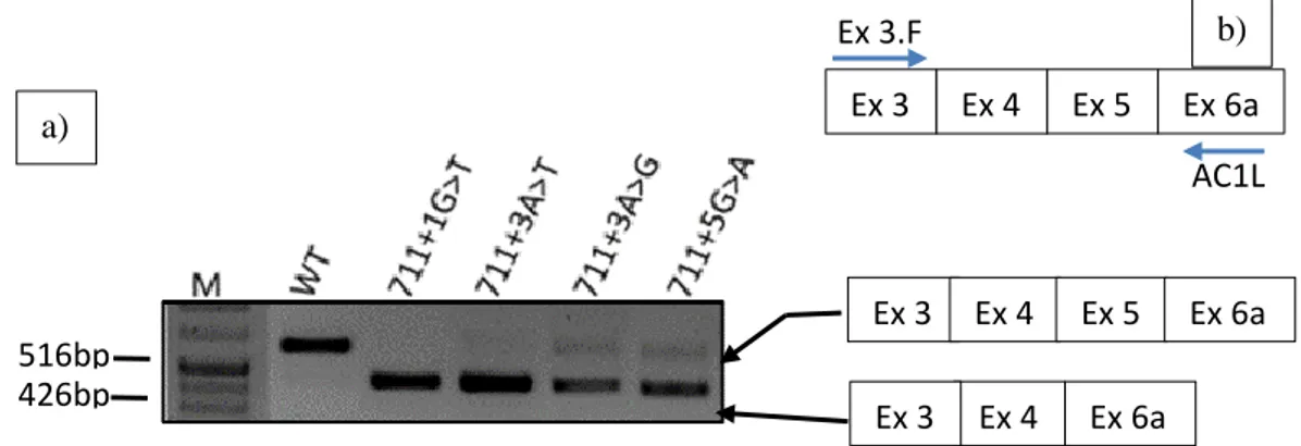
Figure 3.2 – Characterization of CFTR transcripts of HEK 293T cells expressing WT and all four mutants minigenes by RT-PCR………...……20
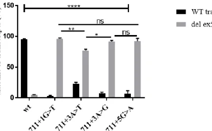
Figure 3.3 - Quantitative analysis of CFTR transcripts with exon 5 inclusion (WT transcripts) and exon 5 exclusion (del ex5 transcripts) from HEK 293T cells transiently expressing different minigenes (WT and mutants)………..………..21
Figure 3.4 - Quantitative RT-PCR analysis of CFTR transcripts (normal and aberrant transcript with no exon 5) from individuals with cystic fibrosis (711+1G>T/F508del) and healthy control………21
Figure 3.5 - Schematic representation of splicing elements located in the 5’ss consensus, that can be disrupted if any mutation occur………..………22
Figure 3.6 - Analysis of the protein obtained from CFBE cells transiently transfected with wt, 711+1G>T, 711+3A>T, 711+3 A>Gand 711+5G>A minigenes………23
Figure 3.7 - Characterization of localization of the CFTR protein of HEK 293T cells expressing the wt and all four mutants minigene by immunofluorescence………..24
Figure 3.8 - Schematic representation of the position of AON in IVS5………24
Figure 3.9 - Correction of alternative splicing accessed by RT-PCR………..25
Figure 3.10 - Correction of alternative splicing accessed by qPCR………25
Figure 3.11: Correction of four splicing mutations in the same splicing consensus by AON strategy at the protein level……….………… ………..………...26
Figure 3.12: Correction of four splicing mutations in the same splicing consensus by AON strategy at the protein level accessed by immunofluorescence………....………..………...27
Figure 3.13: Schematic representation of the ci-acting splicing regulatory elements and trans-acting splicing factors………27
VI
List of tables
Table 2.1: List of primers used to amplify cDNA to clone into pLVX-Puro……….10
Table 3.1: List of the 9 most frequent mutations in Portugal………...15
Table 3.2: Summary of mutations identified in different respiratory illness (non-CF) and CF patients………..………..16
Table 3.3: Association between the number of mutations identified and Genotypes of CFTR found among each group analysed…………..………..………...………..18
Table 1: Description of all primers used for complete sequencing of CFTR………...…..………..37
Table 2: Description of mutagenic primers for splicing mutations introducing……….……….37
VII
Acknowledgments/Agradecimentos
Ao concluir este trabalho, além do meu agradecimento à Deus, pelo dom da vida, e ao projecto Caribu do programa Erasmus Mundus, por ter financiado os meus estudos em Portugal, não posso deixar de agradecer às pessoas que de forma directa e indirecta contribuíram para a realização deste trabalho.
Em primeiro lugar, quero agradecer à Professora Margarida Amaral, minha supervisora, por me aceitar no seu grupo e me ter recebido de braços abertos desde o primeiro momento. Agradeço também por me ter dado a oportunidade de desenvolver o meu trabalho, pela disponibilidade e confiança depositada.
Meu muito obrigada à Professora Rita Zilhão, minha mãe de coração, sem ela meus estudos em Portugal, meu trabalho, nada disto teria acontecido. Obrigada por ter apostado em mim, por ter movido mundos e fundos para que eu conseguisse fazer o meu mestrado em Portugal. Obrigada pelo apoio incondicional, pelo carinho e amor com que sempre me tratou, e por estar sempre disponível quando mais precisava. Agradeço também por me ter ajudado a resolver todos os problemas que tive durante a realização das experiências, principalmente nas clonagens e tranfecções.
Agradeço aos meus pais, que mesmo distantes sempre fizeram-me sentir amada e apoiaram todas as minhas decisões. Obrigada as minhas irmãs, Esperança e Rosiménia pelo apoio e simplesmente por existirem, sei que posso contar sempre convosco – porque somos as três mosqueteira: uma por todas
e todas por uma.
Obrigada a todos os meus colegas de laboratório, com os quais passei momentos felizes de festas, jantares e muito trabalho, e claro momentos stressantes também, quando as experiências não corriam bem. Todos vocês ajudaram-me a crescer pessoal e intelectualmente. Agradeço a todos pelos puxões de orelha, pelas chamadas de atenção quando fazia algo mal, graças a todos vocês comecei a redobrar o cuidado em tudo e a ser mais atenciosa. E como num grupo existem sempre pessoas especiais, quero agradecer de maneira muito especial à Verónica, que mais do que uma colega de trabalho e vizinha de bancada, tornou-se numa grande amiga e quase irmã, por me ter ensinado tudo o que sei sobre genotipagem de pacientes e clonagens, por estar sempre disponível, mesmo quando ocupada. Agredeço pela paciência, atenção e carinho que sempre teve comigo (saiba que para onde eu for levarei comigo os anjinhos de natal que me deste, que são a representação do que tens sido para mim). Agradeço a Sara Canato e ao João Santos pelas brincadeiras, e por me suportarem nos momentos de loucura, e também pelas conversas filosóficas. Agradeço à Íris Lameiro e ao Nikhil pelo apoio nos momentos de crise e desânimo, quero que saibam que deram-me forças para continuar, muito obrigada pela amizade. Muito obrigada à Susana Igreja pelas correcções do trabalho, por ter se esforçado muito que toda história fizesse sentido, e por não ter desistido de mim. Obrigada ao Simão pelas conversas beatas e pelos conselhos sábios que sempre me deu. Agradeço à Joana Lérias pela amizade e pelos conselhos. E claro agradeço à Sofia Correia, nossa lab manager, pelo esforço em manter a ordem no laboratório, pelo apoio moral e pela amizade incondicional.
Meu muito obrigada aos meus colegas de mestrado, de maneira especial ao Clemente, meu amigo de longa data, por me ajudar a traçar objectivos de longo prazo, pelo apoio incondicional e pelo esforço que faz para que eu melhore a cada dia. Obrigada também à Carolina Sousa que mais que uma colega tornou-se a minha miga do estoiro do orçamento, obrigada pela amizade, companheirismo e por estar sempre disposta a ouvir-me, muito obrigada port udo.
E finalmente agradeço às meninas das Lanjeiras, minha primeira residência, minha grande família portuguesa por me ensinarem a organizar melhor os meus estudos e vida no geral, de maneira muito especial à Isabel Coimbra e a Marta Antunes, pela amizade, pelo carinho com que sempre me trataram. Muito obrigada à família Goes e ao padre José de Melo por me terem acolhido quando mais precisei, e foi graças a isso que consegui escrever esta dissertação.
VIII
Summary
Cystic Fibrosis (CF) is the most common autosomal recessive disorder among Caucasians caused by mutations in the CF Transmembrane Conductance Regulator (CFTR) gene, which encodes a protein localized in the apical plasma membrane (PM) of epithelial cells that functions as a chloride (Cl
-) and bicarbonate (HCO3-) channel.
Previous studies have shown the occurrence of at least one CFTR mutation also in non-CF patients with asthma, chronic bronchiectasis and chronic obstruction pulmonary disease (COPD) with unknown etiopathology.
Until now about 2,000 CFTR variations have been found, in which 11% of them are known to be splicing mutations. A novel antisense oligonucleotide (AON) therapy approach corrects splicing defects and is giving some hope for patients carrying this type of mutations.
The objective of the current MSc project was two-fold, namely:
1) To investigate the possible involvement of the 9 most common Portuguese CFTR mutations in causing non-CF respiratory disorders: asthma, COPD and chronic bronchiectasis.
2) To characterize the impact of four splicing mutations located in the same splicing consensus-711+1G>T, 711+3A>T, 711+3A>G and 711+5G>A and correct them using a single AON.
Our results showed that at least 3 mutations out of 9 most common Portuguese CFTR mutations were found in our cohort of patients with respiratory disorders.
Concerning the study of splicing mutations, in vitro experiments were performed using a reporter splicing minigene. We showed that all four splicing mutations located in the same splicing consensus one IVS5 caused skipping of exon 5, which produce a smaller protein and lead to a mislocalization of the CFTR protein in the PM. In addition, the same AON potentially corrected all four splicing mutations.
Altogether these data suggest that there is an involvement of some CFTR mutations commonly found in Portugal in causing non-CF chronic airway diseases and that patients carrying the splicing mutations studied here can benefit from the same AON for therapy.
IX
Resumo
A fibrose quística (FQ) é a doença autossómica recessiva mais comum na população Caucasiana, afetando 1 em cada 6000 portugueses. Esta doença afeta vários órgãos e os doentes apresentam variados sintomas, os mais comuns sendo: elevadas concentrações de cloreto no suor, infeções bacterianas recorrentes devido ao muco espesso formado nos pulmões e malnutrição. A FQ é causada por mutações no gene da CFTR (do inglês Cystic fibrosis transmembrane conductance
regulator) que codifica uma glicoproteína localizada na membrana apical de células epiteliais e funciona
como um canal de cloreto (Cl-) e bicarbonato (HCO 3-).
Tradicionalmente, o diagnóstico da FQ baseia-se nas manifestações clínicas, história familiar e dois testes de suor positivos (>60mM), porém quando esta abordagem é inconclusiva, são feitos testes genéticos para identificar mutações nos dois alelos e medições de corrente para verificar a função ou disfunção da CFTR.
Estudos prévios reportaram uma parcial disfunção da proteína CFTR em doentes sem nenhuma manifestação clínica de FQ, mas com outras doenças como pancreatite, asma, bronquiectasias crónicas e doenças pulmonares de obstrução crónica (DPCO), de origem desconhecida, sugerindo uma associação entre mutações no gene da CFTR e outras doenças que não sejam FQ. Adicionalmente, foi demonstrado que alguns desses indivíduos possuíam apenas uma mutação no gene da CFTR. Contudo, as mutações encontradas são raras e portanto não incluídas entre as mutações mais frequentes encontradas em Portugal. Além disso nem todas as variantes na CFTR estão descritas como causadoras de FQ.
Atualmente são conhecidas mais de 2.000 variações no gene da CFTR, e apenas aproximadamente 250 mutações estão descritas como sendo causadoras de FQ. Estas mutações são classificadas de acordo com o defeito causado na proteína e estão agrupadas em sete classes (I-VII). As mutações de splicing pertencem à classe V (mutações que diminuem os níveis da proteína na membrana apical) e compreendem uma fração de 11% das variações conhecidas no gene da CFTR. Estas mutações resultam num splicing anormal destruindo as junções normais dos RNAs ou criando um novo local de
splicing no interior de um intrão ou de um exão. Tais alterações levam à produção de uma proteína
disfuncional ou truncada o que diminui os níveis da proteína normal que é expressa.
É previsível que a percentagem de mutações de splicing sejam superiores a 11%, devido às mutações missense, que não estão totalmente caracterizadas e que também podem levar a um splicing anormal. Os doentes que possuem esta classe de mutações podem beneficiar apenas de um fármaco que potencia a função da proteína resultante de transcritos normais, o Ivacaftor (VX770). Porém, como algumas mutações de splicing diminuem drasticamente os níveis de proteína funcional, potenciar a proteína não é suficiente, sendo necessário corrigi-la. Para este efeito uma nova abordagem tem sido adotada nomeadamente, o uso de terapias de RNA que corrigem o defeito a nível do RNA mensageiro (mRNA). Uma dessas terapias é o uso de AONs (do inglês antissense oligonucleotides), que bloqueiam ou aumentam o processo de splicing, de acordo com a sequência a que se ligarão.
No presente trabalho de Mestrado tivemos dois objetivos principais, nomeadamente:
1) Investigar o envolvimento das nove mutações do gene da CFTR mais comuns entre os Portugueses no desenvolvimento de doenças respiratórias, tais como asma, doença pulmonar obstrutiva crónica (DPOC) e bronquiectasias.
2) Caracterizar o impacto de quatro mutações -711+1G>T, 711+3A>T, 711+3A>G e 711+5G>A localizadas na mesma sequência de consensus do intrão 5 e finalmente corrigi-las usando um único AON desenhado no nosso laboratório.
Para o primeiro objetivo, foi extraído DNA genómico de amostras de sangue dos indivíduos em estudo e, com técnicas de PCR (do inglês polymerase chain reaction) modificado, foram procuradas as nove mutações no gene da CFTR mais comuns para pacientes FQ portugueses.
X Os resultados mostram que três das nove mutações mais comuns foram encontradas em pacientes com doenças respiratórias, onde a mutação mais comum foi a F508del, a mais comum na população mundial. Adicionalmente constatou-se que a G576A, descrita como sendo um polimorfismo, também parece estar envolvida na suscetibilidade de aparecimento de doenças respiratórias mesmo quando presente individualmente, sem que o outro alelo do indivíduo esteja mutado, porém este polimorfismo não foi encontrado entre a população FQ. Estes resultados sugerem que os polimorfismos no gene da CFTR podem estar envolvidas no aparecimento de asma, bronchiectasias e DPCO, e ainda que mutações num só alelo deste gene podem aumentar a suscetibilidade de um indivíduo portador desenvolver doenças respiratórias, sem que haja manifestação clínica típica de FQ.
Para o 2º objetivo, utilizámos um mini-gene de CFTR consistindo num construto de cDNA de wt-CFTR previamente clonado no plasmídeo pcDNA5, contendo dois intrões: o intrão quatro (IVS4) e o intrão cinco (IVS5) entre os exões 4 e 5 e os exões 5 e 6a, respetivamente, e dois marcadores (eGFP em N-terminal e Flag-tag no 4º loop extracelular) . Introduzimos cada uma das mutações de splicing em estudo por mutagénese direcionada neste mini-gene repórter. De seguida, cada um dos construtos foi clonado num vetor lentiviral para a produção de novas linhas celulares (experiência por concluir para comparação de resultados). Toda a caracterização apresentada ao longo deste trabalho, foi feita em células que expressavam cada um dos construtos por transfeção transitória. O impacto de cada mutação expressa pelas células foi caracterizado a nível do mRNA, expressão e localização da proteína na membrana. Resultados de PCR quantitativo (qPCR) e semi-quantitativo revelaram que as quatro mutações provocam a remoção total do exão cinco e que a mutações 711+1G>T e 711+5G>A diminuem drasticamente os níveis de transcritos normais, mais do que as mutações 711+3A> G e 711+3A>T. Com a técnica de Western blot (WB) verificou-se que a proteína resultante dos construtos com as mutações 711+1G>T, 711+3A>T, 711+3A>G e 711+5G>A é menor do que a proteína normal. Para além disso, quando caracterizadas por imunofluorescência, notou-se uma redução drástica dessas proteínas na membrana plasmática (PM), sugerindo um défice no processamento e possivelmente no tráfego da mesma. Na tentativa de corrigir todas as quatro mutações com o mesmo AON, este foi desenhado à distância de 20 bases a jusante do primeiro nucleótido do intrão 5, e verificou-se que um único AON aumentou o nível de transcritos normais produzidos e a expressão da proteína na PM.
Na sua globalidade, os resultados obtidos ao longo deste trabalho indicam que as mutações no gene da CFTR mais comuns encontradas entre portugueses poderão estar envolvidas no desenvolvimento de doenças respiratórias sem que haja manifestação clínica de FQ, e ainda que um único AON pode beneficiar pacientes com pelo menos uma das mutações estudadas.
Estudos futuros, expandindo a lista das mutações no gene da CFTR de pacientes com doenças respiratórias são importantes para potenciar e comprovar a associação entre a proteína CFTR e essas doenças. Além disso a determinação do genótipo e a caracterização do efeito das mutações ajudará a relacionar o genótipo com o fenótipo, melhorando assim o prognóstico tanto de pacientes com FQ como também a avaliação da suscetibilidade de um indivíduo portador desenvolver outras doenças respiratórias.
A correção das mutações de splicing em estudo por este AON precisa ainda de ser avaliada em sistemas celulares derivados de materiais de doentes ex vivo para tornar os resultados mais robustos e fisiologicamente relevantes. Com efeito são já extremamente encorajadores e comprovar em materias de pacientes a eficácia dum mesmo AON para corrigir 4 mutações de splicing diferentes, aumentará a sua potencialidade para uso futuro in vivo em pacientes com pelo menos uma destas mutações de
splicing.
XI
List of abbreviations
3’ss 3’ splice site 5’ss 5’ splice site
AON Antisense Oligonucleotides ARMS Amplification Refractory System ATP Adenosine Triphosphate
BI Body-mass Index
BSA Bovine Serum Albumin
CABVD Bilateral Absence of Vas Deferens
CF Cystic Fibrosis
CFBE Cystic Fibrosis Bronchilial Epithelia
CFTR Cystic Fibrosis Transmembrane Conductance Regulator COPD Chronic Obstruction Pulmonary Disease
C-terminal Carboxyl Terminal
DB Disseminated Bronchiectasis DNA Deoxy Ribonucleic Acid dNTP Dinucleotide Triphosphate
EMEM Minimum Essential Medium Eagle EMEM Minimum Essential Medium Eagle ENaC Epithelium Sodium Chloride
ER Endoplasmic Reticulum
ESE Exonic Splicing Enhancer ESS Exonic Splicing Silencer
Ex Exon
Fwd Forward
gDNA Genomic DNA
HBE Human Bronchial Epithelia HNE Human Nasal Epithelia IRT Immunoreactive Trypsinogen
XII ISE Intronic Splicing Enhancer
ISS Intronic Splicing Silencer
IVS4 Intron 4
IVS5 Intron 5
MVCC Mutation Varying Clinical Consequence NBD Nucleotide Binding Domain
NBS New-born Screening
NMD Nonsense-Mediated Decay NPD Nasal Potential Difference N-terminal Amino Terminal
PAGE Polyacrylamide Gel Electrophorese PCR Polymerase Chain Reaction
PFA Paraformaldehyde
PI Pancreatic Insufficiency
PM Plasma Membrane
PS Pancreatic Suffiency PTC Premature Stop Codon PVDF Polyvinylidene qPCR Quantitative PCR
RFLP Restriction Fragment Length Polymorphism
RNA Ribonucleic Acid
RT Room Temperature
RT-PCR Semi-Quantitative PCR
RV Reverse
SnRNA Small nuclear RNA SnRNP Small Ribonucleoprotein TMD Transmembrane Domain WHO World Health Organizatio
1
Section I: Introduction
1.1. Cystic Fibrosis (CF) and CF Diagnosis
Cystic Fibrosis (CF) is the most common autosomal recessive disorder among Caucasians, affecting about 75,000 individuals worldwide, around 32,000 individuals in Europe and approximately 300 individuals in Portugal with an estimated incidence of 1:6000 individuals in Portugal.1,2 In medieval
folklore, infants with salty skin, were considered “bewitched” because they died at an early age. Nowadays it is known that these infants had CF.3
The frequency of this disorder is highly variable and is often a function of ethnic and geographic origin of the affected patients.4
CF is an inherited life-threatening disease, and it is caused by mutations in a single gene, the Cystic Fibrosis Transmembrane Conductance Regulator (CFTR), which encodes CFTR protein, a chloride and bicarbonate channel responsible for regulation of ion transport across the apical membrane at the surface of certain epithelia.5–7 The hallmark of the mutational spectrum in the CFTR gene is the
very high frequency of the F508del mutation in the Caucasian population, responsible for about two-thirds of all CF chromosomes.8
Clinically this disease is characterized by elevated levels of Cl- in sweat, meconium ileus,
pancreatic insufficiency (PI), low body-mass index (BMI), infertility in males mostly because of bilateral absence of vas deferens (CBAVD), and undescended testicles or hydrocele, amenorrhea in females, severe nutritional and pulmonary involvement.3,6,7,9Furthermore, patients have a chronic or a
recurrent cough, producing mucoid and purulent sputum. Recurrent wheezing and pneumonia, digital clubbing, nasal polyposis and sinusitis may also occur as CF manifestations.10,11
In summary, CF is a multi-organ disorder. However, the dominant cause of morbidity and mortality is lung disease. There is a significant variability of CF symptoms among different patients, thus posing some difficulties in the clinical diagnosis.12
Diagnosis
The diagnosis of CF has traditionally relied on recognition of characteristic clinical symptoms and a family history of CF.12 However, to confirm a diagnosis of CF evidence of CFTR dysfunction is
required. Approaches to demonstrate the latter include: (i) two consecutive sweat tests showing a high Cl-concentration in sweat (≥60mmol/L) (ii) two CFTR mutations previously described as CF-causing
(mutations which are undefined or defined as a "mutation of varying clinical consequence" (MVCC), are not valid to confirm a diagnosis of CF); (iii) abnormal transepithelial Nasal Potential Difference (NPD) measurements; or (iv) absence or defective CFTR function determined in native colonic epithelium.13–15 Although each test mentioned above is recommended, there is no need to perform them
all. Thus, a diagnosis of CF can be hierarchically established (see Fig 1.1), being the sweat Cl- the first
one, secondly CFTR genetic analysis, and finally CFTR physiologic tests.16 Therefore, when the
concentration of Cl- in sweat test is in the intermediate range (30-59mmol/L for infants less than six
2
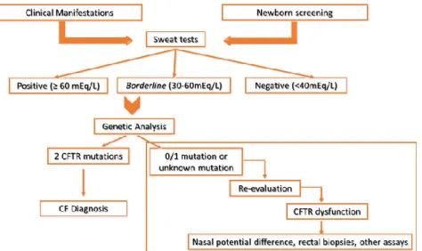
Figure 1.1 - Cystic Fibrosis Diagnosis Algorithm. The diagnosis of CF is firstly relied in clinical manifestations
and in newborn screen results, secondly, it is necessary to have two positive sweat tests and finally two known CFTR mutations. If the results of sweat tests are inconclusive, for instance, borderline results, and if it is found only one CFTR mutation it is necessary to re-evaluate the results and perform functional assays. [Figure from MD Amaral, included with permission]
Several kits to detect CFTR mutations are commercially available (using a discrete group of mutations, with the 50 most frequent mutation of each region), and they are advantageous because are fast and relatively cheap, which will detect 99% of affected individuals in the most populations.16–18
Though these commercially available kits are limited because the diagnosis will be missed if patients carry mutations which are not included in the kit.16–20 To overcome this limitation, full CFTR gene
sequencing is recommended, and it will detect most CFTR mutations, but, it may also detect novel mutations with unknown functional effect and undetermined CF disease prognosis. If such, it is necessary to confirm the CFTR (dys)function by electrophysiologic tests such as NPD and measurements of CFTR-mediated Cl- currents in colonic epithelial tissue (rectal biopsies).7,12,13,16 In
addition, it is important to identify CFTR mutations in CF patients with none or just one mutation previously identified using other approaches such as for example DNA or mRNA analyse (routine technique used in our laboratory) and to characterize their effects ex vivo and in vitro, to establish better the diagnosis and prognosis of the CF disease.
Recently the prenatal population screening for maternal CF carrier status and the newborn screening (NBS) have been widely used in the USA and Europe, which is helpful because early diagnosis and treatment reduce symptoms, improve health, and lower costs associated with disease complications.12 In Portugal NBS consists in a 1st test for immunoreactive trypsinogen (IRT), an assay
for Pancreatitis-Associated Protein (PAP) in case of positive IRT, followed by a 2nd IRT and if all
positive a final genetic analysis. All these strategies have resulted in increased life expectancy, as the predicted survival age of a CF patient in the past, was only six months,21 and now the predicted age is
40 years old in the USA,2243.5 in the UK,23and 30.7 years old in Portugal according to CF adult
follow-up in specialized centres.24
All these diagnosis methods have been advancing due to a better understanding of the CFTR gene, the protein structure and its function.25
3
1.2. CFTR: from Gene to Protein and Function
CFTR gene and expression
The CFTR gene was cloned in 1989 by using chromosome walking and jumping, and linkage disequilibrium studies.26 The gene comprises 27 coding exons (showed in figure 1.2) spanning over
190Kb on the long arm of chromosome 7 (7q31.2), and the transcript is 6.5Kb.27
The expression of CFTR is very complex and involves multiple tissue-specific transcriptions start sites, alternative first exons and alternatively spliced transcripts. The development, the pathologic conditions, the cell type and tissue, regulate the expression of CFTR. The sites of the CFTR expression are the epithelial surface throughout the body, such as submucosal glands, airways (the site of developmental regulation of CFTR expression), pancreas, the crypt in the intestinal tract, sweat glands, vas deferens, salivary glands.28,29
The proper transcription of the CFTR gene gives rise a multi-domain glycoprotein of 1,480 amino acids with a molecular weight of approximately 170 kDa. This protein belongs to the superfamily of the ATP-binding cassette (ABC) transporters.26,28,30It is composed by five domains, as shown in Fig.
1.2: two transmembrane domains (TMD1 and TMD2, each comprising six transmembrane segments) which anchor the protein in the membrane and form the translocation pathway, two cytoplasmatic nucleotide binding domains (NBD1 and NBD2) which bind and hydrolyze ATP, and a central regulatory domain (RD).28,31,32
Function
The CFTR protein plays different roles according to its system/organ localization. For instance, in the intestine, pancreas and sweat gland secretory coil, CFTR plays a key role in fluids and electrolyte secretion, and in the sweat gland duct and the airway epithelia, it participates in fluid and electrolyte absorption.33 It functions as a Cl- and HCO
3- channel and it also works as a regulator of other channels,
for instance, Epithelium Sodium (Na+) Channel (ENaC), that is downregulated by CFTR, potassium
(K+) channels and outwardly rectifying Cl- channels (ORCCs),33–35 or as a tumour suppressor gene in
the intestinal tract.36
Figure 1.2: Schematic representation of transcription, translation and localization of CFTR. The CFTR gene is composed by 27 coding exons overspanning 190Kb. When every intron are skipped during the transcription (maturation of mRNA), it has 6.5Kb. The proper translation, trafficking and folding give rise a multi-domain glycoprotein composed by five domains: two transmembrane domain (TMD1 and TMD2), two nucleotides binding domain (NBD1 and NBD2) and one regulatory domain (RD). Both C- and N- terminus are localized in the cytoplasm. It is also composed by six extracellular loops and four intracellular loops. [Figure from MD Amaral, included with permission]
4 The production of defective CFTR protein results in abnormal transport of salt, not just due to abnormal Cl- but also due to an enhancement of Na+ absorption, caused by ENaC upregulation thus
causing a decrease in the water content and dehydration of epithelia of different organs, namely in the airways. This reduction causes thicker secretions in airway tract, which clog small airways, becoming a favourable environment for bacterial infections from the air (inhalation, for instance). Accumulation of the thick mucus leads to persistent infection and chronic inflammation. Because of chronic inflammation, the bronchi dilate, and their walls weaken, setting up bronchiectasis that results in further airflow obstruction. The cycle of airway obstruction, inflammation, and persistent infection leads to a progressive decline in lung function and eventually causes respiratory failure and lastly to death (Fig.1.3).37–39
When CF was first described, the incidence of death among children in infancy or early childhood was very high. However, progresses in symptomatic therapies, such as mucolytics to dissolve the thick mucus, antibiotics to treat or prevent infections, anti-inflammatory agents to ameliorate chronic inflammation, pancreatic enzymes to compensate exocrine pancreatic insufficiency, fat-soluble vitamins and high caloric intake to overcome the deficiency of vitamins and malabsorption of fats, respectively, have significantly improved the life expectancy of CF patients.6,10,27,40
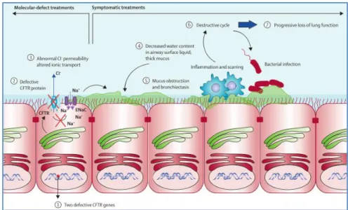
Figure 1.3: Pathophysiologic cascade in Cystic Fibrosis that leads to lung disease, the main cause of high mortality
rate among CF patients. Normally, CFTR proteins are located on the surface of the epithelial membrane and act as Cl- channels that in turn regulate the ENaC (epithelial sodium channel). The complex interplay of these channels regulates the electrochemical gradient that allows appropriate airway surface liquid depth and mucus viscosity. When the CFTR protein is defective or missing (caused by mutations in both alleles) it alters the ionic transports (abnormal Cl- permeability and high rate of Na+ absorption into the cells), which leads to decrease of water content in the airway surface liquid, which in turn leads to production of viscous mucus. ENAC – Epithelium Sodium Channel Adapted from De Boeck & Amaral (2015)6
1.3. CFTR mutations: classification according to their functional defect and class-specific therapies
Until now 2,019 variations have been found,41 with the following distributions: missense (39%);
frameshift (16%); splicing (11%); nonsense (8%); large (3%) and in-frame (2%) deletions/ insertions; and promoter (1%); sequence variation (14%), unknown effect (6%).27,40,41 However, only about 250
have been clearly defined as CF-causing, where approximately 20 mutations occur worldwide with frequency about 0.1% among CF patients, becoming very difficult to define the disease liability of such infrequent mutations.5,27,40,41
Although treatment advances over the past several decades have raised the median predicted survival age, the treatment costs of CF are very high, because of the amount of medicines taken daily by CF patients, which many drugs that prevent and treat pulmonary complications are costly.7,10,42
5 Consequently, a new approach that corrects the basic defect of CF, in this case at the molecular level is needed.
The basic defect in CF can be treated using small molecules to modulate defective CFTR protein and restore functional ion transport. There are already in the market compounds that restore CFTR trafficking and function such as, lumacaftor (VX809 – a corrector), ivacaftor (VX770 - potentiator), orkambi(VX809+VX770).43,44 Ivacaftor increases the time that CFTR channel is open, allowing Cl- ions
to flow through the CFTR proteins on the surface of epithelial cells, and lumacaftor facilitate the trafficking of CFTR protein, thus allowing this protein to reach the membrane and transport Cl-.45–48
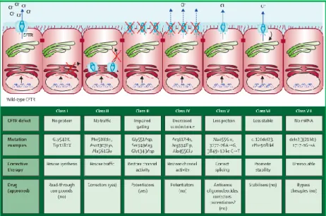
However, these compounds do not restore all CFTR mutations currently described, being necessary a mutation-specific or mutation-class-specific approach to correct the protein defect. Indeed, CFTR mutations have been conventionally grouped into seven classes according to their functional defect (Figure 1.4):5,12,27,40,48,49
❖ Class I mutations – result in no protein production, mostly nonsense or premature stop codon (PTC) mutations, which results in a truncated mRNA, leading to its degradation by nonsense-mediated decay (NMD), an mRNA surveillance mechanism. In this class, CFTR production could be obtained by using read through compounds to mask the PTC, leading to the NMD scape and allowing the cell to produce a full-length protein21,42.
❖ Class II mutations – affect CFTR protein traffic to the correct cellular localization as a result of protein misfolding and retention in the endoplasmic reticulum (ER), resulting in premature degradation of the protein.50 The most common mutation, F508del belongs to
this class. Small molecules, such as correctors can be used to correct this class of mutations, allowing the defective protein to reach the PM.46,51–53 Recently, Orkambi (a
mix of XV809 and VX770) was approved by the Food and Drug Administration for some of the patients with this class of mutations.54
❖ Class III mutations – lead to impaired gating of the CFTR channel, allowing the traffic of CFTR to the apical membrane, but causing poor regulation of the Cl- channel. They
result in a very poorly functional protein. Potentiators, such as VX770 are used to increase the CFTR channel gating, enhancing then the Cl- transport.27
❖ Class IV mutations – cause a significant decrease in CFTR channel conductance, notwithstanding the CFTR protein is present at the apical surface. Potentiators could improve the CFTR function.6,49,55
❖ Class V mutations – lead to a major reduction of normal CFTR protein levels, mainly because of aberrant splicing. These mutations produce normal and aberrant mRNA and the proportion of mRNA variants may vary among patients in different organs of each patient. Patients with this class of mutations can beneficiate with potentiators that will enhance the function of the normal protein and RNA-based therapies that can enhance the normal mRNA production.49,56–58 Recently VX770 has been approved for some of this
class of mutations.59
❖ Class VI mutations – lead to a high turnover of CFTR at the cell surface, either by increasing CFTR endocytosis or by decreasing its recycling back to the cell surface. Stabilizers could be used for this class of mutations.6,27
❖ Class VII mutations – lead to a non-functional and rescuable protein because of large deletions or insertions. Mutations in this class cannot be pharmacologically rescued, and therefore the promising therapies are Bypass therapies, i.e., by activating other alternative Cl- anion channel.6,60
6
Figure 1.4: Classification of CFTR mutations accordingly their functional defect, and its potential target therapies.
Adapted from 6
Patients with the same mutations have different responses to the same therapy, being crucial to study the response of these CFTR modulators in materials from each individual CF patients in a new strategy of personalised medicine. Thus primary cultures of human bronchial epithelial (HBE) cells and of nasal epithelial (HNE) cells as well as intestinal organoids produced from stem cells obtained from rectal biopsies directly from CF patients have been used as models for tests, such as measurements of the functional responses of CFTR and evaluation of drug efficacy.61,62
1.4. Class V mutations: splicing mutations and correction by RNA-based therapies
Alternative splicing is not always deleterious, is a fundamental element in eukaryotic gene expression that increases the coding capacity to the human genome. However, if an intronic or exonic variant disrupts canonical splice motifs or creates a new cryptic splice site, it will lead to aberrantly spliced mRNAs, usually encoding nonfunctional proteins. These, however, do not always replace totally the normal protein expression, as often some levels of normal mRNA splicing also occur in parallel with the abnormal one.63–65
The splicing process is very complex, and several reactions are taken in place. The spliceosome, the major effector of the splicing reaction, is a complex of hundreds of interacting proteins and small nuclear RNAs (snRNAs) including the five small nuclear ribonucleoproteins (snRNPs) U1, U2, U4, U5 and U6.66
First of all, U1 binds to the 5’ splice site (5´-ss) by complementarity and U2 binds to the branch point. The triple snRNPs (U4, U5 and U6) complex moves-in to associate with the assembling spliceosome. Then, the U4 leaves the complex, allowing the replacement of U1 by U6 that interacts with U2 to bring the branch point into proximity to the 5´-ss. At this point, the first transesterification reaction cleaves the 5'-ss of the intron of the downstream exon and attaches it to the branch point. U5 then brings the 3'-ss of the upstream exon and 5'-ss of the downstream exon into proximity with each other, allowing a second transesterification reaction that cleaves the 3'-ss of the upstream exon (as shown in Appendix 1- Fig.1S). The splicing accuracy does not depend only on the mechanisms described above, but it also depends on more discrete elements, splicing regulatory elements which direct the splicing machinery to use the correct splice site. 66,67
Patients carrying one splicing (class V) mutation can have mild CF disease, and most of the times there are considered pancreatic sufficient (PS). It is reported that 11% of the CFTR variations are known to be splicing defective. However, it is predicted that the proportion of these splicing defects can be
7 higher because of the false missense mutations that have not been identified as causing abnormal splicing.40,68,69 In this way each nucleotide modification, including nonsense, missense and silent
modifications, may potentially impact the splicing pattern, resulting in production of aberrant spliced transcripts.70 Thus, it becomes crucial to study splicing mutations, which allow a better understanding
of splicing processes and the mechanism of disease-causing, which may help to develop the most relevant therapeutic strategy and thus contributing to the increase of life expectancy of patients carrying these class of mutations.
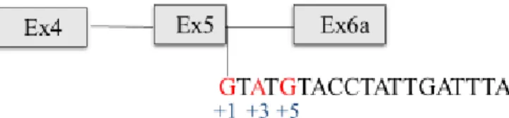
When the alteration is in the invariant donor splice site, +1, it is most likely that the correct spliced transcripts will strongly be reduced.53,71
For clarity, when it is referred to intronic splicing mutations, for example, 711+1G>T or 3272-26A>G, the symbols "+" and "-" indicate nucleotides downstream and upstream of the exon/intron boundary, respectively.
RNA-based therapies
RNA-based therapies are therapies use RNA as a target to potentially tread diseases, caused by aberrant splicing. Targeting the RNA is an advantageous strategy because it avoids many of the risks and concerns associated with (DNA) gene therapy, such as random gene insertion. Furthermore, when an in vivo cellular basis is needed the presence within the cell of multiple different RNA processing pathways means that there is much scope for influencing its control at different levels. The dynamic nature of RNA turnover also implies that therapeutic interventions can be time-limited, dose titrated and modified according to response, adding further levels control.66
Novel therapeutic approaches to correct splicing mutations have been described using antisense oligonucleotides (AONs), that correct splicing defects, indicating that CF patients carrying such mutations may benefit from AON treatment.57
These AONs can be synthesised to be complementary and specific to a particular RNA sequence transcribed from the CFTR gene, meaning that only the RNA sequence of interest will be targeted. By designing AONs that bind to splice sites or to enhancer or silencer elements within the transcript, the splicing mechanism can be manipulated in a precise and reproducible way. Blocking splice sites and regulatory sequences prevents snRNPs and splicing factors such as SR proteins and hnRNPs from binding to the mutated site at the RNA transcript, allowing a directed exon skipping or inclusion, depending on the sequence blocked.72
Although RNA repair is transient and only modestly effective, studies of CFTR splicing polymorphisms suggest that 8% of normal CFTR message might be sufficient for normal lung function, whereas 5% is associated with relatively mild CF lung disease.73,74
8
1.5. Objectives of the present work
This project had two main goals, namely:
1) Firstly, to investigate the possible involvement of the CFTR in other respiratory diseases namely, COPD, DB and asthma.
2) Secondly, to characterize the impact of four mutations localized in the same splicing consensus- 711+1G>T, 711+3A>T, 711+3A>G and 711+5G>A and subsequently modulate them using an AON based strategy.
To achieve these goals we proposed the following specific tasks:
1.1. To screen non-CF individuals with other respiratory disorders for the most common CFTR mutations found in Portuguese CF patients. Control groups included: i) a group of patients known to have CF or suspicious of a CF diagnosis; ii) a group of carrier individuals; and iii) a group of healthy individuals with no respiratory phenotype.
2.1. To generate cells lines expressing CFTR minigenes carrying the splicing mutations 711+1G>T, 711+3A>G, 711+3A>T and 711+5G>A.
2.2. To characterize the impact of four splicing mutations (711+1G>T, 711+3A>G, 711+3A>T and 711+5G>A) at the mRNA, protein and functional levels.
2.3. To use an AON strategy for modulation of the deleterious effect of these mutations at the mRNA and protein levels;
9
Section II: Materials and Methods
Part I: Methods to analyse patients’ materials
2.1. The screen of mutations in CF and non-CF patients
Before the studies, all individuals signed an informed consent. The patients’ materials in this study were irreversibly anonymised as stipulated in artº 19, in the Portuguese law 12/2005 from 26th of
January.
Initially, it was proposed to analyse the nine most common Portuguese CFTR mutations (panel obtained from World Health Organization (WHO), 2004 – shown in table 3.1) in individuals with suspicion of CF (who had two borderline sweat tests or just one identified mutation) and individuals with a CF familial history. However, for this study, the objective was extended to individuals who had two negative sweat tests and no CF familial history but have diagnosis of COPD, DB or asthma, to verify if the most common Portuguese CFTR mutations play a role in these respiratory diseases. It was also included healthy control, in order to discriminate better the CFTR sequence variation from CFTR mutations.
In order to achieve that, 249 Portuguese individuals divided into 4 four groups, namely, patients with suspicion of CF, individuals with CF sibling or with a CF familial history, non-CF with other respiratory diseases and healthy controls (individuals without a CF familial history, without clinical manifestations of any respiratory disease) were analysed.
Genomic DNA (gDNA) was extracted from blood samples of 249 Portuguese individuals, using the Wizard® Genomic DNA Purification kit following the manufacturers’ instructions performed by Verónica Felicio and further analysed for 9 most common CFTR mutations (table 3.1 ) extracted from a report of word Healthy Organizaton.
The CFTR mutations were detected by Amplification Refractory Mutation System (ARMS), or Tetra-ARMS and Restriction fragment length polymorphism (RFLP) techniques, which were previously optimized in our laboratory.
The ARMS technique is a simple, rapid and reliable method for the detection of any mutation involving single base changes or small deletions. This PCR modification consists of two complementary reactions. The first reaction contains an ARMS primer specific for the normal gDNA sequence and cannot amplify mutant DNA and the second one contains a mutant-specific primer and cannot amplify normal gDNA. The genotype of an individual can then be determined by analysis of the amplification products. An individual with no mutations generates a PCR product only in the normal reaction, an heterozygous individual gives products in both reactions, and a homozygous mutant individual does so only in the mutant reaction.75,76
The tetra-ARMS technique is an ARMS modification using tetra-primers in the same reaction. Thus, the allele-specific amplification is achieved in a single PCR reaction using two outer primers and two allele-specific inner primers.77,78 Then PCR reactions were run on a 1%-3% agarose gel to confirm
amplification and product pattern. Mutation detection was confirmed by sequencing.
Part II: Methods to study splicing mutations in cellular systems 2.2. Generation of models to study splicing mutations in IVS5
2.2.1. Plasmid vectors
A pcDNA5/FRT/eGFP/CFTR/IVS4art/IVS5/Flag mini-gene was previously constructed in our lab (the insert will be called here as eGFP/IVS4_5/wt/flag or the respective mutants). The construct carried a complete wildtype (wt) CFTR cDNA, containing an artificial intron 4 (IVS4art) with 351 nucleotides of the 5’ of the human IVS4 sequence joined with 351 nucleotides 3’ of the human IVS4 sequence inserted between exon 4 and exon 5, and a full length of intron 5 (IVS5) sequence from human gDNA
10 inserted between exon 5 and exon 6a. It is also composed by eGFP in the N-terminal and flag in exon 15 (4th extracellular loop), which was previously inserted in our lab. This mini-gene was then used to
generate four splicing mutations (711+1G>T; 711+3A>T; 711+3A>G and 711+5G>A) by-site directed mutagenesis.
Afterwards, each mini-gene was used to transiently transfect HEK293T cells and CFBE parental cells and was also used to stably transfect HEK FLP-In cells. The pcDNA5 mini-gene was also used to amplify the insert and clone into pLVX-puro to stably transfect CFBE parental cells.
2.2.2. Site-directed mutagenesis
Mutations on the same splicing consensus on IVS5 (711+1G>T, 711+3A>G, 711+3A>T and 711+5G>A) were introduced into pcDNA5/FRT/eGFP/CFTR/IVS4art/IVS5/FLAG using the KOD Hot Start DNA Polymerase Protocol (Novagen). Where 10ng of plasmid were mixed with 1.5mM of MgSO4;
1X of 10X Buffer for KOD Hot Start DNA Polymerase; 0.2µM of each dNTPs; 0.6µM of complementary pairs of mutagenic primers (described in Appendix 1 - Table 2); 0.02U/µl of KOD Hot Start DNA Polymerase and nuclease-free water to perform 50µl of reaction, using the PCR program described in Appendix 1 - Table 3.
5µl of the reaction product were run by electrophorese in agarose 0.5% agarose gel to confirm the amplification of the plasmid. The PCR product was then incubated with DpnI (a restriction enzyme that hydrolyses methylated and hemimethylated DNA) for 1hour at 37ºC. After that, 200µl of competent bacteria were transformed with the hydrolysis product and grown on LB agar plates with 100µg/mL of Ampicillin (Sigma-Aldrich) followed by the extraction of the plasmid. The plasmids were then sent for automatic DNA Sequencing (Stabvida, Costa Caparica, Portugal) to confirm the introduction of each mutation. All these steps are in detail described in the section of appendices (Appendix 1: 1-3).
2.2.3. Cloning into pLVX-Puro
The inserts from each of the above pCDNA constructs were subcloned into pLVX-puro in order to generate stable cell lines. The constructs of eGFP/IVS4_5/wt; eGFP/IVS4_5/711+3A>G/flag; and eGFP/IVS4_5/711+5G>A/flag were cloned into pLVX-Puro using In-Fusion® HD Cloning Kit (Clonetch), and the constructs eGFP/IVS4_5/711+1G>T/flag; and eGFP/IVS4_5/711+3A>T/flag were cloned into pLVX-Puro using T4 ligase (ThermoFisher Scientific). Then lentiviral particles were produced by human cells and thus create stable cell lines.
The cDNAs of the constructs were amplified through PCR reactions from the
pcDNA5/FRT/eGFP/CFTR/IVS4art/IVS5/FLAG with wt and the different mutants, creating 15bp
extensions with XhoI local restriction sites in both C- and N- terminal (eGFP/IVS4_5/WT; 711+3A>G; and 711+5G>A), and creating 15bp extensions with XmaI local restriction in N- terminus and SpeI local restriction in C- terminus (eGFP/IVS4_5/711+1G>T; and 711+3A>T) with primers described in table 2.1.
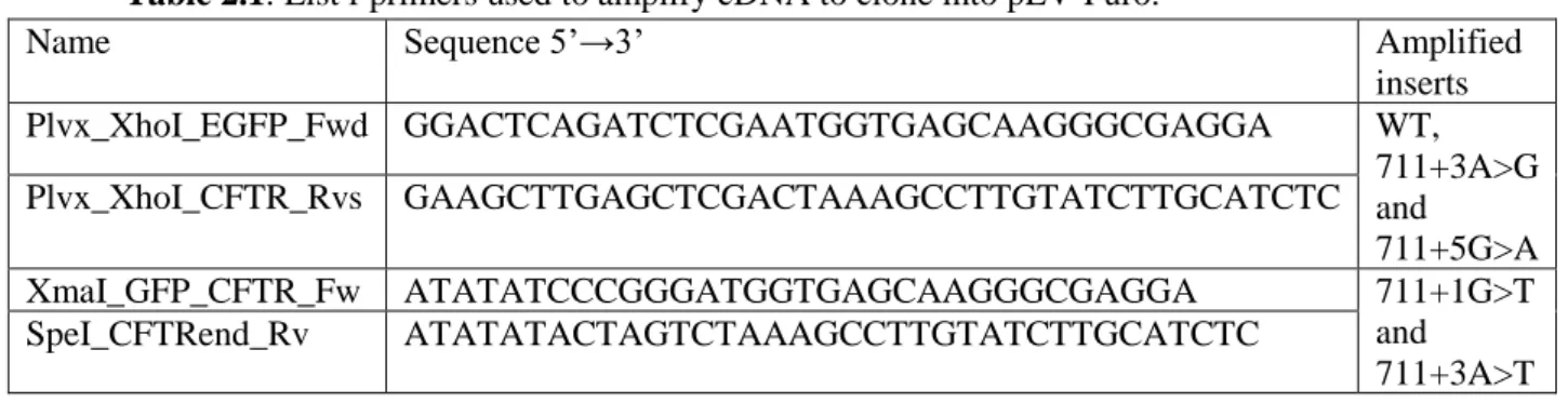
Table 2.1: List f primers used to amplify cDNA to clone into pLV-Puro.
Name Sequence 5’→3’ Amplified
inserts Plvx_XhoI_EGFP_Fwd GGACTCAGATCTCGAATGGTGAGCAAGGGCGAGGA WT, 711+3A>G and 711+5G>A Plvx_XhoI_CFTR_Rvs GAAGCTTGAGCTCGACTAAAGCCTTGTATCTTGCATCTC XmaI_GFP_CFTR_Fw ATATATCCCGGGATGGTGAGCAAGGGCGAGGA 711+1G>T and 711+3A>T SpeI_CFTRend_Rv ATATATACTAGTCTAAAGCCTTGTATCTTGCATCTC
11 Separately, 3µg of the cDNAs resulting from amplification with XmaI and SpeI primers were mixed with 3U SpeI (NZYTech) and 3U XmaI (ThermoFisher Scientific), 1X NZYbuffer U (10 mM Tris-HCl, pH 7.4, 100 mM KCl, 0.1 mM EDTA, 1 mM DTT, 500 μg/mL BSA, 50% (v/v) glycerol) and deionized water to make up the 20µl total reaction. This mixture was then incubated for 3h at 37ºC, followed by enzyme inactivation for 20min at 65ºC.
5µg of pLVX-Puro were linearized using 5U of XhoI (NEB) restriction enzyme mixed with 1X NEBbuffer 3.1 (100mM NaCl, 50mM Tris-HCl, 10mM MgCl2, 100μg/ml BSA, pH 7.9) and deionized
water to make up the 25µl total reaction. The reaction was then incubated for 3h at 37ºC, followed by enzyme inactivation for 20min at 80ºC, to create ends equal to the XhoI primers extensions, used to amplify the cDNAs of eGFP/IVS4_5/WT; 711+3A>G; and 711+5G>A, allowing recombination between the vector and the construct. 3 µg of pLVX-Puro was linearized using 3U of XmaI (ThermoFisher Scientific) and 3U of XbaI (NZYTech) mixed with 1X NZYbuffer U (10 mM Tris-HCl, pH 7.4, 100 mM KCl, 0.1 mM EDTA, 1 mM DTT, 500 μg/mL BSA, 50% (v/v) glycerol) and deionized water to make up the 20µl total reaction. . The reaction was then incubated 3h at 37ºC, followed by enzyme inactivation 20min at 65ºC, to create compatible ends of XmaI and SpeI primers extensions, used to amplify the cDNAs of eGFP/IVS4_5/711+1G>T; and 711+3A>T, allowing the ligation between the construct and vector. It is important to highlight that XbaI and SpeI generate compatible ends.
The linearized vector, as well as the cDNAs extended, were spin-column purified using the NzyGelPure kit (NZYTech). The In-Fusion cloning was performed mixing 2µl of the enzyme, 200ng of cDNA extended, 50ng of linearized vector and nuclease water free up to 20µl, and let it react 15mi at 50ºC. The ligation reaction was performed mixing 1U of T4 ligase (ThermoFisher Scientific), 213ng of cDNA extended and purified, 50ng of linearized vector and nuclease water free up to 20µl, and let it react 10min at 22ºC.
10 µl of the infusion product or the ligation product were then used for bacteria transformation as described in Appendix 1: 3.
‘Colony PCR’ was used to confirm the insertion of the construct into the pLVX. After confirming the insertion, the recombinant plasmid was extracted from the positive colonies using NZYMiniprep kit (NZYTech, MB010) as described previously in Appendix 1: 3, and the recombinant plasmid was then digested with specific restriction enzymes EcoRI (ThermoFisher scientific) to confirm the correct insertion of the gene into the vector. If it was correct, the recombinant plasmids were sent for sequencing in StabVida.
After confirming the correct insertion of the insert into pLVX-Puro, the new lentiviral vector was used to produce lentiviral particles to transduce CFBE cells.
2.2.4. Production of lentiviral particles
Lentiviral particles with pLVX/eGFP/IVS4_5/mut/flag or pLVX/eGFP/IVS4_5/WT/flag were produced in Human Embryonic Kidney 293T (HEK 293T) cells.
The cells were seeded at density 5x10⁵ per well on a 6well plate, in Minimum Essential Medium
Eagle with L-Glutamine (EMEM, Biowhittaker®) supplemented with 10% of Fetal Bovine Serum (FBS, GIBCO Life Technologies) and incubated 24h at 37ºC and 5% CO2. The day after the medium was
changed 3h before the transfection.
The cells were transfected using Calcium phosphate transfection which involves mixing DNA with calcium Cl- in a buffered saline/phosphate solution to generate calcium-phosphate-DNA
co-precipitate, which is then dispersed onto the cultured cells. Calcium phosphate facilitates the binding of the condensed DNA in the coprecipitate to the cell surface, and the DNA enters the cell by endocytosis. 5µg of pLVX (with the desired construct – WT or Mut); 5µg of packaging plasmid pCMV-dR8.74.psPAX2 and 2.5µg of enveloping plasmid VsV-G/pMD2.G were mixed 1/10 of TE (1mM tris-Cl; 0.1mM EDTA; pH 7.60) and 2.5mM of CaCl2 and this mixture was vortexed for 20 seconds and
12 incubated for 5 minutes. After that 250µl of HBS (50mM HEPES; 280mM NaCl; 1.5mM Na2HPO4)
were added to another tube and while vortexing, the previous mixture was added dropwise and then incubated for 30 min. Afterwards, the mixture was added to the cells.
The cells transfected were incubated for 24h at 37ºC, 5% CO2. The medium was then changed
to EMEM supplemented with 10% FBS to remove the transfection reagent, and the cells were incubated for more 42 hours at 37ºC, 5%CO2.
The lentiviral media were harvested, and the packaging cells were discarded. The lentiviral media were immediately used to transduce cystic fibrosis bronchial epithelial (CFBE 41o for short CFBE) cells or were stored at -80ºC for further use.
2.2.5. Generation of stably transduced cells: lentiviral infection
CFBE parental cells were plated in a 6 well plate at a concentration of 3x10⁵ cells per well in 2
ml of EMEM supplemented with 10% FBS a day before of infection with lentiviral media harvested from HEK 293T cells and were incubated at 37ºC, 5% CO2.
The cells were infected with 1ml of lentiviral media and 1ml of EMEM supplemented with 10% FBS with 8µg/ml of Polybrene (Hexadimethrine bromide, Sigma-Aldrich) infection enhancer. The plate was centrifuged at 220rpm for 1h at 25ºC and then incubated for 24h at 37ºC, 5% CO2. The medium
was then changed 24 hours after the transfection to EMEM supplemented with 10% FBS. 42 hours post infection the media was changed to EMEM supplemented with 10% FBS and 2.5µg/ml of Puromycin, which selected the infected cells and killed the non-infected cells. Afterwards, the media was replaced 24 hours after the first selection with increased concentration of Puromycin (5µg/ml). The medium was then changed every 48 hours, and the cells were kept in culture at 37ºC, 5%CO2.
To analyse the expression of the desired proteins in the stably transduced cells we performed biochemical analysis as described in section 2.4.
The cells were then sorted by flow cytometry to achieve a homogeneous expression of the protein.
2.3. Cell culture
The characterization of the splicing mutations was made in different cell types and different conditions.
2.3.1. Cell lines and culture conditions
Human Embryonic Kidney 293T (HEK 293T) parental cells were used for transient transfections and to produce wt and the mutants (711+1G>T, 711+3A>T, 711´3A>G and 711+5G>A) lentiviral particles. These cells were cultured in EMEM supplemented with 10% of FBS.
Human Embryonic Kidney293 Flp-In (HEK293 Flp-In) parental cells (Invitrogen, Carlsbad, CA) were used for stable transfections and were cultured in EMEM supplemented with 10%, selection antibiotic Zeocin 100µg/ml (Sigma-Aldrich, St. Louis, MO) before stable transfections and Hygromycin after transfections. The Flp-In system allows insertion of the construct in only a single integrated FLP recombination target (FRT) site.
CFBE parental cells, were cultured in EMEM supplemented with 10% FBS. These immortalised cells were developed from bronchial epithelial cells from an F508del-CFTR homozygous CF patient and did not express endogenous CFTR because it loses the expression with passages.
All cell lines were maintained at 37ºC in a humidified atmosphere of 5% (v/v) CO2. All cells
were tested for mycoplasma infection, being mycoplasma free.
13 2.3.2. Transient transfections
HEK293T cells were submitted to liposomal transfection. The liposomal transfection, commonly known as lipofection, is based on the ability of cationic lipidic to form unilamellar liposomes, which adsorb nucleic acids molecules to their surface and are capable of being internalised by the cells. We used Lipofectamine 2000 (Invitrogen) to transiently transfect HEK 293T cells with each pcDNA5 minigene
For that 5x10⁵ cells were grown in a 12-well plate 24 hours before the transfection, and
60%-80% of confluence was desired for transfection. Lipofectamine 2000 (3µl) and 1µg of DNA (pCDNA5 plasmids with the different constructs) were separately incubated for 5min in 50µl of OPTIMEM (Invitrogen), then mixed and allowed to incubate for 15min at RT. The mixture was then added to the cells in EMEM without FBS.
The cells were incubated for 48 hours at 37ºC in 5% CO2, and the RNA and protein were
extracted to perform RT-PCR, qPCR and Western blot, respectively as described Appendix 1: 1, 2 and 4 and in this section in 2.5.1, respectively.
2.3.3. Stable transfections in HEK Flp In cells
Lipofectamine 2000 was used to stably transfect HEK Flp In cell. A vector containing the flipase that allow the directed recombination, pOG44, was cotransfected with each desired constructs (pCDNA5/FRT/eGFP/IVS4_5/WT or mut/flag) in proportion 3:1 for a total 2µg of DNA, cell selection started 48h after transfection by changing the medium to a medium supplemented with 100µg Hygromycin B.
2.3.4. Treatment with an AON
The AON was synthesized by Integrated DNA Technologies, Inc. (IDT, Leuven, Belgium). The AON was designed 20 base pairs of distance from the first nucleotide of IVS5 with the following sequence: mA* mT* mC* mT* mT* mT* mT* mA* mG* mG* mC* mA* mC* mT* mA* mT* mT* mG* mT* mT*. The letter ‘m’ represents an O-methyl modification at the second position of a sugar residue, and the asterisk represents a phosphorothioate modification of the backbone. This AON was used to transiently transfect cells by Lipofectamine 2000 (Invitrogen).
48h after transfections mRNA was extracted to perform semi-quantitative and quantitative analysis by RT-PCR and qRT-PCR, respectively as described in the appendix section. Biochemical analysis were also performed by Western blot and Immunofluorescence to characterize the impact of splicing mutations under study.
2.4. Biochemical
2.4.1. Western Blot
Cells were washed twice with cold PBS and lysed with sample buffer (1.5% (w/v) SDS; 0.01% (w/v) bromophenol blue; 5% (v/v) glycerol; 0.05 dithiothreitol (DTT); 0.095 M Tris, pH 6.8). DNA was sheared using benzonase 25U/mL (Sigma-Aldrich) in the presence of 2.5 mM of MgCl2.
Total protein was quantified using the Bradford assay. Briefly, 10µl of the protein extract was added to 990µl of BioRad Protein Assay Reagent (Bio-Rad, 500-0006EDU) diluted in water (200µl:800µl), incubated for 5min. The absorbance was measured at 595nm using the spectrophotometer (Jasco V-560 UV/Vis spectrophotometer). A regression equation for protein concentration was determined using bovine serum albumin (BSA) as a standard protein.
25µg of protein were loaded on each lane for SDS-polyacrylamide gel electrophoresis (PAGE) on 7% separating and 4% stacking gels, and run at 100 V for three hours in a 1X running buffer (25mM Tris; 192mM Glycine, 0.1%(w/v) SDS; pH 8.3, Bio-Rad). Subsequently, proteins were transferred onto
14 Polyvinylidene (PVDF) membranes (Millipore) at 400mA for 1.5h in a 1X transfer buffer (25mM Tris, 192 mM Glycine; pH 8.3, Bio-Rad).
After the transfer, the membrane was blocked 5% (w/v), non-fat milk in phosphate buffered saline (PBS, NaCl 137 mM; KCl 2.7mM; KH2PO4 1.5mM; Na2HPO4 6.5mM, pH 7.4) containing
0.1%(v/v) Tween (PBS-T) for 1h. The membranes were then probed overnight at 4ºC with primary antibodies (596 anti-CFTR mice (1:3000 (BD Transduction Laboratories®) and anti-calnexin mouse (BD Transduction Laboratories®) - as a loading control) diluted in 5% (w/v) skimmed milk in PBS-T. The membrane was washed three times during 10 min in PBS-T, followed by incubation for one hour at Room Temperature(RT) with horseradish peroxidase-conjugated secondary anti-mouse IgG antibody (BIO-RAD, goat, 1:5000) in 5% (w/v) skimmed milk-PBS-T and washed another three times during 10 min with PBS-T.
Chemiluminescent detection was performed using Chemidoc XRS+ analyser (BIO-RAD), and the signal was detected with the Clarity Western ECL substrate (BIO-RAD). Finally, the quantification was performed with the ImageLab software (BIO-RAD).
2.4.2. Immunofluorescence
Cells were grown on coverslips in 24-well plates or 8well- Lab-Tek (Sigma-Aldrich) coated with 0.001% (w/v) poly-L-lysine (Sigma) to reach 40-60% confluence was reached.
48hours after transient transfections the cells were washed once with PBS++ (PBS with components described in 2.4.1 enriched with 0.7mM of CaCl and 1.1 mM of MgCl2) and incubated with
mouse anti-flag (Sigma-Aldrich) diluted in PBS++ (1:500) supplemented with 1% (w/v) BSA for 1h at 4ºC. Afterwards, the cells were rinsed three times with PBS++ and fixed with 4% (w/v) Paraformaldehyde (PFA) for 20 min at 4ºC. The cells were then washed three times and incubated with a rabbit anti-mouse Cy5 secondary antibody (Life Technologies) in PBS++ (1:500) supplemented with 1% (w/v) of BSA, one hour at RT. After, the cells were washed three times with PBS and incubated one hour in the dark with Hoechst 33342 Fluorescent Stain Life (Life Technologies). The coverslips were then mounted on glass slides with Vectashield mounting medium (Vector Laboratories) and sealed.
Immunofluorescence staining was observed in the Leica DMI 6000B fluorescence microscope, which was also used to acquire the images.
2.5. Statistical analysis
Results are expressed as means ± S.E.M for n observations. Students t-test (GraphPad Prism software) for paired and unpaired samples was used as appropriate. Differences were considered statistically significant when P<0.05.
15
Section III: Results and Discussion 3.1. Analysis of Patients
3.1.1. Patients’ screen studies
This study was carried out in order to determine whether patients with non-CF respiratory diseases have at least one mutation in the CFTR gene. Previous studies have shown that patients with, CB/AVD,79 asthma80, disseminated bronchiectasis (DB) and idiopathic pancreatitis with unknown
etiopathology81,82,83 have at least one CFTR mutation. However, there are also some studies showing
that the incidence of CFTR mutations in patients with these non-CF respiratory diseases,84 is not
different from that in the general population,85 thus being contradictory. This study will help to clarify
these contradictory findings.
Herein, we tested the occurrence of the 9 most common Portuguese CFTR mutations (outlined in Table 3.1) in non-CF respiratory diseases Portuguese patients.
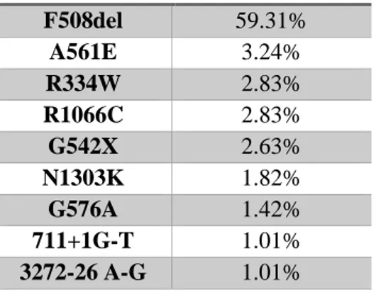
Table 3.1: List of the 9 most frequent mutations in Portugal adapted from86
Mutation Incidence F508del 59.31% A561E 3.24% R334W 2.83% R1066C 2.83% G542X 2.63% N1303K 1.82% G576A 1.42% 711+1G-T 1.01% 3272-26 A-G 1.01%
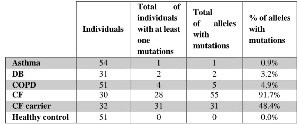
For that purpose, gDNA samples were obtained from 249 individuals including (see Table 3.2): 1) patients with non-CF respiratory diseases (136); 2) patients with CF or with suspicion of a CF diagnosis (30); 3) CF carriers (32); and 4) a control group of 51 healthy individuals with no respiratory phenotype. These gDNA samples were screened for the above mutations. Samples from the above 2-4 groups were included in this study as they were used to validate the pattern of the bands in the ARMS and Tetra ARMS PCRs used to determine each patient genotype (see Methods, section 2.1). In addition, they will be included in further studies aimed to correlate the occurrence of CFTR mutations with the severity of respiratory diseases and with the CFTR-mediated ionic transport. Whenever there was no clear genotype identified, samples were sent for sequencing to confirm the mutation(s).
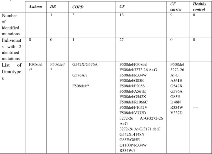
The results revealed that 66 (26.5%) individuals had at least one CFTR allele mutated We were able to find only 7 out of the 9 most common Portuguese CFTR mutations described in Table 3.3, namely: F508del, 3272-26A>G, G542X, R334W, G576A, A561E and R1066C. However, we were also able to identify other 7 mutations that were not included in our cohort, namely, I148N, G85E, P205S, Q1100P, F1052V, V332Q and 3171del C. From the 14 different mutations that we found, F508del was the most common, and it was present in 62% of individuals with at least one mutation, and 14.6% of these individuals were homozygous for this mutation.
Looking into non-CF sub-group with respiratory diseases, the DB patients presented the highest incidence of the F508del mutation (6.5%), followed by the COPD patients (4%).