Tiago Martins da Costa Duarte
Dissertation presented to obtain the Ph.D degree in Technological and Engineering Sciences, specialization in Systems Biology
Instituto de Tecnologia Química e Biológica António Xavier | Universidade Nova de Lisboa
Tiago Martins da Costa Duarte
Dissertation presented to obtain the Ph.D degree in Technological and Engineering Sciences, specialization in Systems Biology
Instituto de Tecnologia Química e Biológica António Xavier | Universidade Nova de Lisboa
Oeiras, June, 2016
B
RIEF
C
ONTENTS
Acknowledgements ... v Jury ... ix Thesis Publications ... xxiii Summary ... xxv 1 Introduction ... 1
2 Metabolic signatures of GS CHO cell clones associated with butyrate treatment and culture phase transition ... 27
3 Exploring GS-CHO metabolism in control and butyrate-treated conditions through parallel 13C-labeling experiments ... 59 4 Metabolic responses of CHO cells to limitation of key amino acids ... 87 5 Discussion ... 115 Appendix ... 133 Curriculum Vitae ... 141
I would like to express my deepest gratitude to the people who have contributed directly or indirectly to the content of this thesis.
To my supervisor Prof. Paula Alves, for the opportunity to carry out my thesis in your group, for your encouraging words and for your commitment to make the Animal Cell Technology Unit a place to do high quality research.
To my co-supervisor Dr. Ana Teixeira, for your vision and guidance throughout this journey. For always pushing me to overcome myself and my limitations. To Prof. Manuel Carrondo, for your words on leadership, hard work and resilience. To Dr. Nuno Carinhas, for your important contributions to this work, for sharing your insights in metabolic flux analysis and your solid scientific writing style. To Laura Barreiro, for your important contribution to this work during your research training and for your high spirit inside and outside the lab. To my colleagues at the ACTU, former and present, especially to Marcos Sousa for your support in bioprocesses and for the camaraderie in the lab, to Dr. Ana Barbas for our fruitful discussions about antibodies, to Dr. Cristina Peixoto for your help in purifying those antibodies, and to Daniel Pais for being such a curious and enthusiast colleague and helping me with my last experiments in the lab. To the iBET Analytical Services Unit, to António Ferreira, Dr. Rosário Bronze, Cláudia Duarte, and especially to Cláudio Almeida, for sharing your knowledge and expertise with GC-MS.
To the Centro de Ressonância Magnética António Xavier (CERMAX) team, Dr. Pedro Lamosa, Dr. Helena Matias, Dr. Ana Rute Neves and Dr. Manolis Matzapetakis for your support and for our fruitful discussions concerning NMR. To the Cell Systems Engineering group at TU Delft, especially to Prof. Joseph Heijnen for being such a good host, to Dr. Aljoscha Wahl and Dr. Reza Seifar for your support and guidance during my stay, to Angela ten Pierick and Cor Ras for teaching me the tips and tricks of mass spectrometry.
Dias.
To Dr. Francisca Monteiro and Dr. Filipa Rodrigues for your endless help and our always interesting discussions about science and philosophy.
To my Science Runners team, (besides the ones mentioned above) Sofia Rebelo, Daniel Simão, Marta Silva, Catarina Pinto, Marta Estrada, João Sá, Rita Costa, Ana Oliveira and Paulo Fernandes, who still give me every day the motivation to always go faster and further to reach my goals – in life as well as in running.
To the InTeraQB team for giving me an excuse to make funny paper hats and to interact with awesome people and scientists across ITQB and beyond.
To the financial support provided by Fundação para a Ciência e a Tecnologia (FCT) (SFRH/BD/81553/2011 and PTDC/BBB-BSS/0518/2012). The NMR spectrometers are part of the National NMR Facility supported by FCT (RECI/BBB-BQB/0230/2012).
To my friends for our discussions about engineering, arts and life, in particular, João Henriques, Ana Espiga and João Chaves. To Cecília and Rita for you continuous support. To an inspiring Iron(wo)man and “office” partner, Ranna, in sports and in business. To my second encouraging “office” partner, Laura Moya. To a team of advisors who helped me in the last stage of my PhD, preparing me for the Viva: Francisca Monteiro, João Dias and Rodolfo Marques. To my family for your support and for handling all the twists and turns inherent to this journey, especially my father Leonel, mother Graça, brother Leonel, and Tia Manuela. To my cousins Pedro and Fernando for your continuous support and encouragement. To James Lanham, for being my special advisor. With special jubilation, to my beloved wife Ana.
J
URY
Prof. Rui Oliveira, Associate Professor with Habilitation, Chemistry Department at Faculdade de Ciências e Tecnologia, Universidade Nova de Lisboa, Caparica, Portugal Prof. Joana Azeredo, Associate Professor with Habilitation, Biological Engineering Department at Universidade do Minho, Braga, Portugal
Prof. Maria do Rosário Bronze, Associate Professor at Faculdade de Farmácia, Universidade de Lisboa, Lisboa, Portugal
Dr. Pedro Cruz, Chief Scientific Officer (CSO) at ECBio, Oeiras, Portugal
Prof. Paula Marques Alves, Principal Investigator and Head of the the Animal Cell Technology Unit at IBET/ITQB-NOVA, Chief Executive Officer (CEO) at IBET, Oeiras, and Invited Associated Professor at Faculdade de Ciências e Tecnologia, Universidade Nova de Lisboa, Caparica, Portugal (Supervisor)
Dr. Ana Palma Teixeira, Auxiliary Investigator and Head of the Bioengineering & Systems Biology Lab at the Animal Cell Technology Unit, IBET/ITQB-NOVA, Oeiras, Portugal (Co-Supervisor)
“We are drowning in information but starved for knowledge.”
John Naisbitt
List of Figures ... xvii List of Tables ... xviii List of abbreviations and acronyms ... xix Thesis Publications ... xxiii Summary ... xxv Resumo ... xxix 1 Introduction ... 1 1.1 Mammalian cells as production factories of biopharmaceuticals ... 3 1.2 Chinese Hamster Ovary Cells ... 5 1.3 Performance of mammalian cell bioprocesses ... 8 1.3.1 Culture medium and fed batch processes ... 8 1.3.2 Classical approaches to improve specific productivity ... 9 1.3.3 Cell line engineering ... 10 1.4 Systems biotechnology ... 13 1.4.1 Transcriptome and proteome layers ... 13 1.4.2 Metabolome and Fluxome layers ... 15 1.5 Thesis motivation and outline ... 19 1.6 Author contribution ... 21 1.7 References ... 21 2 Metabolic signatures of GS CHO cell clones associated with butyrate treatment and culture phase transition ... 27 2.1 Introduction ... 30 2.2 Materials and Methods ... 33
2.2.4 Total protein quantification ... 35 2.2.5 SDS-PAGE and western blot analysis ... 35 2.2.6 Exometabolome analysis ... 35 2.2.7 Metabolic network ... 36 2.2.8 Metabolic flux analysis ... 37 2.3 Results and Discussion ... 39 2.3.1 Culture behaviour and exometabolomic trends of GS CHO cells ... 39 2.3.2 Metabolic flux analysis of GS CHO cell cultures ... 43 2.4 Conclusions ... 55 2.5 Acknowledgements ... 56 2.6 Author contribution ... 56 2.7 References ... 56 3 Exploring GS-CHO metabolism in control and butyrate-treated conditions through parallel 13C-labeling experiments ... 61 3.1 Introduction ... 65 3.2 Materials and Methods ... 67 3.2.1 Cell culture conditions ... 67 3.2.2 Labeling experiments ... 67 3.2.3 Sampling ... 68 3.2.4 Quenching and extraction of intracellular metabolites ... 68 3.2.5 IgG quantification ... 68 3.2.6 Total protein quantification ... 69 3.2.7 Exometabolome analysis ... 69
3.2.11 Quantification of intracellular total metabolite pools ... 72 3.3 Results and discussion ... 72 3.3.1 Cell growth and antibody production ... 72 3.3.2 13C-labeling dynamics of GS-CHO cells under control conditions ... 73 3.3.3 Impact of butyrate on metabolome and 13C label distribution ... 81 3.4 Conclusions ... 85 3.5 Acknowledgements ... 86 3.6 Author contribution ... 86 3.7 References ... 86 4 Metabolic responses of CHO cells to limitation of key amino acids ... 89 4.1 Introduction ... 92 4.2 Materials and Methods ... 94 4.2.1 Cell culture ... 94 4.2.2 Asparagine and serine manipulation ... 95 4.2.3 Cultures with 1-13C-pyruvate supplementation ... 95 4.2.4 Exometabolome analysis ... 95 4.2.5 IgG quantification ... 96 4.2.6 Sampling and extraction of intracellular metabolites ... 96 4.2.7 Derivatization of intracellular metabolites ... 97 4.2.8 GC-MS analysis ... 97 4.3 Results and Discussion ... 98 4.3.1 Modulating asparagine availability ... 98 4.3.2 Modulating serine availability ... 104
4.5 Acknowledgements ... 113 4.6 Author contribution ... 113 4.7 References ... 113 5 Discussion ... 117 5.1 Discussion ... 119 5.1.1 GS-CHO-K1 characterization ... 121 5.1.2 Advantages of using isotopic tracers ... 124 5.1.3 Butyrate effects on GS-CHO cell metabolism ... 125 5.2 Outlook for future research ... 127 5.3 Final Remarks ... 129 5.4 Author contribution ... 129 5.5 References ... 130 Appendix ... 133 Curriculum Vitae ... 141
Figure 1.1 GS and DHFR expression systems ... 7
Figure 1.2 Classical and cell engineering approaches to improve the specific productivity of CHO cells ... 12
Figure 1.3 Diagram of systems biology and state-of-the-art technologies and methods used to improve the performance of CHO cells ... 14
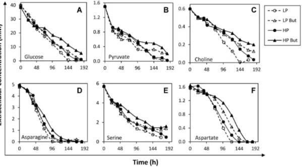
Figure 2.1 Network representation of metabolic reactions used for fluxome analysis 38 Figure 2.2 Cell growth and IgG4 production under control and butyrate-treated
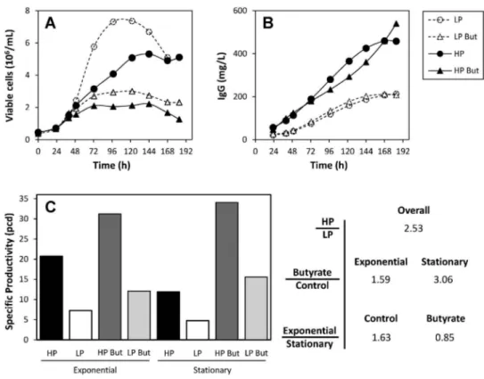
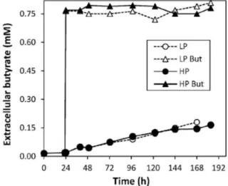
conditions of HP and LP clones ... 40 Figure 2.3 Extracellular butyrate concentration profiles under control and butyrate-treated conditions for culture set I ... 41 Figure 2.4 Extracellular concentration profiles of main substrate metabolites quantified in the exometabolome of culture set I ... 43 Figure 2.5 Extracellular concentration profiles of main secreted metabolites quantified in the exometabolome of culture set I ... 46 Figure 2.6 Metabolic flux distributions during exponential growth and stationary phase ... 52 Figure 2.7 Main modulations in central carbon metabolism associated with culture phase transition and butyrate treatment ... 53 Figure 3.1 (A) Timeline of experimental setup. Viable cell (B) and antibody concentration (IgG4
) (C) profiles, and specific productivity (D) in control and butyrate-treated cultures ... 73 Figure 3.2 -
Mass isotopomer distribution profiles for [1,2-13C]-glucose experiment,
during 24 h after isotopic tracer feed ... 76
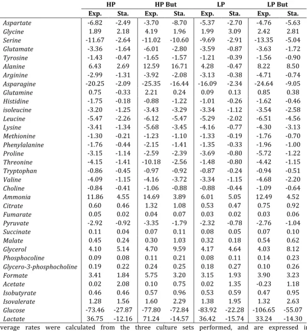
Figure 3.3 Simplified model for GS-CHO cell metabolism in cultures with fully labeled [U-13C/15N]-asparagine, in control condition ... 77
Figure 3.4 Mass isotopomer distribution profiles for [U-13C/15N]asparagine experiment,
during 24 h after isotopic tracer feed ... 79
Figure 3.5 Mass isotopomer distribution profiles for [U-13C/15N]serine experiment,
during 24h after isotopic tracer feed ... 80 Figure 3.6 Total intracellular metabolite pools ratio butyrate/control for amino acids,
after 24 h ... 84
Figure 4.1 Cell growth (A), asparagine (B) and serine (C) profiles of control and modulated cultures ... 100 Figure 4.2 Time profile of selected nutrients and by-products in the supernatant of control and asparagine modulated cultures ... 101 Figure 4.3 Overview of uptake/secretion rates in control and asparagine modulated cultures ... 103 Figure 4.4 Time profiles of selected metabolites in the supernatants of control and serine modulated cultures ... 105 Figure 4.5 Overview of uptake/secretion rates in control and serine modulated cultures ... 106 Figure 4.6 Cellular growth, IgG4 and formate profiles in cultures of clone II,
with/without serine or asparagine, and with/without glycine addition ... 108 Figure 4.7A Schematic of carbon atom transitions in pyruvate metabolism ... 109 Figure 4.7B/C Time profiles of intracellular metabolites after the introduction of 1-13 C-pyruvate in cultures of clone II, in the absence of serine, or asparagine ... 110 Figure 5.1 Thesis aims, strategies adopted, major outcomes and future studies ... 121
L
IST OF
T
ABLES
Table 1.1 Monoclonal antibody (mAb)-based biopharmaceuticals produced in mammalian cells, approved in the United States and Europe after 2010 ... 5 Table 1.2 Compilation of recent studies performed on CHO cells using isotopic tracers ... 18 Table 2.1 Average specific metabolic secretion and consumption rates for HP and LP cultures under control and butyrate-treated conditions ... 45 Table 2.2 Overview of metabolic phases studied ... 47
Table A1 List of CHO metabolic reactions considered in the model ... 135
αKG α-ketoglutarate 3PG 3-phosphoglycerate 6PG 6-phosphogluconate 6PGDH 6-phosphogluconate dehydrogenase AcCoA acetyl coenzyme A ADCC antibody-dependent cellular cytotoxicity Ala alanine Amm ammonia Arg arginine Asn asparagine Asp aspartate ATP adenosine-3-phosphate BHK baby hamster kidney But butyrate-treated CHO chinese hamster ovary Cit citrate CO2 carbon dioxide Cys cysteine DHAP dihydroxyacetone-phosphate DHFR dihydrofolate reductase DMF dimethylformamide DMSO dimethylsulfoxide DNA deoxyribonucleic acid DoE design of experiment DSS-d6 deuterated 3-(trimethylsilyl)-1-propanesulfonic acid sodium salt, 2,2-Dimethyl-2-silapentane-5-sulfonate-d 6 sodium salt EPO erythropoietin F6P fructose-6-phosphate FBA flux balance analysis FSH follicle-stimulating hormone Fum fumarate G6P glucose-6-phosphate GC gas chromatography GEM genome-scale model GIDH glutamate dehydrogenase Glc glucose GLDH glutamate dehydrogenase Gln glutamine Glu glutamate GLUT5 fructose transporter Gly glycine Glyc3PC glycerol-3-phosphocholine
His histidine HP high producer hPRL human prolactin IgG immunoglobulin Ile isoleucine INST isotopically non-stationary Lac lactate LC liquid chromatography LDH-A lactate dehydrogenase A Leu leucine LP low producer Lys lysine mAb monoclonal antibody Mal malate MDH II malate dehydrogenase II ME malic enzymes Met methionine MFA metabolic flux analysis MID mass isotopomer distributions miRNA micro RNA MOX O-methoxyamine hydrochloride mRNA messenger RNA MS mass spectrometry MSTFA N-Methyl-N-(trimethylsilyl)trifluoroacetamide MSX methionine sulfoximine MTBSTFA N–Methyl-N-(t-Butyldimethylsilyl) trifluoroacetamide MTX methotrexate NaBu sodium butyrate
NAD+/NADH nicotinamide adenine dinucleotide
NADP+/NADPH nicotinamide adenine dinucleotide phosphate
NMR nuclear magnetic resonance O2 oxygen OAA oxaloacetate PC pyruvate carboxylase PCMV human cytomegalovirus promoter PDH pyruvate dehydrogenase PEP phosphoenol-pyruvate PEPCK phosphoenolpyruvate carboxykinase Phe phenylalanine PPP pentose phosphate pathway
SDS-PAGE sodium dodecyl sulphate-polyacrylamide gel electrophoresis SEAP secreted alkaline phosphatase Ser serine Suc succinate TCA tricarboxylic acids THF tetrahydrofolate Thr threonine TMB tetramethylbenzidine TMCS tetramethylchlorosilane tPA tissue plasminogen activator Tyr tyrosine Val valine ZFN zinc-finger nuclease
Duarte, TM*, Carinhas, N*, Barreiro, LC, Carrondo, MJT, Alves, PM, Teixeira, AP, 2014, Metabolic responses of CHO cells to limitation of key amino acids. Biotechnol. Bioeng 111(10):2095–2106 *equal contributions
Carinhas N*, Duarte TM*, Barreiro LC, Carrondo MJT, Alves PM, Teixeira AP, 2013, Metabolic signatures of GS CHO cell clones associated with butyrate treatment and culture phase transition, Biotechnol. Bioeng. 110(12):3244-3257 *equal contributions Duarte TM, Seifar RM, Wahl AS, Alves PM, Teixeira AP, Exploring GS-CHO metabolism
under control and butyrate-treated conditions through parallel 13C-labeling
experiments (manuscript in preparation)
B
OOKC
HAPTERS Sá JV, Duarte TM, Carrondo MJT, Alves PM, Teixeira AP, 2014, Metabolic Flux Analysis: A Powerful Tool in Animal Cell Culture, Animal Cell Culture, Springer, Cell Engineering 9:521-539Duarte TM, Carinhas N, Silva AC, Alves PM, Teixeira AP, 2013, 1H-NMR protocol for
exometabolome analysis of cultured mammalian cells, Animal Cell Biotechnology-Methods and Protocols, Springer, 3rd edition, Part III:237-247
O
THERP
UBLICATIONSDuarte TM, Carrondo MJT, Alves PM, Teixeira AP, 2011, Fluorescence-based tools to improve biopharmaceutical process development, BMC proceeding 5(Suppl 8):O5 Teixeira AP, Duarte TM, Carrondo MJT, Alves PM, 2011, Synchronous Fluorescence Spectroscopy as a Novel Tool to Enable PAT Applications in Bioprocess, Biotechnol. Bioeng 108(8):1852-61
Teixeira AP, Duarte TM, Oliveira R, Carrondo MJT, Alves PM, 2011, High-throughput analysis of animal cell cultures using two-dimensional fluorometry, J. Biotechnol 151(3):255-260
S
UMMARY
Chinese hamster ovary (CHO) cells are preferred mammalian hosts for industrial production of therapeutic glycoproteins such as monoclonal antibodies (mAbs), used to treat cancer and immunological disorders. In these cells, the glutamine synthetase (GS) expression system has been widely used for efficient selection of high-yielding clones. However, fundamental knowledge on the metabolic behavior of GS-CHO cells in culture and its impact on product yields have not been yet systematized. The overall objective of this thesis was to comprehensively analyse the metabolome and fluxome of CHO cells exploring how the metabolism is affected by clonal variability and culture conditions. This information is then used to generate hypotheses on how metabolic wiring impacts mAb production.
We started by implementing a 1H-NMR metabolomics platform to assess the metabolic
footprints of two GS-CHO cell clones expressing IgG4 under control and
butyrate-treated conditions. With this method, we could accurately quantify the dynamic profiles of about 40 metabolites, including some not usually analyzed in mammalian cells cultures, such as acetate, pyruvate, formate, TCA cycle intermediates, lipid and nucleotide precursors. Noteworthy, several metabolic by-products accumulated in the cell supernatant over culture time, suggesting that different metabolic nodes could be targeted to decrease overflow metabolism. Overall, most carbon and nitrogen sources were consumed at higher rates during the exponential phase than during the stationary phase.
Then, the exometabolome data collected for the different clones and conditions was contextualized in a network model of 117 intracellular and transport reactions to estimate the corresponding fluxomes through metabolic flux analysis (MFA). While the two clones displayed generally similar specific metabolic fluxes, butyrate treatment had a marked effect on sustaining high nutrient consumption along culture time. This
resulted in a significantly increased energetic state and biosynthetic activity during the stationary phase compared to control conditions, coinciding with the largest effect on specific productivity. Furthermore, the intracellular concentration pools of most metabolites measured in butyrate-treated cultures were 2.5-fold higher (on average) when compared to control cultures. Higher differences were observed for glutamine, alanine and citrate (up to 4-fold), hence these metabolites should be further explored in future experiments to study the impact on recombinant protein production.
In order to have a more in-depth view of cell metabolism, namely the relative activities of parallel pathways and substrate contributions to intracellular metabolites, we next performed tracer studies using labelled versions of the three most consumed nutrients ([1,2-13C]glucose, [U-13C/15N]asparagine and [U-13C/15N]serine). Upon incubation of a
high producing GS-CHO cell clone with each isotopic tracer, we collected samples over the exponential growth phase to follow the transient labelling profiles of intracellular metabolites by GC-MS and LC-MS. The patterns produced from the [1,2-13C]glucose
tracer revealed a high diversion of glucose to the pentose phosphate pathway when compared to that previously published for non-producing CHO cells during the same growth phase, suggesting this route as an important source of NADPH for recombinant protein synthesis. Another potential source of NADPH is the flux through malic enzyme, the high activity of which was suggested by the labelling pattern of pyruvate after incubation with [U-13C/15N]asparagine. Noteworthy, asparagine was essential for
replenishing TCA cycle intermediates as well as for glutamine synthesis in GS-CHO cells. Together with asparagine, serine was confirmed to be a significant nitrogen source in these cells, with 15N from both amino acid tracers propagating to many non-essential
amino acids.
Finally, with the goal of better evaluating the importance of asparagine and serine in GS-CHO metabolism, we modulated the availability of these amino acids by culturing cells in the absence, or maintaining a low concentration, of each amino acid. The absence of asparagine in the medium caused growth arrest, and was associated with a
dramatic increase in pyruvate uptake, a higher ratio of pyruvate carboxylation to dehydrogenation and an inability for de novo asparagine synthesis. The release of ammonia and amino acids such as aspartate, glutamate and alanine were deeply impacted. This confirms asparagine to be essential for GS-CHO cells as the main source of intracellular nitrogen as well as having an important anaplerotic role for TCA cycle activity. In turn, serine limitation also negatively affected culture growth while triggering its de novo synthesis, confirmed by label incorporation coming from pyruvate, and reduced glycine and formate secretion congruent with its role as a precursor in the metabolism of one-carbon units. The results obtained suggest that feeding schemes of asparagine or serine should be tightly tuned to minimize by-product formation while assuring biosynthetic needs.
Overall, the work performed in the scope of this thesis contributes to unfold important insights into the metabolic operation of the industrially relevant GS-CHO cell expression host. An in-depth study of the metabolic network of GS-CHO cells allowed to uncover potential targets for minimizing overflow metabolism, reduce the diversion of resources to by-product accumulation or minimize their impact on recombinant protein production. Also allowed to couple PPP and ME activities, important sources of NADPH, with a high productive phenotype in GS-CHO cells. We believe that these markers represent an opportunity for future cell engineering efforts aiming the improvement of the production of biopharmaceuticals.
R
ESUMO
As células do ovário de hamster chinês (CHO, do inglês Chinese Hamster Ovary) são o hospedeiro mamífero preferido para a produção industrial de glicoproteínas terapêuticas, tais como os anticorpos monoclonais (mAbs, do inglês monoclonal antibodies) usados para o tratamento de cancro e doenças imunológicas. Nestas células, o sistema de expressão através da glutamina sintetase (GS) tem sido frequentemente utilizado para a selecção eficiente de clones de alto rendimento produtivo. No entanto, o conhecimento fundamental sobre o comportamento metabólico de células GS-CHO em cultura e o seu impacto sobre o rendimento na produtividade não tem sido sistematizado. O objectivo geral desta tese foi o de fazer uma análise exaustiva do metaboloma e do fluxoma de células CHO, explorando a forma como o metabolismo é afectado pela variabilidade clonal e condições de cultura. A informação resultante poderá então ser utilizada para formular hipóteses sobre o impacto que determinados rearranjos metabólicos podem ter sobre a produção de mAbs.
Começámos por implementar uma plataforma baseada em metabolómica através de RMN de protão para avaliar os perfis metabólicos de dois clones GS-CHO com diferentes níveis de expressão de uma imunoglobulina (IgG4) em condições controlo e
tratada com butirato. Com este método, conseguimos quantificar rigorosamente os perfis dinâmicos de cerca de 40 metabolitos, incluindo alguns que normalmente não são analisados em culturas de células de mamífero, tais como o acetato, piruvato, formato, intermediários do ciclo dos ácidos tricarboxílicos (ATC), e precursores de lípidos e nucleótidos. De salientar, vários sub-produtos metabólicos foram acumulados no sobrenadante das culturas ao longo do tempo, indicando que vários centros metabólicos podem ser alvo de acção no decréscimo de um metabolismo exagerado. De uma forma geral, a maior parte das fontes de carbono e azoto foi consumida durante a fase exponencial de crescimento celular com taxas de consumo superiores às observadas na fase estacionária.
Posteriormente, os dados do exo-metaboloma, recolhidos das culturas controlo ou tratadas com butirato, foram integrados num modelo metabólico com 117 reacções intracelulares ou de transporte celular para se poder estimar os fluxomas correspondentes através de uma metodologia clássica de análise de fluxos metabólicos. Apesar de ambos os clones apresentarem fluxos metabólicos específicos semelhantes, o tratamento com butirato teve um efeito significativo na manutenção de um elevado consumo de nutrientes ao longo do tempo de cultura. Isto resultou num aumento significativo do estado energético e da actividade biossintética durante a fase estacionária, quando o efeito sobre a produtividade específica é igualmente superior. Para além disto, as concentrações intracelulares obtidas para os metabolitos analisados foram em média 2.5 vezes superiores quando comparadas com as culturas controlo. Diferenças mais substanciais foram detectadas para a glutamina, alanina e citrato (cerca de 4 vezes superiores), pelo que estes metabolitos deverão ser deverão ser explorados em experiências futuras no estudo dos seus efeitos na produção de proteínas recombinantes.
De forma a obter uma visão mais detalhada sobre o metabolismo celular, nomeadamente sobre as actividades relativas de vias metabólicas paralelas e a contribuição de certos substratos para vários metabolitos intracelulares, decidiu-se fazer estudos com a incubação de compostos marcados dos três metabolitos mais consumidos (glucose-[1,2-13C], asparagina-[U-13C/15N] and serina-[U-13C/15N]). Após a
incubação de culturas com o clone de células GS-CHO com mais elevada produtividade com cada um dos isótopos durante a fase exponencial do crescimento celular, recolhemos amostras ao longo do tempo para podermos seguir os perfis transientes de incorporação da marca nos metabolitos intracelulares por GC-MS e LC-MS. Os perfis obtidos através da incubação com [1,2-13C]glucose, revelam um desvio significativo de
glucose através da via das pentoses fosfatadas quando comparadas com culturas de células CHO não-produtivas durante a mesma fase de crescimento, sugerindo que esta via pode ser uma importante fonte de poder redutor (NADPH), necessário para a
síntese de proteínas recombinantes. Outra fonte de NADPH é o fluxo através da reacção das enzimas málicas, como sugerido pela incorporação de isótopos observada no piruvato obtido através da asparagina-[U-13C/15N]. De salientar, a asparagina foi
essencial para o reabastecimento de intermediários do ciclo ATC, tal como para a síntese de glutamina em células GS-CHO. Juntamente com a asparagina, a serina foi confirmada como sendo uma fonte de nitrogénio importante nestas células, tendo-se observado azoto (15N) derivado de ambas as fontes em vários amino ácidos
não-essenciais.
Finalmente, com o objectivo de avaliar a importância da asparagina e da serina no metabolismo de células GS-CHO, foi feita uma análise da disponibilidade da asparagina e da serina: através da cultura de células na ausência de cada um dos amino ácidos, ou através da cultura de células na presença de cada amino ácido em baixas concentrações. A ausência de asparagina no meio basal interrompeu o crescimento celular, e foi associado a um aumento acentuado do consumo de piruvato, um elevado rácio entre as reacções de carboxilação e desidrogenação de piruvato, e à incapacidade das células para a síntese de novo de asparagina. A secreção de amónia e de amino ácidos como o aspartato, glutamato e alanina foi igualmente afectada. Isto confirma que a asparagina é essencial para as células GS-CHO como principal fonte de nitrogénio, assim como tendo uma função anaplerótica na actividade do ciclo ATC. Por outro lado, a limitação de serina também afectou negativamente o crescimento celular tendo despoletado a síntese de novo de serina, confirmada pela incorporação do isótopo observada no piruvato, e reduziu a secreção de glicina e formato, de acordo com o seu papel como precursor no metabolismo de unidades com um carbono unitário. Estes resultados sugerem que as estratégias de alimentação com asparagina ou serina devem ser rigorosamente reguladas para se poder reduzir a formação de sub-produtos, e ao mesmo tempo manter as necessidades biossintéticas.
Em suma, esta tese contribui para o desvendar de importantes fenómenos biológicos no metabolismo de células GS-CHO com alto relevo industrial. O estudo aprofundado da rede metabólica das células GS-CHO permitiu identificar alvos de estudo para minimizar o metabolismo exagerado, reduzir o consumo de recursos dirigidos para a acumulação de produtos secundários ou o impacto sobre a produção de proteínas recombinantes. Da mesma forma, permitiu associar as actividades da via das pentoses fosfatadas e das enzimas málicas, importantes fontes de NADPH, a um fenótipo de produção elevada em células GS-CHO. Julgamos que estes elementos representam uma oportunidade para esforços futuros em engenharia celular no sentido de melhorar a produção de produtos farmacêuticos.
C
HAPTER
1
Introduction
C
ONTENTS
1.1
Mammalian cells as production factories of biopharmaceuticals ... 3
1.2
Chinese Hamster Ovary Cells ... 5
1.3
Performance of mammalian cell bioprocesses ... 8
1.3.1
Culture medium and fed batch processes ... 8
1.3.2
Classical approaches to improve specific productivity ... 9
1.3.3
Cell line engineering ... 10
1.4
Systems biotechnology ... 13
1.4.1
Transcriptome and proteome layers ... 13
1.4.2
Metabolome and Fluxome layers ... 15
1.5
Thesis motivation and outline ... 19
1.6
Author contribution ... 21
1.7
References ... 21
1.1 Mammalian cells as production factories of biopharmaceuticals
Mammalian cells are the dominant biological system for the production of recombinant therapeutic proteins mainly due to their ability to perform human-like post-translational modifications. The first therapeutic protein produced from recombinant mammalian cells, tissue plasminogen activator (tPA), obtained market approval thirty years ago (reviewed by Butler and Spearman 2014; Wurm 2004). Nowadays, monoclonal antibodies (mAbs) constitute the main class of biopharmaceuticals produced from mammalian cell cultures. The development of antibody-based products has increased significantly over the years, representing 27 % of all biologic products approved as of 2014 (Walsh 2014). Global sales revenue for all mAb products in 2013 was nearly US$75 billion, representing the most lucrative single product class (Ecker etal. 2015).
Table 1
summarizes the mAb-based products approved after 2010 in Europe and in the USA.Among common targets of mAbs are various types of cancer, and several inflammatory and cardiovascular diseases. The high incidence of these diseases, associated to an increasing and aging worldwide population and the standard of living in emerging markets increases the pressure to develop more efficient bioprocesses (Ecker et al. 2015).
The prevalence of mammalian cells over other expression systems, such as insect cells or Escherichia coli, goes parallel with the increasing proportion of approved molecules that show post-translational modifications, in particular glycosylation. It is crucial that therapeutic proteins display human-like glycosylation patterns to ensure their optimal efficacy and higher stability in the human serum (Butler and Spearman 2014). Although several mammalian cell lines are used for production of complex biopharmaceuticals (such as murine myeloma cells (NS0 and SP2/0), human embryonic kidney (HEK)-293 and baby hamster kidney (BHK)), nearly 70 % of all recombinant therapeutic proteins are produced in Chinese hamster ovary (CHO) cells (Jayapal et al. 2007).
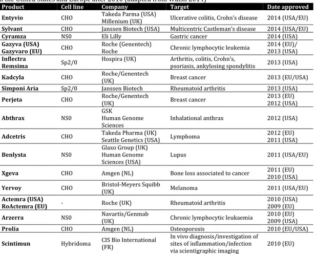
Table 1.1 Monoclonal antibody (mAb)-based biopharmaceuticals produced in mammalian cells, approved
in the United States and Europe after 2010 (adapted from Walsh 2014)
Product Cell line Company Target Date approved
Entyvio CHO Takeda Parma (USA) Millenium (UK) Ulcerative colitis, Crohn’s disease 2014 (USA/EU)
Sylvant CHO Janssen Biotech (USA) Multicentric Castleman’s disease 2014 (USA/EU)
Cyramza NS0 Eli Lilly Gastric cancer 2014 (USA)
Gazyva (USA)
Gazyvaro (EU) CHO Roche (Genentech) Roche Chronic lymphocytic leukemia 2014 (EU)/ 2013 (USA)
Inflectra
Remsima Sp2/0 Hospira (UK) Arthritis, colitis, Crohn’s, psoriasis, ankylosing spondylitis 2013 (USA)
Kadcyla CHO Roche/Genentech (UK) Breast cancer 2013 (EU/USA)
Simponi Aria Sp2/0 Janssen Biotech Rheumatoid arthritis 2013 (USA)
Perjeta CHO Roche/Genentech (UK) Breast cancer 2013 (EU) 2012 (USA)
Abthrax NS0 GSK Human Genome
Sciences Inhalational anthrax 2012 (USA)
Adcetris CHO Takeda Pharma (UK) Seattle Genetics (USA) Lymphoma 2012 (EU) 2011 (USA)
Benlysta NS0 Glaxo Group (UK) Human Genome
Sciences (USA) Lupus 2011 (USA/EU)
Xgeva CHO Amgen (NL) Bone loss associated to cancer 2011 (EU) 2010 (USA)
Yervoy CHO Bristol-Meyers Squibb
(UK) Melanoma 2011 (USA/EU)
Actemra (USA)
RoActemra (EU) - Roche (UK) Rheumatoid arthritis 2010 (USA) 2009 (EU)
Arzerra NS0 Navartis/Genmab (UK) Chronic lymphocytic leukaemia 2010 (EU) 2009 (USA)
Prolia CHO Amgen (NL) Osteoporosis 2010 (EU/USA)
Scintimun Hybridoma CIS Bio International (FR) In vivo diagnosis/investigation of sites of inflammation/infection
via scientigraphic imaging 2010 (EU)
1.2 The Chinese Hamster Ovary Cells
The initial CHO cell strain was isolated at the laboratory of Theodore Puck, after culturing cells derived from the ovarian tissue of a Chinese hamster (Puck et al. 1958). After an unknown amount of culture time the morphology of some cells changed and these underwent spontaneous immortalization (reviewed by Wurm 2013). Several laboratories across the world have propagated the number of CHO cell lines being manipulated, considerably increasing the genetic and phenotypic diversity of the CHO cell system.
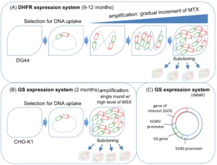
CHO cells are not only used to produce biopharmaceutical products that generate over US$50 billion annually, but the pipelines of pharmaceutical companies are also full of CHO-derived products in different phases of clinical trials (Wurm 2013). The success of these cells is explained by i) their robustness to growth to high cell densities in serum-free, chemically-defined suspension cultures, ii) their safety profile (they are unable to replicate most human pathogenic viruses), iii) their capacity to easily integrate foreign genes, and iv) the powerful selection systems (such as dihydrofolate reductase (DHFR) and glutamine synthetase (GS)) (Durocher and Butler 2009). From all CHO-derived cell lines, the CHO-DG44 with the DHFR selection system and the CHO-K1 in combination with the GS selection system are the two most used industrially. The CHO-DG44 cells are DHFR deficient, thus they are incapable of reducing dihydrofolate to tetrahydrofolate, a reaction that is crucial for the de novo synthesis of nucleotides. As a result, dhfr negative cells have to be cultured in medium containing hypoxanthine and thymidine (HT) in order to survive. Therefore, by transfecting these cells with a plasmid encoding the gene of interest (GOI) and a functional dhfr gene, successfully transfected cells can be selected in culture media without HT. Additional selection pressure can be introduced using stepwise increases in the concentration of methotrexate (MTX), an antagonist of DHFR, which results in amplified copies of the transfected dhfr gene together with the GOI, consequently increasing the productivity of the GOI (Hacker et al. 2009; Wurm 2013) (Fig. 1.1A).
GS-CHO-K1 cells are obtained from the transfection with a functional glutamine synthetase gene together with gene of interest on the same plasmid, followed by culture selection in medium without glutamine (Wurm 2013). Nevertheless, CHO cells possess endogenous GS activity which must be specifically inhibited by low levels of methionine sulphoximine (MSX) to ensure that any cell that survive on gln-deficient medium possess the GOI. Gene amplification can be achieved by further application of higher levels of MSX. However, as opposed to the DHFR amplification system, a single round of amplification is sufficient to achieve high expression levels of the GOI (Noh et
al. 2013) (Fig. 1.1B/C). Importantly, the GS-system has the advantage of reducing the accumulation of ammonia in the medium, a toxic byproduct resultant of glutaminolysis in other cell lines (Wurm 2013).
Figure 1.1 GS and DHFR expression systems. DHFR system (A) requires five to six fold more time to attain
a high expression rate, when compared to the GS system (B); (C) detail of the GS-expression system (adapted from Noh et al. 2013).
The development and identification of a highly-productive cell line generally involves a high-throughput selection step, which is usually low efficient and thus generates large numbers of low and non-productive cells, like in the case of GS selection in CHO cell lines (Cacciatore et al. 2010). It has been supposed that the basal expression from the endogenous GS gene may decrease the GS-system selection stringency (Fan et al. 2012). In order to maximize the number of desirable and rare highly-productive candidates, which is to say improving selection stringency, while reducing timeline and costs, Fan and colleagues knocked out the endogenous CHO GS gene. This was achieved through specific zinc-finger nuclease (ZFN)-based technology, and translated into a six-fold efficiency increase in cell line generation, when compared to GS-CHO cells (expressing both endogenous GS activity and the GS system) (Fan et al. 2012). Posteriorly, the
same group used a weakened promoter to drive GS expression (an engineered version of the SV40), in combination with the GS-knockout CHO cells, resulting in improved productivities associated to higher selection efficiencies and eliminating the need to add MSX, a toxic and expensive chemical GS inhibitor, to the culture medium (Fan et al. 2013).
1.3 Performance of mammalian cell bioprocesses
In order to meet the growing demand for mAb products, there is a persistent need to develop robust, cost-effective and efficient cell culture processes (Sheikholeslami et al. 2013; Wurm 2004). The mammalian cell culture field has matured significantly over the years, and today it is possible to deliver gram per litre titres of product in fed-batch cultures, representing over 100-fold improvement since the 1980s (Mead et al. 2012). This yield improvement has been the result of better expression vector designs, improved host cell lines, development of medium composition and feeding regimes, and advanced screening techniques.
1.3.1 Culture medium and fed batch processes
Medium development for mammalian cells has progressed enormously since the first formulations that required serum supplementation (Bruhlmann et al. 2015). Fully chemically defined media consisting of amino acids, vitamins, trace elements, inorganic salts, lipids and insulin or insulin-like growth factors, have been developed for mAb production processes (Pasupuleti et al. 2008). In general, medium development is labor-intensive and time-consuming, and it is based in statistical design of experiment (DoE) (Kim and Lee 2009).
Not all antibody production cell lines can achieve high yield in chemically defined media. Therefore, the addition of non-animal hydrolysates (composed of amino acids, small peptides, carbohydrates, vitamins and minerals) became a common strategy in order to maximize cell density, culture viability and productivity (Kim and Lee 2009; Michiels et al. 2011). However, because of its composition complexity and lot-to-lot variations, hydrolysates can be a significant source of medium variability.
Bioreactors operating in fed-batch mode are the most common processes used to produce monoclonal antibodies (Wurm 2004). High yields of up to 10 g/L have been achieved through feeding optimization, which increase cell productivity, maintain cell viability, and extend culture longevity (Kim et al. 1998; Lee et al. 1999; Liu et al. 2001). Perfusion has been used also like fed-batch mode for the production of recombinant proteins in mammalian cell cultures. In fact, the highest volumetric productivities have been achieved using continuous stirred tank bioreactors in perfusion mode, using internal retention systems. However, due to the limited scalability of these systems, novel biopharmaceuticals have been produced in fed-batch cultures (Lee et al. 2012). While the most common approach to develop a feed medium is to concentrate the basal medium, a sophisticated optimization of feed composition and feeding strategy would require to take into consideration: nutrient consumption, by-product accumulation and the balance between promoting growth versus volumetric productivity. Nevertheless, step-wise bolus addition of the feed solution to the production bioreactor is still the most widely used technique in the industry due to its simplicity and scalability (Li et al. 2014).
1.3.2 Classical approaches to improve specific productivity
Further strategies for process optimization often included modulations of temperature, or addition of chemicals (Fig 1.2). Temperature shifts from 37 °C to 30-34 °C allowed improvements in cell specific and volumetric productivity, protein folding and product quality (Al-Fageeh et al. 2006). For instance, Yoon et al showed a significant improvement in specific productivity of erythropoietin (EPO) and follicle-stimulating hormone (FSH) when decreasing culture temperature from 37 to 32 °C (Yoon et al. 2006). Several chemicals have been applied as productivity enhancers for recombinant protein production in cultured mammalian cells, including dimethylsulfoxide (DMSO) (Allen et al. 2008), sodium butyrate (NaBu) (Yee et al. 2008) and rapamycin (Balcarcel and Stephanopoulos 2001). In particular, sodium butyrate has been used to improve the production of tPA (Palermo et al. 1991), EPO (Wang et al. 2011), mAb (Mimura et al.2001) and human prolactin (hPRL) (Goulart et al. 2010). NaBu has several effects on cultured cells, namely growth inhibition, induction or repression of gene expression and induction of apoptosis (Jiang and Sharfstein 2008). NaBu regulates gene transcription by improving gene accessibility through the inhibition of histone deacetylases (HDAC) (Jiang and Sharfstein 2008). When hyper acetylated histones become more relaxed complexes with DNA, genes are more accessible to regulatory factors and thus transcribed at higher rates.
Although these strategies managed to boost productivities and yields, they have to be optimized for each cell line, being time consuming and frequently with little or no understanding of the actual reasoning behind the improvements obtained (Selvarasu et al. 2012).
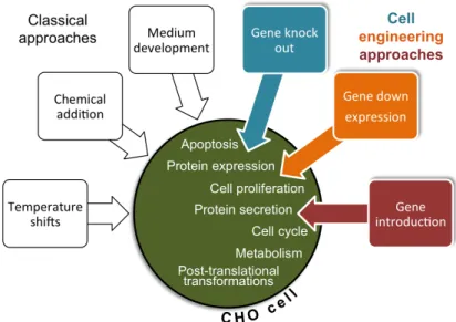
1.3.3 Cell line engineering
A significant amount of work has been done to genetically engineer host cells to improve their robustness, enhance their specific productivity or improve product quality (Le et al. 2015). Cell engineering efforts usually target the machinery of several cellular processes, such as: apoptosis, protein expression, cell proliferation, metabolism, cell cycle, protein secretion and post translational modifications (Balcarcel and Stephanopoulos 2001; Fischer et al. 2015; Le et al. 2015) (Fig. 1.2). Most common approaches include the knockout and downregulation of key genes that are involved in these mechanisms, or the introduction of other genes (Fischer et al. 2015). A lot of attention has been given to anti-apoptosis engineering as a main strategy of cell line development for protein production (Majors et al. 2007). Anti-apoptosis approaches rely, at a first instance, on the overexpression of anti-apoptotic genes held by mammalian cells. These genes encode for anti-death proteins that prevent the activation of the apoptosis cascade. Among these, with positive results in terms of cell density and protein titre are Bcl-2, Bcl-xL and 30Kc6 (Figueroa et al. 2007; Mastrangelo et al. 2000; Wang et al. 2011). A second approach to avoid apoptosis has been the inhibition of caspase expression, since the activation of members of the caspase family
is part of the cellular processes that lead to apoptosis (Laken and Leonard 2001; Mimura et al. 2000). Thirdly, the inhibition of cell cycle progression as this significantly increases cell density and productivity in mammalian cell cultures. These strategies led to a slower cell cycle that lead to an extended cell viability through time and, consequently, an increased specific mAb productivity (Balcarcel and Stephanopoulos 2001). Recently, Tan et al (2015) introduced heat shock protein 27 in CHO cells in order to improve cell density and viability with a 2.3-fold increase in mAb titre, affecting apoptosis signaling pathways and delaying caspase activities.
CHO cells and other continuous mammalian cell lines have low efficiency in completely oxidizing glucose to CO2, producing high levels of lactate. This can cause acidification of
culture medium and lead to high osmolarity and low viability due to the base added to control the medium pH. A significant amount of work has been performed to reduce lactate and ammonia accumulation; strategies include the partial disruption of lactate dehydrogenase A (LDH-A) gene, or the overexpression of pyruvate carboxylase (PYC2), increasing the flux of glucose into TCA cycle (Kim and Lee 2007b), or the overexpression of urea cycle enzymes, such as carbamoyl phosphate synthetase I and ornithine transcarbamoylase (Chen et al. 2001; Kim and Lee 2007a; Park et al. 2000; Wilkens and Gerdtzen 2015).
Figure 1.2 Classical and cell engineering approaches to improve the specific productivity of CHO cells.
Inside the CHO cell representation are presented the common targets of cell engineering approaches (Fischer et al. 2015; Le et al. 2015).
Very recently, Wilkens and Gerdtzen (2015) compared different engineered CHO cell clones with overexpression of several metabolic genes, namely PYC2, MDH II (malate dehydrogenase II) and GLUT5 (fructose transporter), in terms of their effect on IgG production performance. Although previous studies had reported improved specific productivities when overexpressing these genes (Chong et al. 2010; Irani et al. 1999; Le et al. 2013), Wilkens study did not corroborate those studies. They propose that the modifications promote an unbalanced redox state which may have a negative impact on IgG assembly and, consequently, reducing its secretion.
Glycosylation pathway engineering has also received considerable attention because glycan structures on antibodies can have substantial impact on clearance rate and bioactivities. For instance, gene deletion technologies have been used to decrease or eliminate the fucose on antibodies to dramatically improve antibody-dependent cellular cytotoxicity (ADCC) (Malphettes et al. 2010). In particular, non-fucosylated antibodies present a substantially higher efficacy than their fucosylated counterparts, in most cases, due to a higher ADCC activity (Konno et al. 2012). However, these
approaches are often host cell dependent, as each host has its own specific post-translational modifications fingerprint.
1.4 Systems biotechnology
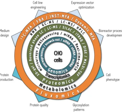
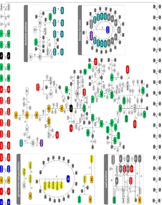
With the progress made in Omics technologies in last 10 years, it became possible to study the molecular traits that characterize a high-producer cell (reviewed by Carinhas et al. 2012). High-throughput experimental techniques and superior computer power have permitted obtaining great amounts of Omics data, allowing to gain in-depth knowledge of biological processes. Although we cannot integrate yet all levels of information, we can use parts of it to determine new targets for host improvement (Lee et al. 2005). The recent sequencing of CHO genome, combined with advances in technologies that allow a better characterization of the proteome and metabolome of cells under different environments, may now permit a comprehensive understanding of these industrially relevant cell lines (Datta et al. 2013; Xu et al. 2011). Figure 1.3 summarizes most of the state-of-the-art tools used in systems biology studies.
1.4.1 Transcriptome and proteome layers
Transcriptomics, the study of all RNA forms encoded by the genome, has been widely exploited for cell line and bioreactor process development in the last decades (Chen et al. 2016). DNA microarrays have revolutionized the quantitative analysis of gene expression as they allow covering a large number of genes simultaneously in a single assay. This genome-scale technology has been applied to i) investigate gene expression under culture conditions that enhance productivity, including temperature shifts, butyrate treatment, and high osmolality (Gatti et al. 2007; Kantardjieff et al. 2010), ii) to compare CHO cell lines expressing different levels of recombinat protein (Clarke et al. 2011; Doolan et al. 2010; Kang et al. 2014), or iii) at different culture stages. For instance, Wong and co-workers applied comparative transcriptomic analysis to find key factors regulating the apoptosis, which occurs at the later stages of batch and fed-batch cultures (Wong et al. 2006a; 2006b). Overexpressing the identified anti-apoptotic genes (Fadd and Faim), or knocking down the pro-apoptotic genes (Alg-2, and
Requiem), allowed to increase the yields of the recombinant protein up to 2.5-fold in CHO cells.
Figure 1.3 Diagram of systems biology and state-of-the-art technologies and methods used to improve the
performance of CHO cells. Main goals for Omics approaches (upper arrows) and main insights (lower arrows) to be obtained or predicted (Chen et al. 2016; Kildegaard et al. 2013; Lewis et al. 2016).
MicroRNAs are a class of small non-coding RNAs that can regulate global gene expression at the post-transcriptional level by mRNA cleavage or translational repression, or both. The ability of microRNAs to bind multiple target genes and regulate their abundance has made them significantly interesting for CHO cell engineering (Barron et al. 2011). There are several studies focused on improving productivity, either through miRNA overexpression or knockdown (Fischer et al. 2015). Jadhav and co-workers managed to improve cell growth and enhance the specific protein productivity of CHO cells (3-fold) through the stable overexpression of miR-17 (Jadhav et al. 2014). The stable miR-30 overexpression in CHO cells allowed to improve
secreted alkaline phosphatase (SEAP) productivity or maximum cell density by 2-fold (Fischer et al. 2014). Kelly and co-workers successfully improved productivity of SEAP-producing CHO cells through miR-23 knock-down, without affecting cell growth (Kelly et al. 2015). Interestingly, the improved productive phenopype was associated with an enhanced mitochondrial function and TCA cycle activity. Comparatively, proteomics has been less explored than the transcriptional analysis for cell culture engineering (Griffin et al. 2007). Proteomics has been applied to identify differentially expressed proteins that may have an impact in cell growth and death, metabolism and protein processing, including glycosylation (Baycin-Hizal et al. 2012). Since the transcriptome and the proteome might not be directly correlated, combined approaches are often chosen in order to deepen our understanding of cellular physiology (Baycin-Hizal et al. 2012). For instance, Doolan and co-workers combined microarray and proteomic expression profiling of CHO cells to identify genes and proteins involved in high cell growth rates and protein production (Doolan et al. 2010). 21 targets were identified and associated to high growth rate, from which 5 were validated by siRNA knockdown and considered feasible for further studies. More recently, multiple mAb-producing CHO cell lines were compared through a multiomics approach, including microarray-based transcriptomics and LC-MS/MS shotgun proteomics, in order to identify features associated to a high productivity (Kang et al. 2014). This approach allowed identifying several potential genes and proteins associated to productivity, cell size and proliferation.
1.4.2 Metabolome and Fluxome layers
Although metabolomics had been shadowed by genomics, transcriptomics and proteomics, it has reached a “golden era” potentiated by analytical techniques and computational power (Idle and Gonzalez 2007). The comprehensive quantification of the cellular metabolome (the collection of metabolites at any given time) can be carried out by state-of-the-art analytical approaches based on nuclear magnetic resonance (NMR) or mass spectrometry (MS) (Fig. 1.3).
Obtaining an accurate snapshot of cellular metabolite levels is dependent of several delicate steps, which address significant challenges inherent to physicochemical diversity, variety and rapid turnover of metabolites in mammalian cells. The sampling and arrest of metabolic activity (usually achieved by the use of a quenching solution at low temperatures) should be fast to prevent significant changes in metabolite concentrations due to ongoing metabolic activity. As opposed to extracellular metabolites that can be easily quantified through supernatant analysis, the accurate quantification of intracellular metabolite pools is dependent also of an extraction step. Furthermore, MS analysis is usually preceded by separation and ionization steps that allow reducing the complexity of a biological sample and the analysis of a smaller number of metabolites at different retention times (Alonso et al. 2015). Liquid and gas chromatography (LC and GC, respectively) are the most common separation techniques. Additionally, after data acquisition through NMR or MS, spectral data must be processed through several sequential steps aimed at accurately identifying and quantifying the metabolites present in a culture sample, including baseline correction, peak detection or spectral alignment. Peak overlapping issues require deconvolution methods; several software are currently available to facilitate spectra integration, such as Chenomx NMR Suite or Agilent MassHunter.
Metabolomics tools have been used to support animal cell process development. For instance, Sellick et al. (2011b) developed a feeding approach based on the identification of limiting nutrients in a mAb-producing CHO cell culture by GC-MS analysis. By supplying the limiting nutrients, the biomass could be increased by 35 % and the antibody titre could be doubled (Sellick et al. 2011a). In another MS-based study, Chong et al. (2010) found a high accumulation rate of malate in the medium of CHO cells. Subsequent cell engineering to overexpress malate dehydrogenase II resulted in significant growth improvement.
On the other hand, it is the fluxome (collection of metabolic fluxes inside the cell) the actual target of considerable attention as it reflects the final output of the interactions between genome, transcriptome, proteome and metabolome layers; and it is closely related to the cells phenotype (Sauer 2006). As opposed to transcripts, proteins and metabolites, fluxes cannot be measured directly and they have to be inferred from data through model-based interpretation. Mathematical models became essential to quantify the cellular metabolism under different culture conditions, and generate the knowledge needed to optimize the performance of cell factories.
When metabolic steady-state can be assumed, metabolic flux analysis (MFA) became the preferential technique to obtain quantitative information about in vivo cellular metabolism (Wiechert 2001). Thus, MFA can provide the knowledge to allow a manipulation of metabolic fluxes, in order to obtain improved process yields and better quality attributes in biopharmaceuticals production (Sá et al. 2015).
Classical (or stoichiometric) MFA is based on the flux balancing of intracellular metabolites within a stoichiometric matrix, in which extracellular measurements such as nutrient uptake, by-product secretion and growth rate are used as constraints to determine intracellular fluxes (Antoniewicz 2015). Such stoichiometric flux analysis can provide a good snapshot of the fluxome state, allowing for instance a broad comparison of different culture conditions. However, it cannot resolve fluxes through cyclic pathways, reversible and parallel reactions; only the net fluxes can be calculated (Goudar et al. 2010).
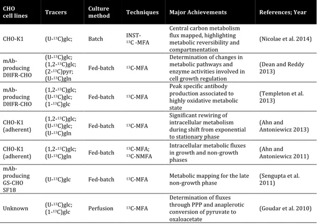
Table 1.2 Compilation of recent studies performed on CHO cells using isotopic tracers (Ahn and
Antoniewicz 2012; Lewis et al. 2016)
CHO
cell lines Tracers Culture method Techniques Major Achievements References; Year
CHO-K1 (U-13C)glc; Batch INST-
13C -MFA Central carbon metabolism flux mapped, highlighting metabolic reversibility and compartmentation (Nicolae et al. 2014) mAb-producing DHFR-CHO (U-13C)glc; (1,2-13C)glc; (2-13C)pyr; (U-13C)gln Fed-batch 13C-MFA Determination of changes in metabolic pathways and enzyme activities involved in cell growth regulation (Dean and Reddy 2013) mAb-producing DHFR-CHO (1,2-13C)glc; (U-13C)glc; (1-13C)glc Fed-batch 13C-MFA Peak specific antibody production associated to highly oxidative metabolic state (Templeton et al. 2013) CHO-K1 (adherent) (1,2-13C)glc; (U-13C)glc; (U-13C)gln Fed-batch 13C-MFA Significant rewiring of intracellular metabolism during shift from exponential to stationary phase (Ahn and Antoniewicz 2013) CHO-K1 (adherent) (1,2-13C)glc; (U-13C)gln Fed-batch 13C-MFA; 13C-NMFA Intracellular metabolic fluxes in growth and non-growth phases (Ahn and Antoniewicz 2011) mAb-producing GS-CHO SF18
(U-13C)glc Fed-batch 13C-MFA Metabolic mapping for the late
non-growth phase (Sengupta et al. 2011)
Unknown (U-(1-1313C)glc C)glc; Perfusion 13C-MFA
Determination of fluxes through PPP and anaplerotic conversion of pyruvate to oxaloacetate
(Goudar et al. 2010)
(mAb – monoclonal antibody; glc – glucose; pyr – pyruvate; gln – glutamine; INST – isotopically non-stationary; NMFA – non-stationary MFA; GS – glutamine synthetase; DHFR – dihydrofolate reductase)
Complementing extracellular data with isotopic labeling information in 13C-MFA
studies provides higher resolution, allowing to detect even the more subtle differences in flux phenotypes. An isotopic tracer is a molecule where a specific atom has been replaced with a different stable isotope, usually 13C, 15N or 2H. When labeled versions of
nutrients are added to cells, the label propagates through the network as a function of metabolic activity, producing labeling patterns in the backbone of metabolic intermediates over time. Mathematical models describing this propagation of the label are then used to calculate the in vivo fluxes from measured isotopic patterns and extracellular transport rates. The accumulation of a specific labeled metabolite allows to identify preferential pathways or their activity through time. Isotopic tracers became essential for providing insights on intracellular pathways, in particular on the presence
of parallel, reversible or cyclic pathways (Crown and Antoniewicz 2012; Quek et al. 2010). These approaches have been applied primarily to support metabolic engineering of simpler biological systems (Crown et al. 2015; Leighty and Antoniewicz 2012; Toya and Shimizu 2013), but are starting to be applied more and more to animal cell cultures as well. Table 1.2 shows a compilation of recent MFA studies done on CHO cells, using isotopic tracers.
A recent trend to resolve intracellular pathways has been the use of parallel labelling experiments. This technique has been addressed as COMPLETE-MFA – short for complementary parallel labelling experiments technique for metabolic flux analysis. This method results from fitting several data sets from several experiments into a single flux model (Antoniewicz et al. 2007). The main advantage is that it allows producing results with increased accuracy over 13C-MFA (Antoniewicz 2015). This
approach minimizes the issue of selecting the best single tracer that would allow a good screening of the whole network and with a good resolution (Crown and Antoniewicz 2012).
1.5 Thesis motivation and outline
Up to now, cell engineering has encompassed intuitive manipulations of enzyme activities, allowing slight improvements in yields and quality of therapeutic protein production. More efficient production requires deeper understanding of cellular metabolism and its regulation, which provides the building blocks and energy for cell growth and recombinant protein production.
This thesis aims at studying the central metabolism of producer cells in order to identify markers for posterior metabolic engineering and ultimately to improve the production of target proteins. We combined experimental and computational tools for quantitative dissection of the metabolome and fluxome of GS-CHO cell lines expressing a model monoclonal antibody. In chapter 2, the exometabolome of two IgG4-producing
through 1H-NMR spectroscopy, allowing a comprehensive characterization of medium
composition, in particular the identification of i) the most consumed substrates, and ii) several metabolic by-products secreted over culture time besides the common lactate and ammonia. This data was then integrated into an extended metabolic network to estimate the in vivo fluxes in different productivity states using MFA, to identify metabolic features correlated with higher productivity. In chapter 3, we performed short-term parallel labelling experiments using 13C and 15N labeled versions of the
three most consumed substrates (glucose, asparagine and serine), to explore with more detail the intracellular metabolism of GS-CHO cells in control and butyrate-treated cultures. The incorporation of 13C/15N from each substrate into intracellular
metabolites, including amino acids and intermediates from glycolysis, pentose phosphate pathways and TCA cycle, was followed over time by GC-MS and LC-MS. In chapter 4, modulations of asparagine and serine availability in cell culture media were performed, in order to study their essentiality and effect on cell physiology, namely their inter-relationships in nitrogen and carbon metabolism, biosynthesis and the impact on culture performance. Furthermore, we used 13C labeled pyruvate and GC-MS
analysis to discern intracellular partitioning of this substrate in the presence or absence of asparagine or serine.
Chapter 5 presents a global discussion of the results obtained.
Overall, this thesis provides an in-depth study of the metabolic network of GS-CHO cells that allowed identification of potential targets for minimizing overflow metabolism, reducing the diversion of resources to by-product accumulation, or minimising their impact on recombinant protein production. Also, it allowed the association of PPP and ME activities, important sources of NADPH, with a high productive phenotype in GS-CHO cells. These biological markers can represent an opportunity for future process and cell engineering efforts aiming towards the improvement of biopharmaceuticals production.