MESTRADO EM ONCOLOGIA
ESPECIALIZAÇÃO EM ONCOLOGIA MOLECULAR
Decoding the usefulness of miRNAs
as biomarkers in breast cancer
patients treated with endocrine
therapy
Maria Rodrigues Amorim
M
2017M
a
ri
a
Ro
d
ri
g
u
e
s
A
m
o
ri
m
D
e
c
o
d
in
g
th
e
u
s
e
fu
ln
e
s
s
o
f m
iR
N
A
s
a
s
b
io
m
a
rk
e
rs
in
b
re
a
s
t c
a
n
c
e
r p
a
tie
n
ts
tr
e
a
te
d
w
ith
e
n
d
o
c
rin
e
th
e
ra
p
y
M
.IC
BAS
2017
D
e
c
o
d
in
g
th
e
u
s
e
fu
ln
e
s
s
o
f m
iR
N
A
s
a
s
b
io
m
a
rk
e
rs
in
b
re
a
s
t c
a
n
c
e
r p
a
tie
n
ts
tr
e
a
te
d
w
ith
e
n
d
o
c
rin
e
th
e
ra
p
y
M
a
ri
a
Ro
d
ri
g
u
e
s
A
m
o
ri
m
IN S TITU TO D E C IÊ N C IA S B IO M É D IC A S A B E L S A LA ZA RDECODING THE USEFULNESS OF
MIRNAS AS BIOMARKERS IN
BREAST CANCER PATIENTS
TREATED WITH ENDOCRINE
THERAPY
Maria Rodrigues Amorim
MESTRADO EM ONCOLOGIA
M
Decoding the usefulness of miRNAs as biomarkers in breast
cancer patients treated with endocrine therapy
Dissertação de Candidatura ao grau de Mestre em Oncologia – Especialização
em Oncologia Molecular submetida ao Instituto de Ciências Biomédicas de Abel
Salazar da Universidade do Porto
Orientadora: Professora Doutora Carmen de Lurdes Fonseca Jerónimo
Professora Associada Convidada com Agregação
Departamento de Patologia e Imunologia Molecular
Instituto de Ciências Biomédicas Abel Salazar - Universidade do Porto
Investigadora Auxiliar e Coordenadora do Grupo de Epigenética e Biologia do
Cancro
Centro de Investigação
Instituto Português de Oncologia – Porto
Coorientador: Professor Doutor Rui Manuel Ferreira Henrique
Professor Catedrático Convidado
Departamento de Patologia e Imunologia Molecular
Instituto de Ciências Biomédicas Abel Salazar - Universidade do Porto
Diretor do Serviço de Anatomia Patológica
Investigador Sénior do Grupo de Epigenética e Biologia do Cancro
Centro de Investigação
“Arranja tempo para a amizade, é o caminho da felicidade. Não corras, não te aflijas. Só estás aqui de passagem, e é curta a tua visita.
O importante é parar e cheirar as flores.”
This study was funded by a grant of the Research Centre of
Portuguese Oncology Institute of Porto (PI 74-CI-IPOP-19-2016)
ACKNOWLEDGEMENTS
A realização desta tese não teria sido possível sem o contributo, pessoal e profissional, de várias pessoas a quem quero demonstrar a minha gratidão.
Em primeiro lugar, à minha orientadora, Professora Doutora Carmen Jerónimo, e ao meu co-orientador, Professor Doutor Rui Henrique, pelo voto de confiança e por me permitirem integrar o grupo de Epigenética e Biologia do Cancro. Agradeço por terem acreditado e apostado em mim, pela partilha de conhecimento e rigor científico, e por me possibilitarem desenvolver novas capacidades na área de investigação em Oncologia.
Ao professor Doutor Manuel Teixeira, na qualidade de Diretor do Centro de Investigação do IPO do Porto, por me ter possibilitado a realização da minha tese neste centro de investigação.
À Professora Doutora Berta Martins, na qualidade de Diretora do Mestrado de Oncologia, por ter aceite a minha candidatura a este metrado.
Ao Serviço de Anatomia Patológica do IPO do Porto, em particular à Técnica Paula Lopes pela realização de toda a análise imunohistoquímica e dos cortes de tecidos parafinados, à Técnica Isa Carneiro, por me ter ensinado a realizar cortes de tecido congelado no criostato, e ao João Lobo, pela disponibilização de tecido congelado de mama normal, pela ajuda na seleção dos blocos de parafina mais representativos, bem como pela delineação das células tumorais nas lâminas de tecido parafinado.
Ao Serviço de Epidemiologia do IPO do Porto na pessoa do Engenheiro Luís Antunes, pela ajuda na realização da análise estatística desta tese.
Ao Serviço de Patologia da Mama desta instituição, em particular à Drª Susana Sousa e ao Dr. Mário Fontes e Sousa, pela ajuda na recolha de informação clínica das pacientes incluídas neste estudo.
Agradeço ainda a todos os membros do Grupo de Epigenética e Biologia do Cancro que foram, sem dúvida, fulcrais para a concretização desta tese. Aos membros do grupo que acabaram a tese pouco depois de eu chegar, obrigada por me terem mostrado logo de início que, em momentos de muito stress, a entreajuda e a boa disposição são pilares essenciais para o sucesso. Às mais velhas, Sofia, Catarina, Inês, Sara e Vera, obrigada por todos os ensinamentos e conhecimentos partilhados. Aos meus “parceiros de guerra”, Danimocas, Angel-O, Laura, Lameirinhas, Bárbara e David, porque, olhando para trás, apercebo-me que não são os p-values que ficam, mas sim os momentos de gargalhadas, asneiras, frustração, e companheirismo que partilhámos ao longo destes meses. Obrigada também às mais novinhas, sobretudo à Helena e à Sandra, porque, melhor do que ser
ajudada, só mesmo ter a possibilidade de ajudar! A todos, obrigada pela disponibilidade e ajuda na realização prática deste trabalho mas, sobretudo, por tornarem os dias de trabalho mais leves e divertidos. Agradeço ainda aos restantes membros do Mestrado em Oncologia, por todos os bons momentos que partilhámos no primeiro ano do mestrado. Agradeço ainda a todos os meus amigos, por todas as vezes que me ajudaram a descomprimir do trabalho.
Por último, mas não menos importante, agradeço à minha família, em particular aos meus pais, irmãos e avó. Por terem sido os que mais sofreram com o meu mau feitio ao fim-de-semana, esta é para vocês!
vii
RESUMO
Introdução: O cancro da mama é a neoplasia mais frequente e a principal causa de
morte por cancro nas mulheres em todo o mundo. Aproximadamente 70% dos cancros da mama são do subtipo luminal e expressam os recetores de estrogénio. As terapias mais comuns e eficazes para as doentes com este subtipo são as terapias endócrinas. No entanto, a eficácia destas terapias é limitada, e cerca de 30-40% das mulheres acabam por ter recorrência da doença. Uma vez que os microRNAs têm sido associados com vários mecanismos de sensibilidade e resistência endócrina, estas moléculas podem servir como biomarcadores preditivos e/ou de prognóstico neste subgrupo de doentes.
Objetivo: O principal objetivo desta dissertação de Mestrado foi investigar se microRNAs
que estão desregulados em tumores da mama com resistência endócrina podem ser clinicamente relevantes como biomarcadores preditivos e de prognóstico em pacientes com tumores da mama luminais tratadas com terapia adjuvante endócrina.
Materiais e Métodos: Começou-se por realizar um ensaio de expressão global com o
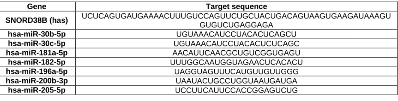
objetivo de identificar microRNAs com expressão diferente entre doentes luminais com e sem recidiva da sua doença após o tratamento com terapias adjuvantes endócrinas. Posteriormente, sete microRNAs - miR-30b-5p, miR-30c-5p, miR-181a-5p, miR-182-5p, miR-196a-5p, miR-200b-3p e miR-205-5p - foram escolhidos para validação num maior número de tecidos por RT-qPCR. Seguidamente, o potencial de diagnóstico e de prognóstico destes microRNAs foi avaliado através da construção de curvas de ROC e de modelos de Regressão de Cox, respetivamente. Realizou-se igualmente análise do status de metilação por PCR quantitativo específico em DNA modificado por bissulfito de sódio. Por último, analisámos igualmente a expressão de microRNAs em tecidos tumorais primários e metástases correspondentes.
Resultados e Discussão: Após o ensaio de expressão global os microRNAs miR-30b-5p,
miR-181a-5p, miR-182-5p, miR-196a-5p, miR-200b-3p e miR-205-5p foram selecionados para validação, tendo o miR-30c-5p sido utilizado como controlo positivo. A combinação dos microRNAs miR-182-5p e miR-200b-3p num painel foi capaz de identificar cancro da mama em amostras de tecido com uma acuidade de 95,55%. Adicionalmente, os microRNAs miR-30c-5p, miR-30b-5p, miR-182-5p, bem como o miR-200b-5p foram identificados como preditores independentes de resposta a terapias endócrinas. Além disso, os microRNAs miR-182-5p e miR-200b-3p são marcadores de prognóstico em doentes com tumores luminais após tratamento adjuvante com terapias endócrinas. Verificou-se ainda que os microRNAs miR-30b-5p e miR-200b-3p estão significativamente
viii mais expressos nas metástases relativamente aos tumores primários correspondentes. Além disso, os nossos resultados sugerem que a menor expressão do miR-200b-3p nos tumores com resistência à terapia endócrina é dependente de outros mecanismos epigenéticos, e não da metilação das suas regiões promotoras.
Conclusões e perspectivas futuras: Os resultados sugerem que um painel específico
de microRNAs poderá ser útil na decisão terapêutica em doentes com tumores da mama luminais. No entanto, estudos adicionais, idealmente estudos multicêntricos, são essenciais para a sua validação. Como principal perspectiva futura, pretendemos avaliar a expressão destes microRNAs em biópsias líquidas, de maneira a avaliar o seu potencial como biomarcadores não-invasivos.
ix
ABSTRACT
Introduction: Breast cancer is the most frequent malignancy and the leading cause of
cancer death among women worldwide. Approximately 70% of BrCa are of the luminal type, expressing the estrogen receptor. One of the most common and effective adjuvant therapies for this BrCa subtype is endocrine therapy. However, its effectiveness is limited, with relapse occurring in up to 40% of patients. Because microRNAs have been associated with several mechanisms underlying endocrine resistance and sensitivity, they may serve as predictive and/or prognostic biomarkers in this setting.
Aims: The major goal of this master dissertation was to investigate whether miRNAs
deregulated in endocrine-resistant breast cancer may be clinically relevant as prognostic and predictive biomarkers in luminal
breast cancer patients treated with adjuvant endocrine therapy.
Material and Methods: We started by performing a global expression assay with the aim
of identifying microRNAs differentially expressed between luminal patients with or without breast cancer recurrence after endocrine therapy. Then, seven microRNAs - miR-30b-5p, miR-30c-5p, miR-181a-5p, miR-182-5p, miR-196a-5p, miR-200b-3p e miR-205-5p - were chosen for validation using quantitative reverse transcription polymerase chain reaction in a larger set of tissue samples. ROC curves and cox-regression models were constructed to evaluate miRNAs diagnostic and prognostic performance, respectively. DNA methylation analysis was also performed by sodium bisulfite modification followed by quantitative methylation-specific polymerase chain reaction. Furthermore, microRNAs expression levels were also analyzed in metastatic tissues and the paired primary tumor tissue.
Results and Discussion: From the initial global expression assay, 30b-5p,
miR-181a-5p, miR-182-5p, miR-196a-5p, miR-200b-3p and miR-205-5p were selected for further validation, and miR-30c-5p was chosen as a positive control. The combination of miR-182-5p and miR-200b-3p accurately detects BrCa in tissue samples with an overall accuracy of 95.55%. MR-30c-5p, miR-30b-5p, miR-182-5p and miR-200b-5p were found to be independent predictors of clinical benefit from endocrine therapy. Moreover, miR-182-5p and miR-200b-3p displayed independent prognostic value for disease recurrence in luminal BrCa patients after endocrine therapy. MiR-200b-3p and miR-30b-5p were significantly higher in metastatic tissues when compared to the paired primary tumor tissues. Furthermore, our results suggest that miR-200b-3p’s downregulation in
endocrine-x resistant tumors might be dependent on other epigenetic mechanisms rather than DNA methylation.
Conclusions and future perspectives: We concluded that selected miRNAs may
constitute clinically useful ancillary tools for management of luminal BrCa patients. Additional validation, ideally in a multicentric setting, is required to confirm our findings. As a future perspective, we intend to assess these miRNAs expression in liquid biopsies in order to evaluate their potential as non-invasive biomarkers.
xi
TABLE OF CONTENTS
FIGURES INDEX ... xiv
TABLES INDEX ... xvi
LIST OF ABBREVIATIONS ... xvii
INTRODUCTION ... 1
Breast Cancer ... 2
Breast Cancer overview ... 2
Epidemiology and Risk Factors ... 2
Diagnosis and screening ... 4
Histological subtypes ... 5
Prognostic and predictive biomarkers ... 5
Molecular Subtypes ... 7
Therapeutic approaches ... 10
Endocrine Resistance ... 11
Epigenetics ... 12
DNA methylation ... 13
Covalent histone modifications and histone variants ... 13
Non-coding RNAs ... 14
MicroRNAs ... 15
Biogenesis and mode of action ... 15
MicroRNAs deregulation in cancer ... 17
MicroRNAs and their use in the clinic ... 18
MicroRNAs and Breast Cancer ... 19
MiRNAs and endocrine resistance ... 19
AIMS ... 22
MATERIAL AND METHODS ... 24
Patients and samples collection ... 25
Breast cancer subtyping ... 25
MicroRNA expression analysis ... 26
RNA extraction from fresh frozen tissues ... 26
RNA extraction from formalin-fixed paraffin-embedded (FFPE) tissue samples ... 26
MicroRNAs cDNA synthesis ... 27
xii
Individual assays ... 29
Methylation Analysis ... 30
DNA extraction from fresh frozen tissues ... 30
Sodium Bisulfite Modification ... 30
Quantitative Methylation-Specific PCR ... 31
Statistical Analysis ... 32
RESULTS ... 34
Characteristics of study populations ... 35
Global expression assay analysis ... 38
Gene-specific assays ... 38
Assessment of miRNA expression in luminal tumor tissues and normal breast tissues 38 Assessment of miRNA expression in non-luminal tumor tissues and evaluation of miRNAs diagnostic performance ... 39
Validation of selected miRNAs in endocrine-resistant and –sensitive luminal tumor tissues ... 42
Association between miRNAs expression and clinicopathological features ... 43
Survival Analysis ... 44
MicroRNAs’ expression analysis in paired metastasis ... 49
Methylation Analysis ... 50
DISCUSSION ... 52
CONCLUSIONS AND FUTURE PERSPECTIVES ... 58
REFERENCES ... 61 SUPPLEMENTARY MATERIAL ... I
Appendix I. Magnitude of risk of BrCa risk and protective factors with different scientific evidence. Adapted from (219). ... II Appendix II. Nottingham combined histologic grade. Adapted from (36). ... IV Appendix III. Tumor-node-metastases (TNM) staging system for carcinoma of the breast. Adapted from (220). ... IV Appendix IV. Stage grouping system for carcinoma of the breast. Adapted from (220). VII Appendix V. Amorim, Maria, et al. "Decoding the usefulness of non-coding RNAs as breast cancer markers." Journal of translational medicine 14.1 (2016): 265. ... VIII Decoding the usefulness of non-coding RNAs as Breast Cancer markers ... VIII
Appendix VI. MiRNAs with fold variation values higher than 1 in the global expression assay... IX
xiii Appendix VII. Univariable cox regression models assessing the association between clinicopathological features and clinical outcome. ... X
xiv
FIGURES INDEX
Figure 1. Estimated Age-Standardized Incidence and Mortality Rates (per 100 000) in Portugal in 2012. Adapted from (3). ... 2 Figure 2. MiRNA biogenesis pathway: canonical and alternative pathway, from nucleus to cytoplasm. Abbreviations: miRNA – microRNA; POL – polymerase; DGCR8 - Di-George syndrome critical region gene 8; Ldbr - lariat debranching enzyme; GTP - Guanosine-5'-triphosphate; TRBP - Trans-activation response RNA binding protein; HSP - Heat shock proteins; UTR - untranslated region; RISC - RNA-induced silencing complex; AGO – Argonaute; Ran - RAs-related Nuclear protein. Amorim, Maria unpublished. ... 16 Figure 3. Examples of oncomiRs and tumor suppressor miRs associated with the
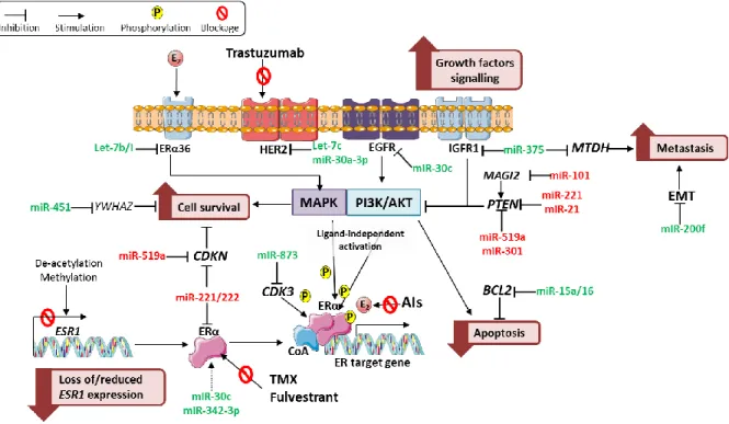
carcinogenesis process, and some of the target genes through which they exert their regulatory function. Abbreviations: MYC – MYC proto-oncogene, bHLH transcription factor; E2F – Transcription factor E2F/dimerisation partner family protein; PTEN – Phosphatase and tensin homolog; CDKN – Cyclin-dependent kinase inhibitor PRKAA1 – Protein kinase AMP-activated catalytic subunit alpha 1; TP53INP1 - Tumor Protein P53 Inducible Nuclear Protein 1; INPP5D – Inositol polyphosphate-5-phosphatase D; TPM1 – Tropomyosin 1; PDCD4 – Programmed cell death 4; CDKN1B – Cyclin dependent kinase inhibitor 1B; PI3KR1 – Phosphoinositide-3-Kinase Regulatory Subunit 1; LATS2 – Large tumor suppressor kinase 2; CDK – Cyclin-dependent kinase; CCNA1 - Cyclin A1; ABL1 – ABL proto-oncogene 1, non-receptor tyrosine kinase; SOX2 – SRY-box 2; KLF4 – Kruppel like factor 4; TCL1A – T-cell leukemia/lymphoma 1A; DNMT – DNA methyltransferase;
IGFBP2 – insulin like growth factor binding protein 2; MERKT – MER proto-oncogene, tyrosine kinase; RHOA – ras homolog family member A; BCL2 – BCL2, apoptosis regulator; CCN – cyclin; E2F3 – E2F transcription factor 3; TNFRSF6B – TNF Receptor Superfamily Member 6b; KRAS – KRAS proto-oncogene, GTPase; HMGA2 – high mobility group AT-hook 2; Amorim, Maria unpublished. ... 18 Figure 4. MiRNAs and their established targets involved in endocrine resistance. The miRNAs and their targets involved in several mechanisms associated with endocrine resistance, along with their functional implication (in pink boxes), including loss of/reduced
ESR1 expression, alternative growth factors signaling, including PI3K/Akt and MAPK
signaling pathways, dysregulation of cell survival and apoptosis pathways, and increased metastasis. MiRNAs that confer sensitivity and resistance to endocrine therapies are depicted in gree and red, respectively. Abbreviations: ER – estrogen receptor; HER2 - Human Epidermal growth factor Receptor 2; EGFR - epidermal growth factor receptor; IGFR1 - insulin-like growth factor 1 receptor; YWHAZ - Tyrosine
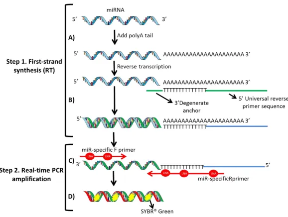
3-Monooxygenase/Tryptophan 5-Monooxygenase Activation Protein Zeta; MTDH – metadherin; MAGI2 - membrane-associated guanylate kinase inverted 2; PTEN - Phosphatase and tensin homolog; EMT - epithelial–mesenchymal transition; CDKN - Cyclin-dependent kinase inhibitor; CDK3 - cyclin dependent kinase 3; BCL2 - BCL2, apoptosis regulator; PI3K/AKT - phosphoinositide 3-kinase/Protein kinase B; ESR1 - estrogen receptor 1; TMX – Tamoxifen; AIs – aromatase inhibitors; E2 – Estradiol; miR – microRNA; miR-200f – miR-200 family. Amorim, Maria unpublished. ... 19 Figure 5. A. A poly-A tail is added to the mature microRNA template. B. cDNA is
xv primer sequence. C. The cDNA template is then amplified using microRNA-specific and LNATM-enhanced forward and reverse primers. D. SYBR® Green is used for detection. Abbreviations: miRNA – microRNA; miR – microRNA; LNA - Locked Nucleic Acid. Amorim, Maria unpublished. ... 28 Figure 6. Box-plots (left panel) and the respective Receiver Operating Characteristic (ROC) Curves (right panel) for 182-5p (A), (B), 196-5p (C), 200b-3p (D) and miR-205-5p (E). A *** denotes p-value <0.001 and a **** denotes p-value < 0.0001 by non-parametric Mann-Whitney U test. Y-axis denotes 2-ΔCT values multiplied by 1000. ... 41 Figure 7. Receiver Operating Characteristic (ROC) Curve for miR-182-5p and miR-200b-3p combined. ... 42 Figure 8. Box-plots of miR-30b-5p (A), miR-30c-5p (B) and miR-200b-3p (C) expression levels in tumor tissues from endocrine-sensitive and –resistant patients. A * denotes p-value <0.05 and a ** denotes p-p-value <0.01 by non-parametric Mann-Whitney U test. Y-axis denotes 2-ΔCT values multiplied by 1000. ... 43 Figure 9. Box-plots of miR-30c-5p (A) expression levels according to PR-status (left) and HER2-status (right), miR-30b-5p (B) expression according to HER2-status, and miR-196a-5p (C) and miR-205-miR-196a-5p (D) expression according to grade. A * denotes p-value <0.05 by non-parametric Mann-Whitney U test. Y-axis denotes 2-ΔCT values multiplied by 1000. .... 44 Figure 10. Endocrine Resistance-free survival curves of miR-30b-5p (A), miR-30c-5p (B), miR-182-5p (C) and miR-200b (D). Abbreviations: P25 – percentile 25. ... 45 Figure 11. Disease-free survival curves (Kaplan–Meier with log rank test) of miR-30b-5p (A), miR-30c-5p (B), miR-182-5p (C) and miR-200b (D). Abbreviations: P25 – percentile 25. ... 48 Figure 12. MiR-30b-5p (A) and miR-200b-3p (B) relative expression levels in primary tumors and the corresponding metastasis. A ** denotes p-value <0.01 by non-parametric Wilcoxon paired sample test. Y-axis denotes 2-ΔCT values multiplied by 1000. ... 49 Figure 13. Comparison of 30b-5p (A), 30c-5p (B), 182-5p (C) and miR-200b-3p (D) in primary breast tumors versus corresponding metastasis. X-axis represents each patient. Y-axis represents -ΔΔCt values; positive values correspond to higher
expression in the distant metastasis versus corresponding primary breast tumor. ... 50 Figure 14. Relative miR-200b-3p promoter 1 (A) and promoter 2 (B) methylation levels in normal breast tissues and tumors. A ** denotes p-value <0.01 and a **** denotes a p<0.0001 by non-parametric Mann-Whitney U test. Y-axis denotes relative methylation values multiplied by 1000. ... 51 Figure 15. Box-plots of promotor 1 (P1) relative methylation levels in HER2-negative and HER2-positive tumors. A * denotes p-value <0.05. Y-axis denotes relative methylation values multiplied by 1000. ... 51
xvi
TABLES INDEX
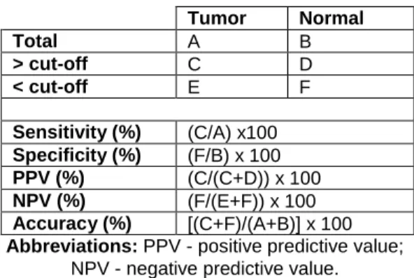
Table 1. Breast Cancer molecular subtypes characterization (1, 30, 32, 48, 49, 53-57). .... 9 Table 2. Non-coding RNAs involved in response (sensitivity/resistance) to endocrine therapies along with their putative targets/mechanism. ... 21 Table 3. Specific target sequence of miRNAs tested. ... 30 Table 4. Primer sequences and qMSP conditions for each gene studied. ... 32 Table 5. Formulas used for the calculation of the biomarkers performance parameters. .. 33 Table 6. Clinical and pathological data of luminal tumors included in the discovery cohort. ... 35 Table 7. Clinical and pathological data of tumors and normal breast samples used in this study. ... 35 Table 8. Clinical and pathological data of luminal tumors and normal breast samples included in the validation cohort. ... 36 Table 9. Clinical and pathological data of primary tumor tissues and paired metastasis tissues used in this study. ... 37 Table 10. MicroRNAs and the respective fold variation values between luminal tumors and normal breast tissues. ... 39 Table 11. MicroRNAs and the respective fold variation values between luminal tumors and normal breast tissues. ... 39 Table 12. Performance of miRNAs expression as biomarkers for breast cancer detection in tumor tissues. ... 40 Table 13. Performance of miR-182-5p and miR-200b-3p expression levels combined as biomarkers for detection of breast cancer in tumor tissues... 42 Table 14. MicroRNAs and the respective fold variation values between in endocrine-resistant and endocrine-sensitive tumors. ... 43 Table 15. Univariable and multivariable cox regression models assessing the association between microRNAs expression levels and clinical outcome. ... 46 Table 16. Cox regression models stratified according to the clinicopathological features with statistical significance in the multivariable analysis. ... 47
xvii
LIST OF ABBREVIATIONS
ABL1 – ABL proto-oncogene 1, non-receptor tyrosine kinase ACTβ - Actin β
ADH1B - Alcohol Dehydrogenase 1B (Class I), Beta Polypeptide AGO – Argonaute
AI – aromatase inhibitor
AJCC – American Joint Committee on Cancer Akt - Protein kinase B
ALCAM - Activated Leukocyte Cell Adhesion Molecule ANA – Anastrozole
AntiE – Anti estrogen Ap1 - Activation protein 1
ARPP19 - cAMP-regulated phosphoprotein 19 ASCO – American Society of Clinical Oncology AUC – Area Under the Curve
BBC3iso-2 - BCL2 Binding Component 3 isoform 2 BCAR4 - Breast cancer anti-estrogen resistance 4 BCL2 – BCL2, apoptosis regulator
BCL2L11 - BCL2 like 11
BMP7 - Bone morphogenetic protein 7 bp – Base pairs
BrCa – Breast cancer
BRCA1 – BRCA1, DNA repair associated BRCA2 – BRCA2, DNA repair associated CA 15-3 - Carbohydrate antigen 15-3 CCN – Cyclin
CDH – Cadherin
CDK – Cyclin-dependent kinase
CDKN - Cyclin-dependent kinase inhibitor CEA - Carcinoembryonic antigen
Cht – Chemotherapy CI – Confidence interval CK – Cytokeratins
xviii CpG - Cytosine-phosphate-Guanine
CTNNB1 - Catenin Beta 1
CYP19A1 - Cytochrome P450 family 19 subfamily A member 1 DCIS - Ductal carcinoma in situ
DFS - Disease-free survival
DGCR8 - Di-George syndrome critical region gene 8 DMFS – Distant metastasis-free survival
DNMT - DNA methyltransferase
DSCAM-AS1 - DSCAM Antisense RNA 1 E2 – Estradiol
E2F3 - E2F transcription factor 3 EDTA – Ethylenediamine teracetic acid EFNA3 - Ephrin A3
EGFR - Epidermal growth factor receptor EMT - Epithelial–mesenchymal transition ER – Rstrogen receptor
ERBB - Erb-B2 Receptor Tyrosine Kinase ERE - Estrogen response element
ERFS - Endocrine resistance-free survival ERK - Extracellular signal regulated kinases ESMO – European Society for Medical Oncology ESR1 - Estrogen Receptor 1
ESRRG - Estrogen related receptor gamma ET – Endocrine therapy
EZH2 - Enhancer of zeste homolog 2 FFPE - Formalin-fixed paraffin-embedded FGFR1 - Fibroblast Growth Factor Receptor 1 FGFRL1 - Fibroblast growth factor receptor-like 1 FOX - Forkhead Box
FULV – Fulvestrant G – Grade
GATA3 - GATA Binding Protein 3
GEMIN4 - Gem (nuclear organelle)-associated protein 4 GnRH - Gonadotropin-releasing hormone
xix GRB7 - Growth Factor Receptor Bound Protein 7
GSTP1 - Glutathione S-Transferase Pi 1 GTP - Guanosine-5'-triphosphate
H&E - Hematoxylin-eosin HAT - Histone acetyltransferase HDAC - Histone deacetylase HDMs - Histone demethylases
HER2 - Human Epidermal growth factor Receptor 2 HMTs - Histone methyltransferases
HNRNPL - Heterogeneous Nuclear Ribonucleoprotein L HPF – High-power field
HR – Hazard ratio
HRT – Hormone-replacement therapy HSP - Heat shock proteins
IDC - Invasive ductal carcinoma IGF-1 - Insulin-like growth factor-1
IGFBP2 – Insulin like growth factor binding protein IGFR1 - Insulin-like growth factor 1 receptor IHC - Immunohistochemistry
ILC - Invasive lobular carcinoma
INPP5D – Inositol polyphosphate-5-phosphatase D IPC - Inter-plate calibration
IRR - Incidence rate ratio
KIT - KIT Proto-Oncogene Receptor Tyrosine Kinase KLF4 – Kruppel like factor 4
KRAS – KRAS proto-oncogene, GTPase HMGA2 – high mobility group AT-hook 2 KRT – Keratin
LAM – Laminin
LATS2 – Large tumor suppressor kinase 2 LCIS - Lobular carcinomas in situ
Ldbr - lariat debranching enzyme LNA - Locked Nucleic Acid lncRNA – Long non-coding RNA Lum – Luminal
xx MAGI2 - Membrane-associated guanylate kinase inverted 2
MAPK - Mitogen-activated protein kinase MBC - Methyl-CpG binding domain
MERKT – MER proto-oncogene, tyrosine kinase MET - Mesenchymal-to-epithelial transition
MET - MET Proto-Oncogene, Receptor Tyrosine Kinase MiR – MicroRNA
MiR-200f – MiR-200 family MiR-30f – MiR-30 family MiRNA – MicroRNA
MKI67 - Marker Of Proliferation Ki-67 MRI - Magnetic resonance imaging mRNA – Messenger RNA
MSP – Methylation Specific Methylation MTDH - Metadherin
MYBL2 - MYB Proto-Oncogene Like 2 MYBL2 - MYB Proto-Oncogene Like 2
MYC – MYC proto-oncogene, bHLH transcription factor n.a. – not applicable
NBr – normal breast tissues ncRNAs - Non-coding RNAs NF-κB - Nuclear factor kappa B NPV - Negative predictive value
NSAID - Nonsteroidal anti-inflammatory drug NST - No special type
nt - Nucleotide OR - Odds ratio
ORF - Open reading frames OS – Overall survival P1 – Promoter 1 P2 – Promoter 2 P25 – Percentile 25
PDCD4 – Programmed cell death 4 PGR - Progesterone Receptor
xxi PI3K - phosphoinositide3-kinase
PI3KR1 – Phosphoinositide-3-Kinase Regulatory Subunit 1 POL – polymerase
PPV - Positive predictive value PR - Progesterone receptor
PRKAA1 – Protein kinase AMP-activated catalytic subunit alpha 1 PTEN - Phosphatase and tensin homolog
qMSP - Quantitative real-time methylation specific PCR R – Resistance
RAD52 - RAD52 homolog, DNA repair protein Ran - RAs-related Nuclear protein
RASSF1A - Ras Association Domain Family Member 1 RB1 – Retinoblastoma 1
Rec – Recurrent
RHOA – Ras homolog family member A RISC - RNA-induced silencing complex RISC - RNA-induced silencing complex ROC - Receiver Operating Characteristic RR -relative risk
rRNAS – ribosomal RNAs RS - Recurrence score RT – Radiotherapy S – Sensitivity
SDS – Sodium Dodecyl Sulfate
SERD - Selective SER down-regulators SLNB - Sentinel lymph node biopsy snoRNAs - Small nucleolar RNAs
SOCS - Suppressor of cytokine signaling SOX2 – SRY-box 2
SRE - Serum response element SSC - Special subtypes carcinomas TCL1A – T-cell leukemia/lymphoma 1A
TGFBR1 - Transforming growth factor, beta-receptor 1 TMX – Tamoxifen
xxii TNBC – Triple-negative breast cancer
TNFRSF6B – TNF Receptor Superfamily Member 6b TNM - Tumor, Node and Metastases
TP53 - Tumor protein p53
TP53INP1 - Tumor Protein P53 Inducible Nuclear Protein 1 TP63 - Tumor Protein P63
TPM1 – Tropomyosin 1
TRBP - Trans-activation response RNA binding protein TRBP - Trans-activation response RNA binding protein tRNA - Transfer RNAs
UNKN – Unknown
UTR - untranslated region VIM – Vimentin
WHO - World Health Organization XBP1 - X-Box Binding Protein 1 XPO5 - Exportin-5
yr – Years
YWHAZ - Tyrosine 3-Monooxygenase/Tryptophan 5-Monooxygenase Activation Protein Zeta
YWHAZ - Tyrosine 3-Monooxygenase/Tryptophan 5-Monooxygenase Activation Protein Zeta
ZEB - Zinc finger E-box-binding homeobox ZNF217 - Zinc finger protein 217
2
Breast Cancer
Breast Cancer overview
Epidemiology and Risk Factors
Breast cancer (BrCa) is the second most common cancer worldwide and the most frequent cancer among women (1). In 2012, the estimated age-adjusted annual incidence of BrCa in 40 European countries was 92.8/100 000 and the mortality 23.1/100 000 (2). In Portugal, BrCa was the leading cancer in 2012 and the first cause of cancer death in women (Figure 1) (3). BrCa in males is rare, contributing to ~1% of cases (4).
Figure 1. Estimated Age-Standardized Incidence and Mortality Rates
(per 100 000) in Portugal in 2012. Adapted from (3).
BrCa incidence increased after the application of mammography screening and it is still increasing with the ageing of the population. On the contrary, due to earlier diagnosis and increased treatment with the implementation of adjuvant chemo-, radio- and endocrine-therapies (ET), the mortality rate has been decreasing in most Western countries, while
3 BrCa prevalence is increasing (3). Indeed, despite the high global incidence, BrCa is the fifth cause of death from cancer for both genders. However, it constitutes the leading cause of cancer-related deaths in European women and in women worldwide (5, 6). Ten-year survival of BrCa exceeds 70% in most European regions, being 89% for local and 62% for regional disease (7). Approximately 5% of patients present with distant metastasis at time of diagnosis and 10-15% of patients develop distant metastasis within the first 3 years (8). Importantly, metastatic BrCa is almost always incurable (9).
Multiple factors have been associated with an increased risk of developing BrCa with distinct scientific evidence (Appendix I); however, it has been estimated that approximately 50% of women who develop BrCa have no identifiable risk factors excepting increased age and female gender (10). Indeed, BrCa incidence rises sharply with age, with the highest rate of BrCa being observed among women aged 75 to 79 (11). In addition, BrCa incidence also differs by race and ethnicity (12, 13). For instance, in women less than 40 years, BrCa incidence is higher in African-american women than in Caucasian women; however, the contrary occurs among those aged 40 years or older (12).
Moreover, some benign breast lesions, e.g., proliferative disease without and with atypia, have been associated with a slight increase in the subsequent risk of developing invasive BrCa (14). Furthermore, women with a family history of BrCa, especially if the affected family member was diagnosed at a younger age, have an increased risk of developing BrCa (15). Mutations in the BrCa susceptibility genes BRCA1, DNA repair associated (BRCA1) and BRCA2, DNA repair associated (BRCA2) were also associated with a significant increase in the lifetime risk of BrCa, that ranges from 26 to 85% (10), however, these account for 5 to 10% of all BrCa, and are most strongly related to BrCa occurring in younger premenopausal women (16). Additional genes such as Tumor Protein p53 (TP53), associated with the Li-Fraumeni syndrome (17) and Phosphatase and Tensin
homolog (PTEN), associated with the Cowden syndrome (18), play a minor role in familial
BrCa syndromes.
Many of the established BrCa risk factors can be attributed to some means of elevated estrogen exposure, as many studies have consistently demonstrated that increased levels of endogenous estrogen are associated with increased BrCa risk in postmenopausal women (19), which might be explained by estrogen’s capacity to stimulate proliferation of both normal and malignant breast cells (20). Reproductive factors linked to an increase in
4 BrCa risk include early age at menarche and late age at menopause. On the other hand, parity and premenopausal oophorectomy have a protective effect on BrCa risk (21). Breastfeeding also appears to contribute to a reduced risk of BrCa, although parity may be a confounding factor (22). Besides reproductive factors, in postmenopausal women, obesity and hormone replacement therapy (HRT) are also associated with increased BrCa risk (23).
There is also a well-established relationship between exposure to ionizing radiation and the risk of developing BrCa, with the risk being inversely associated with age at radiation exposure (24).
Finally, there is also a substantial interest in whether dietary or lifestyle factors modify BrCa risk. For instance, vegetable consumption and physical activity seem to have a moderate protective effect (25), while high-fat diets (26) and alchohol consumption (27) seem to be associated with higher rates of BrCa.
Diagnosis and screening
The diagnosis of BrCa is initially based on clinical examination, which includes bimanual palpation of the breast and locoregional lymph nodes, in combination with imaging (28). The current in vivo diagnostic tools for the detection of early-stage BrCa are mammography, for which accuracy is greatly affected by age and consequently denser breasts (29) and in specific cases magnetic resonance imaging (MRI) (30). A number of circulating tumor markers [e.g., carcinoembryonic antigen (CEA) and carbohydrate antigen 15-3 (CA 15-3)] are sometimes used in the management of BrCa, but due to their low sensitivity they are not for screening intent but only for disease monitoring (31). Indeed, the presence or absence of carcinoma can only be correctly determined by tissue sampling and pathological examination of the primary tumor and cytology/histology of the axillary nodes, if involvement is suspected (32). Thus, biopsy remains the standard technique for diagnosing both palpable and non-palpable breast abnormalities (32). In order to detect BrCa at a pre-clinical stage, several countries have established population-based mammography screening programs (33). Mammography screening, every 2 years, has shown the greatest mortality reduction benefit in women between 50-69 years (32). In women with familial BrCa, annual MRI concomitantly or alternating every 6
5 months with mammography, starting 10 years younger than the youngest case in the family, is recommended (30).
Histological subtypes
BrCa is a highly heterogeneous disease with distinct biological features and clinical outcomes. Currently, the World Health Organization (WHO) identifies more than 20 histological types, using a classification scheme based on the growth pattern and cytological features of the tumor cells, and independent of the site of origin in the breast (34).
The majority of BrCa has origin at epithelial cells and can be subdivided into in situ and invasive carcinomas. In situ carcinomas might be lobular carcinoma in situ (LCIS) or ductal carcinoma in situ (DCIS), and are defined as pre-invasive lesions in which neoplastic epithelial cells proliferate confined to the ductal/lobular tree of the breast without evidence of invasion through the basement membrane (35). DCIS is more frequent than LCIS and, with the implementation of mammographic screening programs, represents about 20-25% of newly diagnoses BrCa (34, 35). Invasive carcinomas are the most common lesions, representing 70-80% of all BrCa malignant neoplasms (35). Invasive carcinomas can generally be grouped in two categories: invasive carcinoma of no special type (NST), also known as invasive ductal carcinoma (IDC), and special subtypes carcinomas (SSC), with IDC representing up to 75% of all invasive carcinomas (34). The most common lesion of the SSC group is the invasive lobular carcinoma (ILC), representing 5-15% of all BrCa. Tumors that have both SSC pattern (10-49%) and NST pattern are categorized as mixed (34).
Prognostic and predictive biomarkers
A prognostic factor is defined as a measurement taken at the time of diagnosis or surgery that is associated with the innate aggressiveness of untreated BrCa and thus outcome, while a predictive factor is a measurement that predicts response or lack of response to a specific treatment (10). In clinical practice, many biomarkers have both prognostic and predictive significance.
Histologic grade, determined by using the Nottingham combined histologic grade proposed by Elston and Ellis (Appendix II), has been shown to have prognostic significance and is a key component for clinical decision-making (34, 36). Moreover, the Tumor, Node and
6 Metastases (TNM) staging system published by the American Joint Committee on Cancer (AJCC)/Union for International Cancer Control (UICC) is based on established clinical and pathological prognostic factors, namely tumor size (T), the extent of axillary lymph node involvement (N) and the spread of distant metastases (M) (34) (Appendix III). This staging system allows the establishment of five stages in order to evaluate the disease extension and the patient’s prognosis (Appendix IV).
However, grade and stage display limited value as sole prognostic factors and other prognostic and therapy predictive biomarkers have been introduced in the daily practice (32). Indeed, estrogen receptor (ER), progesterone receptor (PR) and human
epidermal growth factor 2 receptor (HER2) status assessed by immunohistochemistry
are also evaluated in all breast tumors.
ER and PR are two nuclear transcription factors activated by the hormones estrogen and
progesterone, respectively, and are the most important and useful predictive factors currently available. Indeed, ER expression is a strong ET predictive marker of response (34, 37). Besides, PR-negative patients have a higher relative risk of disease recurrence after ET in comparison to patients with PR-positive tumors. Moreover, ER-negative tumors have a poorer prognosis in the first years after diagnosis, but after 5 to 10 years positive tumors have the poorer outcome (38). Importantly, BrCa may relapse in ER-positive patients more than 20 years after the diagnosis (39).
HER2 gene is an oncogene localized in the chromosome 17 and is amplified in
approximately 15% of BrCa tumors yielding overexpression of its coding protein, a growth factor receptor present in breast epithelial cell surface (40). HER2 status has both prognostic and predictive significance. Although HER2-positive BrCa patients have a worse prognosis, HER2 positivity is predictive of a favorable response to HER2-targeted therapy (e.g. trastuzumab) (34, 37). In addition, HER2 positivity is also a response predictor of anthracycline- and taxane-based therapy (41) and of unresponsiveness to ETs (42).
Proliferation markers such as the Ki-67 may supply additional useful prognostic information (43). Furthermore, patient age has been consistently shown to be a prognostic factor, as very young BrCa patients have a poorer prognosis than older patients (10).
7 Nonetheless, clinical decisions based upon one or small number of genes or their coding proteins in the tumor tissue have failed to predict patients’ outcome, which prompt to the development of other prognostic assays, based in the examination of multiple expressed genes, e.g., Oncotype DX [21‑gene derived recurrence score (RS)], MammaPrint (Amsterdam 70‑gene derived RS) and PAM50 (50‑gene derived RS) (44), for both classification and prognostication of individual tumors. However, widespread use of
gene-expression profiling in clinical practice remains limited, primarily due to the high costs
and technical difficulty encountered when carrying out high-throughput gene-expression profiling, in addition to the invasive diagnostic procedures, since tissue biopsies are required.
Molecular Subtypes
Based on gene expression profiling, BrCa is often classified into four well-established intrinsic subtypes (Table 1), which are associated with distinct biological features and clinical outcomes (45, 46). These intrinsic subtypes can be defined by gene expression profiling using multiparameter molecular tests such as the PAM-50 (47, 48). Luminal tumors typically express luminal cytokeratins (CK) 8 and 18 (44). Luminal tumors can be further subdivided into Luminal A and Luminal B. The major molecular distinctions between luminal A and B tumors are based in the ER‑related genes’ higher expression in luminal A tumors, whereas luminal B tumors exhibit a higher expression of proliferation‑related genes (49). ER-negative tumors encompass two subtypes: the
HER2-enriched subtype, characterized by high expression of several genes in the HER2
amplicon at 17q22.24, including HER2 (48) and the basal-like subtype, characterized by CK 5/6 and CK17 and basal epithelial genes’ expression (48).
However, due to financial constraints, surrogate approaches have been developed for routine clinical practice using more widely available immunohistochemistry (IHC) assays for ER, PR and Ki-67 index, together with IHC and/or in situ hybridization for HER overexpression/amplification (32). Luminal A tumors do not overexpress HER2 and have a low Ki-67 index, while luminal B tumors can be HER2 negative or positive. Commonly, Luminal B negative for HER2 expression has either high Ki-67 value or a negative or low PR expression. Eighty % of Basal-like tumors overlap with triple-negative tumors, negative for both hormone-receptors and HER2 expression (50) (Table 1).
8 These subtypes contribute to insights into cancer initiation and progression and might be of value in assessing prognosis and prediction response to therapy, guiding clinical management (32, 51, 52).
9
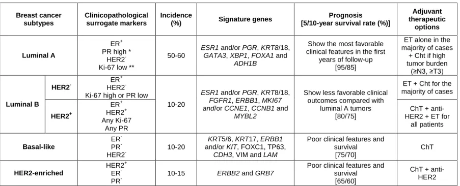
Table 1. Breast Cancer molecular subtypes characterization (1, 30, 32, 48, 49, 53-57). Breast cancer subtypes Clinicopathological surrogate markers Incidence (%) Signature genes Prognosis
[5/10-year survival rate (%)]
Adjuvant therapeutic options Luminal A ER+ PR high * HER2- Ki-67 low ** 50-60 ESR1 and/or PGR, KRT8/18, GATA3, XBP1, FOXA1 and
ADH1B
Show the most favorable clinical features in the first
years of follow-up [95/85] ET alone in the majority of cases + Cht if high tumor burden (≥N3, ≥T3) Luminal B HER2 -ER+ HER2 -Ki-67 high or PR low
10-20
ESR1 and/or PGR, KRT8/18, FGFR1, ERBB1, MKI67 and/or CCNE1, CCNB1 and
MYBL2
Show less favorable clinical outcomes compared with
luminal A tumors [80/75] ET + Cht for the majority of cases HER2+ ER+ HER2+ Any Ki-67 Any PR ChT + anti-HER2 + ET for all patients Basal-like ER -PR -HER2 -10-20 KRT5/6, KRT17, ERBB1
and/or KIT, FOXC1, TP63,
CDH3, VIM and LAM
Poor clinical features and survival [75/70] ChT HER2-enriched HER2+ ER -PR -10-15 ERBB2 and GRB7
Poor clinical features and survival
[65/60]
ChT + anti-HER2 * Suggested cut-off value is 20% (32) ** Ki-67 scores should be interpreted in the light of local laboratory median values (32)
Abbreviations: ER – estrogen receptor; PR – progesterone receptor; HER2 - Human Epidermal growth factor Receptor 2; ESR1 - Estrogen
Receptor 1; PGR - Progesterone Receptor; KRT – keratin; ; GATA3 - GATA Binding Protein 3; XBP1 - X-Box Binding Protein 1; FOX - Forkhead Box;
ADH1B - Alcohol Dehydrogenase 1B (Class I), Beta Polypeptide; FGFR1 - Fibroblast Growth Factor Receptor 1; ERBB - Erb-B2 Receptor Tyrosine
Kinase; MKI67 - Marker Of Proliferation Ki-67; CCN – Cyclin; MYBL2 - MYB Proto-Oncogene Like 2; MYBL2 - MYB Proto-Oncogene Like 2; KIT - KIT Proto-Oncogene Receptor Tyrosine Kinase; TP63 - Tumor Protein P63; CDH – Cadherin; VIM – vimentin; LAM – laminin; GRB7 - Growth Factor
10
Therapeutic approaches
Currently, several treatment strategies available for BrCa patients are based on the tumor burden/localization and biology, as well as age, general health status and patient’s preferences (58). Indeed, BrCa is the pioneer of personalized medicine in oncology.
Neoadjuvant treatment might be performed in multifocal disease, or in order to downsize
locally advanced and large unifocal unresectable primary tumors that would require mastectomy (58). All modalities that will be described for adjuvant systemic treatment might also be used as neoadjuvant therapy.
Regarding local treatment, breast-conserving surgery is amenable in the vast majority of newly diagnosed cancers, while mastectomy is carried out for larger tumor sizes, tumor multicentricity, inability to achieve negative surgical margins after multiple resections, prior radiation to the chest wall/breast or other contraindications to radiotherapy (RT) or even when patient demands (59). The treatment of regional lymph nodes can be performed using two approaches: axillary clearance and sentinel lymph node biopsy (SLNB). Presently, SLNB is the standard care for axillary staging in early, clinically-negative BrCa, due to the associated reduced morbidity (60). Conventional axillary lymph node clearance is mandatory in the presence of macrometastatic spread in the sentinel node (61). After breast-conserving surgery and after mastectomy in node-positive patients RT is highly recommended (62, 63).
Adjuvant systemic therapy, to prevent BrCa recurrence by eradicating micrometastatic
tumor deposits present at diagnosis, comprises three modalities: chemotherapy, anti-HER2 therapy (trastuzumab) and ET.
Most luminal A tumors, except those with the highest risk of relapse, do not require adjuvant chemotherapy, whereas most luminal B tumors, especially those with HER2 overexpression, benefit from this modality (64). Triple-negative tumors and HER2-overexpressing tumors benefit from this systemic therapy. HER2-positive patients are also treated with a monoclonal antibody that interferes with HER2 (e.g. trastuzumab) (64). ET, which blocks ER activation, is indicated for all ER-positive patients to stop or slow the growth of hormone-sensitive BrCa (58). ETs include: selective ER modulators (SERMS), such as tamoxifen, which competes with the estrogen hormone estradiol (E2)for binding ER and inhibit ER transcriptional activity in BrCa cells by recruiting corepressors; selective
11 SER down-regulators (SERDs), such as fulvestrant, that in addition to binding to ER also stimulate its degradation; and aromatase inhibitors (AIs), such as letrozole (non-steroidal/reversible inhibitor) and exemestane ((non-steroidal/reversible inhibitor) that suppress estrogen production in adipose tissue and other peripheral tissues by blocking the activity of the aromatase enzyme that synthesis estrogen via aromatization of androgens. Ovarian suppression with the use of gonadotropin-releasing hormone (GnRH) agonists or ovarian ablation remains controversial (65), and GnRH agonists should only be used as an alternative to cyclophosphamide/methotrexate/fluorouracil-type chemotherapy when this treatment is not tolerated (66) or in patients with contraindications to tamoxifen (32). The choice of the agent is primarily determined by patients’ menopausal status, being AI therapy recommended for postmenopausal women, while in premenopausal women tamoxifen (5-10 years) is the standard ET (67, 68).
Although ET results in substantial improvement of patients’ outcome, treatment resistance has become a major limitation (49), affecting 30-40% of ER-positive BrCa patients, with all those treated in the metastatic setting eventually progressing (69). Among the luminal subtype, luminal A tumors display the best clinical outcomes after ET.
Endocrine Resistance
According to 3rd ESO–ESMO International Consensus Guidelines, endocrine resistance may be defined as primary endocrine resistance when patients relapse within the first 2 years of adjuvant ET, or as secondary (acquired) endocrine resistance, when patients relapse while on adjuvant ET after the first 2 years of treatment or within the 12 months after completing treatment (70). Detailed information on the biology of the several molecules implicated in endocrine resistance in vitro and the means by which they cause resistance is summarized in several recent reviews (71-75).
As mentioned before, ER expression is currently the most important ET response biomarker and altered ER expression contributes to the development of endocrine resistance (76). However, ER has been found to be absent in only 15-20% of resistant cancers, implicating other mechanisms in the development of endocrine-resistance (77).
ER may have a genomic or a non-genomic signaling. Genomic ER signaling includes the classic mode of estrogen signaling, in which E2 binds to ER inducing a conformation
12 change that leads to its activation and binding to DNA sequences denominated estrogen response elements (ERE). Alternatively, the E2-ER complex may interact with other transcription factors to facilitate their binding to serum response elements (SREs) and the activation of transcription (78). Ligand-independent ER activation has been shown to play a role in endocrine resistance, and may be caused, for example, by mitogen-activated protein kinase (MAPK) and protein kinase B (Akt) phosphorylation (79). Furthermore, increased ER splice variants’ expression, e.g., ERα36, has also been linked to endocrine resistance (80, 81).
A small percentage of ER is localized at the plasma membrane and E2 binding to these receptors mediates a non-genomic ER signaling by initiating rapid intracellular phosphorylation cascades’ activation mediated by phosphoinositide3-kinase (PI3K) or Akt (82, 83). Indeed, several mechanisms of endocrine resistance include amplification or abnormal activation of multiple growth factor signaling pathways (84), including HER2 (85), MAPK (86) and PI3K (87). In some cases, deregulation of these signaling pathways occurs as a result of genetic or epigenetic modifications (88), whereas in other cases, deregulation reflects aberrations in upstream regulators, such as the activation of Akt in association with the loss of PTEN expression (89, 90).
In addition to the deregulation of estrogen signaling, unrelated mechanisms that endows tumor cells with alternative proliferative and survival stimuli, e.g. disturbances in the apoptosis pathways, might also play a significant role in endocrine resistance (91). Besides, a link between epithelial-to-mesenchymal transition (EMT) and endocrine resistance has already been reported in BrCa (92, 93).
Epigenetics
The concept of epigenetic has been evolving through the years, and currently is defined as the study of mechanisms that initiate and maintain patterns of gene function and regulation without affecting the nucleotide sequence, in a heritable manner (94). The potential role of epigenetic processes to complement genetic changes in cancer has been already accepted. Indeed, epigenetic aberrations arise early in the carcinogenesis process and are therefore potential targets for early detection (94). In addition, epigenetic alterations may be reversed by drugs, providing the opportunity to design epigenetic therapies (95). Four different mechanisms of gene expression regulation are comprised in the field of
13 epigenetics, namely: DNA methylation, covalent histone modifications, histone variants and non-coding RNAs (ncRNAs) (94).
DNA methylation
DNA methylation commonly occurs with the addition of a methyl group at the 5’ position of a cytosine ring inside CpG dinucleotides (96, 97). CpG sites are concentrated at 5’-gene regulatory regions, denominated CpG islands, which are characterized by having 200 or plus base pairs (bp), CG content of at least 50% and a ratio of observed/expected CpG frequency of at least 0.6 (98, 99). About 60% of human gene promoters contain a CpG island, which are usually unmethylated in normal cells (98, 100). DNA methylation is performed by specific enzymes denominated DNA methyltransferases (DNMTs), either by
de novo (DNMT3A and DNMT3B) or for the maintenance of methylation (DNMT1) (98, 99).
This epigenetic alteration may be removed through demethylation, which can take place as a passive process due to the impairment of DNA methylation maintenance or as an active mechanism that remains controversial (101, 102).
DNA methylation is responsible for gene expression inhibition, directly by avoiding the binding of transcription factors and/or indirectly by recruiting methyl-CpG binding domain (MBC) proteins, which in turn promote the recruitment of histone modifying and chromatin-remodeling complexes that lead to repressive states of chromatin organization (97, 98). Several studies throughout the last two decades have been showing the DNA methylation deregulation in cancer (103). Indeed, tumor cells are characterized by a global loss of DNA methylation and also by hypermethylation at specific CpG islands, especially in promoter regions of tumor-suppressor genes (95, 98). In BrCa, several genes have been identified as being hypermethylated, including genes coding metabolic enzymes [e.g. Glutathione S-Transferase Pi 1 (GSTP1)] and cell cycle inhibitor genes [e.g. Ras Association Domain Family Member 1 (RASSF1A)] (104).
Covalent histone modifications and histone variants
The basic units of chromatin are nucleosomes, core particles that consist of about 147 bp of DNA wrapped around an octamer consisting of two copies of each core histones - H2A, H2B, H3 and H4 - and a molecule of histone H1 that seals the two turns of DNA (105). Chromatin is not a static construction, but varies between two different states: heterochromatin, a highly packaged conformation that commonly contains inactivate
14 genes; and euchromatin, a more relaxed form associated with an active transcription (106).
Histone proteins present an unstructured N-terminal tail that may undergo
post-translation modifications, which affect the chromatin structure (105, 107, 108). The most
well-known modifications are histone acetylation and histone methylation. Histone acetylation is performed by a family of enzymes denominated histone acetyltransferases (HATs), and histone deacetylation is performed by histone deacetylases (HDACs) (108). Acetylation occurs in lysines and leads to the promotion of gene expression (105). Histone methylation, in turn, can occur in lysine (mono-, di- or trimethylation) or arginine (mono- or di-methylation) residues, leading either to transcription repressing or gene activation (105). Histone methyltransferases (HMTs), responsible for the addition of methyl groups to residues, and histone demethylases (HDMs) that can reverse such change, are the two families of enzymes that control histone methylation. The various histone modifications are interpreted by other proteins termed “readers”, which modify local chromatin structure to either stimulate or repress gene expression (108).
Post-translation modifications of histone tails have also been found deregulated in several cancers, including BrCa (109). For instance, Enhancer of zeste 2 (EZH2), a methyltransferase responsible for the H3K27me3 mark, is found overexpressed in BrCa and correlates with poorer prognosis of BrCa patients (110).
Histone variants are non-allelic variants of the major histones that are key players in
shaping chromatin structure and, when deregulated, may contribute to cancer initiation and progression (111). For instance, a direct role for H2A.Z in cancer was found in hormone-dependent breast, where an elevated level of H2A.Z expression significantly associated with metastasis to lymph node and with shorts overall survival (OS) (111-113)
Non-coding RNAs
The most-well studied sequences in the human genome are those of protein-coding genes, which comprises only 2% of the human genome. However, about 90% of the human genome is transcribed into RNA molecules (114). In the last years, it has become increasingly apparent that the non-protein-coding portion of the genome is of crucial importance, and particular emphasis has been given to ncRNAs. NcRNAs are composed of different classes depending on their size, interactions and activity, including some
well-15 known RNAs such as transfer RNAs (tRNAs), ribosomal RNAs (rRNAs) and small nucleolar RNAs (snoRNAs), as well as microRNAs (miRNAs) and long non-coding RNAs (lncRNAs) (31).
As expected by their functional importance within cellular context, ncRNAs are deregulated in human diseases, including cancer (114, 115). Indeed, new potential diagnostic, prognostic and predictive biomarkers, as well as therapeutic targets, have been emerging since the established role of ncRNAs in disease onset and development (116). MiRNAs involvement in cancer has been the most studied throughout the last years (114).
MicroRNAs
MiRNAs are a class of small [~22 nucleotides (nt)] non-coding single-stranded RNAs first
discovered in Caenorhabditis elegans, in 1993 (117). MiRNAs have important roles in a wide range of biological processes at posttranscriptional level (118). Indeed, computational predictions estimate that each miRNA regulates hundreds of different messenger RNAs (mRNAs) and about 50% of the human transcriptome is subject to miRNA regulation (119).
Biogenesis and mode of action
MiRNAs genes are found dispersed across the genome either as single genes or in gene clusters that give rise to polycistronic transcripts from which the individual miRNAs are processed. MiRNAs genes may be located in intergenic regions or in transcription units in either sense or antisense orientation (Figure 2) (120). MiRNAs localized within introns have been denominated “mirtrons” (121). Besides, miRNAs may be transcribed from their own promotor, or be co-transcribed with the host gene in which they reside (120)..
MiRNAs pass through a long maturation process that begins in the nucleus and ends in the cytoplasm (Figure 2). Briefly, in the standard (canonical) miRNA biogenesis pathway, miRNAs genes are transcribed by RNA polymerase II, generating a large miRNA precursor (primary-miRNA) which contains one or more stem-loops or hairpin structures that are recognized and cleaved by the nuclear microprocessor complex consisting of the endonuclease Drosha and the Di-George syndrome critical region gene 8 (DGCR8) protein, originating an RNA hairpin intermediate (pre-miRNA) with two nt 3’ overhang (122). In turn, mirtrons seem to follow an alternative biogenesis pathway, being processed by the splicing machinery and originating debranched introns that mimic the structural features of precursor miRNAs without Drosha-mediated cleavage (123). From this point
16 on, canonical and alternative mechanisms merge following a common pathway. The pre-miRNA is actively transported from the nucleus to the cytoplasm through the Exportin-5 (XPO5), where it is cleaved by the endonuclease Dicer together with the trans-activation response RNA binding protein (TRBP), yielding a transitory miRNA duplex (118). Supported by the HSC70-HSP90 chaperone machinery, this duplex is loaded into an Argonaute (AGO) protein as a dsRNA and subsequent maturation steps expel one of the miRNA duplex strands, producing a mature RNA-induced silencing complex (RISC) (124).
Figure 2. MiRNA biogenesis pathway: canonical and alternative pathway, from nucleus to cytoplasm.
Abbreviations: miRNA – microRNA; POL – polymerase; DGCR8 - Di-George syndrome critical region gene 8;
Ldbr - lariat debranching enzyme; GTP - Guanosine-5'-triphosphate; TRBP - Trans-activation response RNA binding protein; HSP - Heat shock proteins; UTR - untranslated region; RISC - RNA-induced silencing
complex; AGO – Argonaute; Ran - RAs-related Nuclear protein. Amorim, Maria unpublished.
MiRNAs targets are identified through base-paring interactions between a 6-8 nt domain at the 5’ end of the loaded miRNA, denominated “seed sequence”, and an mRNA target [generally the 3’ untranslated regions (UTR)]. MiRNAs can downregulate gene expression by either two posttranscriptional mechanisms: mRNA cleavage when the mRNA has perfect complementarity to the miRNA, or translational repression, when the
17 complementary between the miRNA and the mRNA is only partial (119). Initially, miRNAs binding sites were thought to be exclusively located in the 3’UTR of target mRNAs. Recently, 5’-UTR and open reading frames (ORF) were reported as containing target sequences for miRNAs function, increasing the complexity degree in miRNAs’ research filed (125, 126). Indeed, miRNAs binding to the 5’UTR seems to upregulate the target mRNA translation (127). Recently, the so-called P-bodies were singled out as sites where translational repression occurs. Target mRNAs were found to be sequestered in the cytoplasmatic P-bodies away from the ribosomes thereby precluding their translation (128).
MicroRNAs deregulation in cancer
MiRNAs are known to play a significant role in cellular transformation and carcinogenesis, and several studies have been showing a differential miRNA expression profile and a global miRNA downregulation in human malignancies as compared with normal (129). Many changes occur in cancer cells that might influence, in a direct or indirect manner, miRNA expression, e.g. genomic rearrangements and abnormalities in miRNA processing genes or proteins (130, 131). Besides, recent studies have suggested that disrupted epigenetic alterations may also be involved in the dysregulation of miRNAs, particularly abnormal DNA methylation of CpG islands in the miRNAs promoter regions (131-133). Indeed, a comprehensive bioinformatics analysis found that about 50% of miRNA genes are associated with CpG islands, suggesting that several miRNAs might be candidate targets of the DNA methylation machinery (14).
Figure 3 exemplifies some of the miRNAs that are associated with the carcinogenesis
process, and some of the target genes through which they exert their regulatory function (129). Indeed, the aberrant miRNA expression in human tumors is not just a casual association, but they can exert a causal role, as oncogenes (oncomiRs) or tumor suppressors, in different steps of the tumorigenic process. OncomiRs act by repressing the expression of tumor suppressor genes and are frequently upregulated in cancer.
Tumor suppressor miRNAs act by targeting oncogenes and are frequently
downregulated in cancer (129). However, this miRNA categorization may be inaccurate, as many studies have shown that miRNAs may present a dual function, with oncogenic or tumor suppressive properties based on tumor type and cellular context (134).