S
S
T
T
U
U
D
D
I
I
E
E
S
S
O
O
N
N
A
A
H
H
U
U
M
M
A
A
N
N
M
M
O
O
D
D
E
E
L
L
A
A
N
N
D
D
A
A
M
M
U
U
R
R
I
I
N
N
E
E
M
M
O
O
D
D
E
E
L
L
O
O
F
F
I
I
M
M
M
M
U
U
N
N
O
O
D
D
E
E
F
F
I
I
C
C
I
I
E
E
N
N
C
C
Y
Y
:
:
H
H
I
I
V
V
-
-
2
2
I
I
N
N
F
F
E
E
C
C
T
T
I
I
O
O
N
N
A
A
N
N
D
D
T
T
A
A
C
C
I
I
D
D
E
E
F
F
I
I
C
C
I
I
E
E
N
N
C
C
Y
Y
.
.
Catarina dos Santos Cortesão
Doutoramento em Ciências Biomédicas
Especialidade de Ciências Biopatológicas
S
S
T
T
U
U
D
D
I
I
E
E
S
S
O
O
N
N
A
A
H
H
U
U
M
M
A
A
N
N
M
M
O
O
D
D
E
E
L
L
A
A
N
N
D
D
A
A
M
M
U
U
R
R
I
I
N
N
E
E
M
M
O
O
D
D
E
E
L
L
O
O
F
F
I
I
M
M
M
M
U
U
N
N
O
O
D
D
E
E
F
F
I
I
C
C
I
I
E
E
N
N
C
C
Y
Y
:
:
H
H
I
I
V
V
-
-
2
2
I
I
N
N
F
F
E
E
C
C
T
T
I
I
O
O
N
N
A
A
N
N
D
D
T
T
A
A
C
C
I
I
D
D
E
E
F
F
I
I
C
C
I
I
E
E
N
N
C
C
Y
Y
.
.
Catarina dos Santos Cortesão
Tese orientada pela Prof. Doutora Ana Espada de Sousa
e pela Prof. Doutora Marília Cascalho
Doutoramento em Ciências Biomédicas
Especialidade de Ciências Biopatológicas
A impressão desta dissertação foi aprovada pela Comissão Coordenadora
do Conselho Científico da Faculdade de Medicina de Lisboa em reunião de
14 de Julho de 2009.
As opiniões expressas nesta publicação são da exclusiva responsabilidade
do seu autor.
Albuquerque A*, Cortesão CS*, Foxall R, Soares R, Victorino RM, Sousa AE.
Rate of increase in circulating IL-7 and loss of IL-7Ralpha expression differ in
HIV-1 and HIV-2 infections: two lymphopenic diseases with similar hyper
immune activation but distinct outcomes. J Immunol. 2007;178:3252-9.
* AA e CSC contribuíram igualmente.
Mantchev G*, Cortesão CS*, Rebrovich M, Cascalho M, Bram R. TACI is
required for efficient plasma cell differentiation in response to T-independent
type 2 antigens. J Immunol 2007; 179:2282-8.
* GM e CSC contribuíram igualmente.
Gautier D, Beq S, Cortesão CS, Sousa AE, Cheynier R. Efficient thymopoiesis
participates in the maintenance of peripheral CD4 T-cells during chronic HIV-2
infection. J Virol. 2007; 81(22):
12685-8.
Trabalho realizado na Unidade de Imunologia Clínica do Instituto de
Medicina Molecular, Faculdade de Medicina da Universidade de Lisboa e
na Transplantation Biology Unit da Mayo Clinic, Rochester, MN, USA.
O trabalho aqui apresentado foi co-financiado pelo POCI 2010 e o FSE –
Bolsa de Doutoramento da Fundação para a Ciência e a Tecnologia,
referência SFRH/BD/10373/2002.
1
T
T
a
a
b
b
l
l
e
e
o
o
f
f
C
C
o
o
n
n
t
t
e
e
n
n
t
t
s
s
FIGURE AND TABLE INDEX ...3
ABBREVIATIONS ...7 PREFACE ...9 ACKNOWLEDGMENT RESUMO ...19 S ...13 SUMMARY ...25 PART I TCELL PRODUCTION IN HIV-2INFECTION:INSIGHTS TO AIDSPATHOGENESIS ...33
INTRODUCTION ...35
The Human Immunodeficiency Virus (HIV) ...39
Natural history of HIV-1 infection ...46
HIV Immunopathogenesis ...50
T cell production in HIV disease: relative contributions of thymic output and peripheral T cell homeostasis ...53
Models to study HIV-1 infection ...61
HIV-2 as a model of attenuated disease ...62
2 RESULTS
Rate of Increase in Circulating IL-7 and Loss of IL-7Rα Expression Differ in HIV-1 and HIV-2 Infections: Two Lymphopenic Diseases with Similar Hyperimmune
Activation but Distinct Outcomes. ...85
Efficient Thymopoiesis Contributes to the Maintenance of Peripheral CD4 T Cells During Chronic Human Immunodeficiency Virus Type 2 Infection. ...111
CONCLUSIONS AND FUTURE PERSPECTIVES ...127
PART II TACIREGULATION OF BCELL DIFFERENTIATION AFTER ANTIGENIC STIMULATION ...143
INTRODUCTION ...145
TACI deficiency in humans and in mice ...148
TACI intracellular signaling and ligand interactions ...151
AIM ...157
RESULTS TACI is Required for Efficient Plasma Cell Differentiation in Response to T-Independent Type 2 Antigens ...165
3
F
F
i
i
g
g
u
u
r
r
e
e
a
a
n
n
d
d
T
T
a
a
b
b
l
l
e
e
I
I
n
n
d
d
e
e
x
x
PART I
TCELL PRODUCTION IN HIV-2INFECTION:INSIGHTS TO AIDSPATHOGENESIS
INTRODUCTION:
Figure 1 ...38
Adults and children estimated to be living with AIDS.
Figure 2 ...40
Phylogenetic relationship of primate lentiviruses based on identity of pol gene sequences.
Figure 3 ...40
Representation of the genomic organization of HIV-1 and HIV-2/SIV.
Figure 4 ... 43
The life cycle of HIV-1.
Figure 5 ...47
Typical course of untreated HIV-1 infection
Figure 6 ...54
Schematic representation of changes in IL-7Rα expression during post-thymic T cell development.
Figure 7 ...56
TCR α and β chains gene rearrangement.
Figure 8 ...60
Schematic representation of the effect of intrathymic proliferation on βTREC and sjTREC numbers.
4 RESULTS:
“Rate of Increase in Circulating IL-7 and Loss of IL-7Rα Expression Differ in HIV-1 and HIV-2 Infections: Two Lymphopenic Diseases with Similar Hyper Immune-Activation but Distinct Outcomes”
Table I ...90
Clinical and epidemiological features.
Figure 1 ...94
Strong inverse correlation between circulating IL-7 levels and CD4 T-cell counts in HIV-2 infection.
Figure 2 ...95
Longitudinal analysis of circulating IL-7 levels and CD4 T-cell counts in HIV-2 infected patients.
Figure 3 ...97
No significant correlation between circulating IL-7 levels with age or virological parameters.
Figure 4 ...99
Analysis of CD4 T-cell subsets and IL-7Rα expression.
Figure 5 ...101
Imbalances of CD8 T-cell subsets and IL-7Rα expression.
“Efficient Thymopoiesis Contributes to the Maintenance of Peripheral CD4 T-Cells During Chronic Human Immunodeficiency Virus Type 2 Infection”
Table 1 ...115
Characteristics of the HIV-1- and HIV-2-infected patients and healthy controls studied.
Figure 1 ...117
HIV-2-infected patients demonstrate high thymic function.
Figure 2 ...119
Consequences of the increased thymic function on peripheral RTE population.
Figure 3 ...121
5
PART II
TACIREGULATION OF BCELL DIFFERENTIATION AFTER ANTIGENIC STIMULATION
INTRODUCTION:
Figure 1 ...150
(A) Summary of mutations in the TACI locus, TNFRSF13B, that have been described to date in CVID patients. (B) Schematic representation of TACI structure and ligand dependent recruitment of intracellular signaling (CAML, TRAFs) by TACI ligands (APRIL, BAFF).
Figure 2 ...152
Representation of TACI and its ligands, BAFF and APRIL and their receptors, BCMA and BAFF-R.
Figure 3 ...154
Schematic representation of signaling pathways downstream of BAFF receptors.
RESULTS:
“TACI is Required for Efficient Plasma Cell Differentiation in Response to T-Independent Type 2 Antigens”
Figure 1 ...175
QM TACI-deficient mice have an increased number of B cells in the spleen and maintained marginal zone (MZ)/follicular B cell subset distribution.
Figure 2 ...176
TACI deficiency leads to impaired antibody-secreting cell (ASC) formation after TI-2 immunization.
Figure 3 ... 178-179
TACI-negative QM B cells proliferate longer than TACI-positive B cells following immunization.
Figure 4 ...181
QM B cell autonomous TACI deficiency impairs TI-2 antibody responses.
Figure 5 ...183
deficient QM B cells proliferate equally well but differentiate less than TACI-proficient B cells following stimulation in vitro
7
A
A
b
b
b
b
r
r
e
e
v
v
i
i
a
a
t
t
i
i
o
o
n
n
s
s
γc Common Cytokine Receptor Chain [3H]TdR Tritiated Thymidine
2-ME 2 - Mercaptoethanol Ab(s) Antibody(ies) Ag(s) Antigen(s)
agm African Green Monkeys AICD Activation Induced Cell Death
AIDS Acquired Immunodeficiency Syndrome AP-1 Activator Protein 1
APRIL A Proliferation Inducing Ligand ART Antiretroviral Therapy
ASCs Antibody-Secreting Cells BAFF B Cell Activating Factor
BAFF-R B Cell Activating Factor Receptor Bcl-2 B-Cell Lymphoma 2
BCL-6 B Cell Lymphoma 6 BCMA B Cell Maturation Antigen BCR B Cell Receptor
Blimp-1 B Lymphocyte-Induced Maturation Protein-1 BLyS B Lymphocyte Stimulator (Also Known As BAFF) BSA Bovine Serum Albumin
CAML Calcium Modulating Cyclophilin Ligand CCL Chemokine (C-C motif) Ligand
CCR Chemokine (C-C motif) Receptor CD
CDC
Cluster of Differentiation
Centers For Disease Control and Prevention CDR Complementarity Determining Region CLP Common Lymphoid Precursors CRDs Cysteine-Rich Domains
CVID Common Variable Immunodeficiency CXCL Chemokine (CXC motif) Ligand CXCR Chemokine (CXC motif) Receptor DN Double-Negative
DNA Deoxyribonucleic Acid DP Double-Positive
ELISA Enzyme-Linked Immunosorbent Assays ELISPOT Enzyme-Linked Immunosorbent Spot Assay FCS Fetal Calf Serum
FITC Fluorescein
GALT Gut-Associated Lymphoid Tissue gp Glycoprotein
HIV Human Immunodeficiency Virus HLA Human Leukocyte Antigen
HTLV-III Human T-Lymphotropic Virus Type III
IDCV Imunodeficiência Comum Variável
8 IL Interleukin
IL-7 Interleukin 7
IL-7Rα Interleukin 7 Receptor α (CD127) InsP3 Inositol-1,4,5-Trisphosphate JNK c-Jun NH2-Terminal Kinase
KO Knockout
LAV Lymphadenopathy-Associated Virus LPS Lipopolysaccharide
LTNP Long-Term Non-Progressors LTRs Long Terminal Repeat Sequences MHC Major Histocompatibility Complex MIP-1α Macrophage Inflammatory Protein 1α MIP-1β Macrophage Inflammatory Protein 1β Neu5Ac N-Acetylneuraminic Acid
Neu5GC N-Glycolylneuraminic Acid
NFAT Nuclear Factor of Activated T Cells NF-κB Nuclear Factor Kappa B
NP 4-Hydroxy-3-Nitrophenylacetate Acid PBMCs Peripheral Blood Mononuclear Cells PBS Phosphate Buffered Saline
PCR Polymerase Chain Reaction PE Phycoerythrin
Pen/Strep Penicillin Streptomycin PerCP Peridinin chlorophyll PICs Preintegration Complexes PLC-γ Phospholipase C-γ
PNA Peanut Aglutinin QM Quasi-Monoclonal
RANTES Regulated Upon Activation, Normal T Cell Expressed And rhIL-7
Secreted Recombinant Human IL-7
RNA Ribonucleic Acid
RTE Recent Thymic Emigrants SDF-1 Stromal Derived Factor 1
SIDA Síndrome da Imunodeficiência Adquirida
SIV Simian Immunodeficiency Virus sj Signal Joint
sm Sootey Mangabeys
TACI Transmembrane Activator Calcium Modulator And Cyclophilin Ligand Interactor TCR T Cell Receptor
TI-2 T-Independent Type Ii TNF Tumor Necrosis Factor
TNFR Tumor Necrosis Factor Receptor TRAFs TNF Receptor Associated Factors TREC T Cell Receptor Excision Circles
9
P
11
My main scientific area of interest is lymphocyte biology specifically in the settings of immunodeficiencies. I started working in human T cells at the Unidade de Imunologia
Clínica of the Instituto de Medicina Molecular with Prof. Doutora Ana Espada de Sousa,
where I had the opportunity to develop a project on T cell production during HIV-2 infection. This project focused on a natural model of attenuated HIV disease and generated important results on the role of IL-7 and the contribution of the thymus for the understanding of HIV/AIDS pathogenesis that were published in:
“Rate of increase in circulating IL-7 and loss of IL-7Ralpha expression differ in HIV-1 and HIV-2 infections: two lymphopenic diseases with similar hyperimmune activation but distinct outcomes.”(J.Immunol 2007, 178, 3252-9).
“Efficient thymopoiesis contributes to the maintenance of peripheral CD4 T cells during chronic human immunodeficiency virus type 2 infection”. (J.Virol 2007, 81, 12685-8).
After developing this study in human immunodeficiency, I was offered the possibility to develop research in a murine model of B cell immunodeficiency, at the Transplantation Biology Unit, Mayo Clinic, with Prof. Doutora Marília Cascalho. The work done with a murine model of TACI knockout generated data that contributed to the understanding of the role of TACI in the differentiation of plasma cells upon antigenic stimulation, and resulted in the publication:
“TACI is required for efficient plasma cell differentiation in response to T-independent type 2 antigens.”( J Immunol 2007, 179, 2282-8).
Thus, the present thesis is organized into two Parts. Part I is entitled “T-cell production in HIV-2 infection: insights to AIDS pathogenesis” and Part II is entitled “TACI regulation of B cell differentiation after antigenic stimulation”. Given the distinct topics studied, each part includes an Introduction / State of the Art, a Result section comprising the published papers and a section summarizing the Conclusions and Future Perspectives.
12
In conclusion, it is clear to me that these two different experiences during my Ph.D. were critical for my development as a scientist. I had the opportunity to work in basic and clinical science, in two completely different systems, human studies and mouse knockout models, which allowed me to learn an array of different techniques. Also, it has brought me closer to different forms of scientific reasoning, as well as new ways of posing scientific questions and design of experiments to answer them.
Lisbon, 2009
13
A
15
First, I want to thank my supervisors Prof. Doutora Ana Espada de Sousa (Unidade de
Imunologia Clínica) and Prof. Doutora Marilia Cascalho (Transplantation Biology Unit) and
also Prof. Doutor Rui Victorino (Unidade de Imunologia Clínica) and Prof. Doutor Jeffrey Platt (Transplantation Biology Unit) for their time and effort invested in my project, for the ideas, creativity and scientific input and discussions. Also I want to acknowledge the Fundação para a Ciência e a Tecnologia for the funding of my PhD sholarship.
From the Unidade de Imunologia Clínica I firstly thank Adriana Albuquerque for her infinite patience and for teaching me so much in those first years and Russell Foxall for helping me throughout this period with insightful discussions, most of them related to scientific topics and to both of them for the help with the long lab protocols. Everyone else that helped during the fruitful lab meetings or with new protocols, mainly Rita Cavaleiro and Rui Soares and all others that worked or still work at Unidade de Imunologia Clínica. I also want to thank Jorge Carneiro and his team from Instituto Gulbenkian de Ciência for all the scientific discussions.
I gratefully acknowledge Dr. Remi Cheynier from the Département de Virologie, Institut
Pasteur and his collaborators David Gautier and Stephanie Beq, for the quantifications of sj
and βTREC, fundamental for the work on thymus function on HIV-2 infection.
I also want to thank Prof. Doutora Perpétua Gomes and Prof. Doutora M Helena Lourenço (Departamento de Microbiologia, Faculdade de Farmácia) for the quantifications of HIV-2 viremia; to Dr. Emilia Arranhado and her lab staff from Hospital Curry Cabral for helping me with the first ELISA and to all the doctors and nurses from the Departments of Doenças
Infecciosas and Medicina 2 from Hospital Santa Maria for their help in recruiting patients
and obtaining samples for my lab work.
I have a special thanks to all the patients and healthy subjects recruited for my studies, for their interest and commitment to science.
From the Transplantation Biology Unit, I thank Michelle Rebrovich, Brynn Radtke, Josie Williams and Kim Butters for all the immense help in the lab and with the animal work, Karen Lien for her perfect immuno-histochemical and immuno-fluorescence tissue stainings,
16
Bruce Knudsen for all the mice surgeries and cell transfers, Brenda Ogle and Gregory Brunn for the help in the lab, for always having great solutions for my ideas and for all the very important scientific discussions. Also, I am thankful to the Transplantation Biology Unit team for all the help in the lab and for the time spent with helpful and usually long discussions, Mobolaji Ajao, Pedro Geraldes, Mouhammed Abuattieh, Sam Balin, Shoichiro Tsuji, Zilin Nie, Brian Nam and Esther Liu.
I have already started working on a very promising scientific adventure at the Epigenetics and Soma Lab (Instituto Gulbenkian de Ciência) and I want to thank Vasco Barreto for his support and patience and my co-workers Clara Pereira, Nadine Caratão, Daniel Espadinha and Thiago Guzella for their help in these transition times.
And furthermore, since not all of my time was spent doing science, I want to thank all my friends from around the world that helped me keep my sanity and made my life so much better.
To my friends that I met at Unidade de Imunologia Clínica and that became much more than co-workers, Adriana Albuquerque, Russell Foxall, Rita Cavaleiro, Rui Soares for making long lab hours so much easier, and also Rita Barbosa, Rita Azevedo, Rita Tendeiro, Paula Matoso, Rita Marçal, Susana Lopes da Silva, for always being available to talk about everything.
To my friends that became my family during the time spent in Rochester, Cláudia Ferreira for being so patient and so unique, Cristina Correia for great times of laughter and for morning calls on Saturdays to go hiking, Helene Barcelo, Susanne Lang, Gerlies Bock and Johanna Lampe for the breakfasts at Panera, the skiing lessons, pool hours, dinners and all the great times. And to all the great people I meet and that I had the pleasure of being friends with, Bolaji Ajao, Kostandinos Sideras, Sarwa Darwish-Murad, Noemi Vidal-Folch, Xavier Frigola-Baro, Mouhammed Abuattieh, Michelle Rebrovich, Brynn Radtke, Justo Sierra Johnson, Carolina Garza, Angel Gonzalez, Laura Moreno-Luna, Pedro Geraldes, Rafaela Rego, Filippo Agnesi, Clifton Haider, Misbil Hagi-Salaad, Fatima Kuniyoshi, Carolina Trabuco, Sandra Soares, Josef Korinek, the “table-soccer” team at Kathy’s, and many more… and to my foster-cats Tiger and Squeeky for fun, cuddly times.
17
To my best friend since high school in Amadora, Ana Cláudia Miguel, and to my friends from the heart Joana Borga and Liliana Ribeiro for all the good times of relaxation and all the long distance phone-calls.
I thank with all my heart my family, my Mother Fernanda Santos, my Brother Filipe Cortesão my Father João Cortesão, for helping so much and for always being available. And I have a very special thanks to my better half, Rui Ribeiro, for all the help and the love and the strength, for believing and waiting and for being who you are.
Para o Rui
19
R
21
Neste trabalho foram investigados alguns mecanismos que contribuem para a manutenção de imunocompetência usando dois modelos: um modelo humano natural atenuado da síndrome da imunodeficiência adquirida (SIDA), a infecção pelo HIV-2; e um modelo murino de deficiência de células B por defeito de TACI (Transmembrane Activator and Calcium
Modulating Cyclophilin Ligand Interactor).
A Parte I deste trabalho intitulada “Produção de células T durante a infecção pelo HIV-2” teve por objectivo estudar o impacto da infecção pelo Vírus da Imunodeficiência Humana tipo 2 (HIV-2) na maturação das células T no timo e na sua diferenciação na periferia. A epidemia HIV/SIDA constitui actualmente um dos principais problemas de saúde, estimando-se que entre 1981 e 2007 aproximadamente 65 milhões de pessoas foram infectadas em todo o mundo. Os agentes responsáveis pela SIDA são o HIV-1 e HIV-2. A infecção por HIV-1 é caracterizada por uma inexorável depleção das células T CD4, para a qual contribuem vários factores além de efeitos citopáticos do vírus. Independentemente dos mecanismos envolvidos na depleção das células T CD4, um factor determinante do ritmo de progressão da doença é a capacidade de reconstituição do compartimento de células T. Num indivíduo saudável, a manutenção da população de células T é assegurada por mecanismos homeostáticos que regulam a proliferação e sobrevivência de células na periferia, conjuntamente com a produção de linfócitos no timo, sendo a contribuição tímica dependente da idade. A infecção pelo HIV-1 associa-se a alterações da produção de novo de células T e, por outro lado, a uma activação imunitária persistente que compromete os mecanismos de proliferação homeostática. Na infecção por HIV-2, a progressão da doença é mais lenta do que por HIV-1 associando-se a um impacto limitado na sobrevivência da maioria dos adultos infectados. A infecção por HIV-2 caracteriza-se por um ritmo de perda das células T CD4 menos acentuado e por menor carga viral circulante, embora os níveis de activação imunitária sejam semelhantes nos doentes infectados pelo HIV-1 e pelo HIV-2 com grau de depleção de células T CD4 comparável. Deste modo, a infecção por HIV-2 pode ser considerada um modelo natural de doença HIV “atenuada”.
O primeiro objectivo deste trabalho foi estudar o papel da interleucina 7 (IL-7), uma citocina essencial para a homeostasia das células T, em situações clínicas de linfopenia provocadas pela infecção pelo HIV-2. Os níveis séricos de IL-7 foram quantificados por ELISA e determinou-se a expressão da cadeia α do seu receptor (IL-7Rα) em células T CD4 e CD8
22
por citometria de fluxo, num estudo transversal envolvendo grupos de doentes infectados pelo HIV-2 e doentes infectados pelo HIV-1, equiparados para idade e estadio da doença. Os resultados documentaram uma correlação entre os níveis séricos de IL-7 e o grau de depleção de células T CD4 na infecção pelo HIV-2, que se revelou significativamente mais forte do que a observada na infecção pelo HIV-1, sendo esta semelhante à documentada em outros
cohorts de doentes infectados pelo HIV-1. É importante realçar que na fase inicial da
infecção por HIV-2 os níveis séricos de IL-7 não diferem do grupo controlo de indivíduos saudáveis, ao contrário dos níveis elevados documentados nos doentes infectados pelo HIV-1. Nos estadios mais avançados, os níveis séricos de IL-7 apresentam-se significativamente aumentados em ambas as infecções em comparação com o grupo de controlos saudáveis. Em relação ao receptor IL-7Rα documentou-se uma melhor preservação da sua expressão nas diferentes sub-populações de células T, em particular nas células T CD4 naive, nos doentes infectados pelo HIV-2. Estes resultados sugerem uma maximização da utilização dos recursos de IL-7 disponíveis durante a infecção pelo HIV-2.
O timo é essencial para o desenvolvimento e maturação das células T, contribuindo assim para manter a diversidade do repertório do receptor das células T (TCR). Esta diversidade é considerada fundamental para montar respostas contra novos agentes patogénicos e para prevenir a evasão de microorganismos persistentes às respostas do sistema imunitário. Apesar de o timo involuir com a idade, é actualmente reconhecida a funcionalidade do timo adulto, principalmente em situações clínicas em que há necessidade de reconstituir as populações de células T na periferia. Não existem estudos sobre a função tímica na infecção pelo HIV-2. É plausível admitir que o melhor curso da infecção pelo HIV-2 seja devido, em parte, a uma melhor capacidade do timo de compensar a perda de células T e de manter na periferia uma maior diversidade do repertório do TCR. A função tímica pode ser avaliada através da quantificação de pequenos fragmentos de DNA epissómico designadas T-cell receptor
excision circles (TREC), resultantes dos rearranjos, na timopoiese, dos genes das cadeias que
compõem o TCR. Durante a maturação das células T αβ, ocorre o rearranjo do gene da cadeia β seguido do rearranjo do gene da cadeia α, originando, respectivamente, βTREC e sjTREC entre outros. A proliferação celular que ocorre entre estes dois rearranjos é o principal determinante do número de timócitos gerados no timo. Como os níveis de TREC se diluem progressivamente com a proliferação celular, a quantificação do rácio sj/β TREC nas células do sangue periférico permite estimar a dinâmica proliferativa intratímica e, desta
23
forma, indirectamente a produção de linfócitos T no timo. Assim, no segundo objectivo proposto para esta parte do trabalho, utilizou-se esta metodologia para estimar a função tímica em grupos de doentes infectados com HIV-2 e doentes infectados com HIV-1, correlacionando-a com parâmetros de progressão da doença. Tal como esperado, os indivíduos saudáveis apresentaram uma diminuição do rácio sj/βTREC de acordo com a idade, sendo estes valores significativamente inferiores na infecção pelo HIV-1. Nos doentes infectados pelo HIV-2 documentou-se uma relativa manutenção do rácio sj/β TREC com a idade.
Em conclusão, os resultados aqui apresentados sugerem que na infecção pelo HIV-2, quer a timopoiese quer a homeostasia das células T na periferia estão menos comprometidas, podendo contribuir para um ritmo de progressão da doença mais lento. Estes resultados enfatizam o papel do timo como alvo terapêutico para uma completa reconstituição imunitária em conjugação com terapêutica anti-retroviral na infecção pelo HIV-1.
A Parte II deste trabalho, intitulada "Regulação pelo TACI da diferenciação de células B após estimulação antigénica”, teve por objectivo geral clarificar o papel da molécula TACI na diferenciação de células B. Esta molécula é uma proteína transmembranar tipo III da superfamília dos receptores do TNF (Tumor Necrosis Factor), expressa em células B. A sua importância tem sido evidenciada em estudos de modelos de ratinho com deficiência em TACI onde se observam infecções bacterianas recorrentes do tracto respiratório e hipogamaglobulinemia, designadamente de IgG e IgA, atribuídas a uma disfunção das células B. A deficiência em TACI foi recentemente associada a Imunodeficiência Comum Variável (IDCV), um grupo heterogéneo de doenças caracterizadas por deficiência de produção de anticorpos. Estima-se que 5% a 10% dos doentes diagnosticados com IDCV têm pelo menos uma mutação no locus TNFRSF13B que codifica o TACI.
Com o objectivo de clarificar de que forma o TACI promove a produção de anticorpos, examinou-se o efeito da deficiência de TACI num modelo de ratinho em que a maioria das células B produz um anticorpo especifico para o hapteno 4-hydroxy, 3-nitro-phenylacetic
acid (NP), o modelo Quasi-Monoclonal (QM). Os ratinhos QM TACI KO e QM TACI+
foram imunizados com NP-Ficoll, que activa as células B na ausência de ajuda das células T e por essa razão se denomina um estimulo T-independente. A frequência de células B
24
produtoras de anticorpos foi avaliada por ELISPOT e os anticorpos produzidos foram quantificados por ELISA. Neste ratinho observou-se que a deficiência em TACI nas células B causa uma diminuição no numero de células produtoras de IgM e IgG específicas para NP em resposta a NP-Ficoll. Determinou-se que a origem do defeito na produção de anticorpos reside no estadio precedendo a diferenciação em plasmócitos. De facto, deficiência em TACI não diminui a intensidade e frequência de activação ou proliferação das células B quando estimuladas por NP-Ficoll. Pelo contrário, células B deficientes em TACI permaneceram em ciclo celular durante mais tempo e produziram clones maiores do que células B TACI+ após imunização com NP-Ficoll sugerindo controle da expansão clonal por TACI.
Estes resultados sugerem que TACI exerce duas funções autónomas nas células B: por um lado, controla o número de ciclos de proliferação após activação; por outro lado, estimula a diferenciação das células B em plasmócitos. Assim, este estudo contribuiu para clarificar o aparente paradoxo que envolve o papel simultaneamente activador e inibidor do TACI na função das células B.
Palavras Chave: HIV/AIDS; HIV-2; IL-7/IL7R-α; Timo; TREC; TACI; respostas de células
25
S
27
This work aimed to investigate mechanisms that contribute to the maintenance of immune competence using two models: the HIV-2 associated immunodeficiency and a murine model of B cell immunodeficiency, the TACI (Transmembrane Activator and Calcium Modulating Cyclophilin Ligand Interactor) deficiency.
Part I, entitled “T cell production in HIV-2 infection: insights to AIDS pathogenesis”, was designed to study the impact of infection with HIV-2 on T cell maturation in the thymus and on their differentiation in the periphery in order to provide insights into HIV/AIDS pathogenesis. AIDS epidemic represents one of the most important health problems world-wide. Between 1981, when the first AIDS patients were described, and 2007, approximately 65 million cases of HIV infection have been diagnosed and 25 million people have died of AIDS-related illnesses. The viral agents responsible for AIDS are HIV-1 and HIV-2. HIV-1 infection is characterized by a progressive and relentless decline of CD4 T cells and this depletion cannot be solely attributed to the directed effects of cell infection by the virus per
se. Regardless of the mechanisms involved in CD4 T cell depletion, the replenishment of the
T cell pool is a major determinant of disease progression. In a healthy state, the maintenance of the naive T cell pool is currently thought to be due to homeostatic mechanisms that control proliferation and survival of peripheral T cells combined with an age-dependent contribution of thymic output. Importantly, de novo T cell production in the thymus is impaired by HIV-1 infection. Additionally, the HIV-associated persistent hyper-immune activation is known to increase susceptibility to apoptosis and to impair peripheral T cell proliferation as well as to increase the outflow of naive T cells into the memory-effector compartment. HIV-2 infection is characterized by a progressive CD4 T cell decline that is much slower than in HIV-1 infection, but features similar levels of hyper-immune activation throughout the natural history of the disease, despite the reduced viremia. Therefore, HIV-2 was used in this work as a model of “attenuated” HIV disease to provide insights into the alterations of the mechanisms necessary for the replenishment of the T cell pool during HIV/AIDS pathogenesis.
The first objective was to address the role of interleukin 7 (IL-7), a nonredundant cytokine for T cell homeostasis, in lymphopenic clinical settings associated to HIV-2. It has been previously shown that IL-7 levels correlate with CD4 T cell depletion during HIV-1 infection. Serum IL-7 was quantified by ELISA and the expression of the α chain of the IL-7
28
receptor (IL-Rα) in CD4 and CD8 T cells was assessed by flow cytometry, in a cross-sectional study involving HIV-2 and HIV-1 cohorts of patients paired for disease stage. A strong positive correlation was documented between circulating IL-7 levels and the degree of CD4 T cell depletion in HIV-2 infection. Moreover, in contrast to HIV-1 infection, the levels of circulating IL-7 were not significantly increased in the early stages of HIV-2 infection. On the other hand, at advanced stages, similar levels of circulating IL-7 were found in both HIV-2 and HIV-1 cohorts, which were significantly higher than the ones found in seronegative controls. In relation to the IL-7 receptor, a clear better preservation of IL-7Rα expression was documented in different lymphocyte T sub-populations in the HIV-2 infected cohort, in particular within the naive CD4 T cell pool. Overall, the data suggest a maximization of available IL-7 resources in HIV-2 infection.
The thymus provides a unique environment for T cells to mature and to generate a highly diverse repertoire of T cell receptors (TCR). Diversity of the TCR repertoire is considered necessary for recognition of all possible “invaders” and for mounting adequate specific responses to persistent pathogens in order to prevent escape. There is increasing evidence for the functional capacity of the adult thymus, particularly in clinical settings in which the T cell pool needs to be regenerated, despite thymic involution with age. There are no studies on thymic function in HIV-2 infection. It is conceivable that a better ability of the thymus to compensate the loss of naive T cells and to maintain a diverse TCR repertoire could contribute to the distinct course of the HIV-2 disease. T cell receptor excision circles (TREC) are small episomal DNA molecules excised from the genome of T cells during the rearrangement of their TCR in the thymus. The rearrangements of the loci of β and α chains of the TCR occur sequentially, generating βTREC and sjTREC, respectively. The cellular proliferation that occurs between these two recombination events is a major determinant of thymic output. Since TREC levels are diluted down during cell proliferation, the quantification of the ratio between β and sj TREC in peripheral cells gives an estimative of the intrathymic cellular division history that correlates with thymic output.
The second objective was to estimate thymic activity by measuring intrathymic proliferation history of circulating lymphocytes through the quantification of sj/β TREC ratio in order to evaluate a possible thymic contribution to the long term maintenance of peripheral CD4 T cells during HIV-2 infection. β and sj TREC levels were quantified in peripheral blood T
29
cells from HIV-2 and HIV-1 chronically infected patients without antiretroviral treatment. In seronegative controls there was a reduction in the sj/β TREC ratios with age in agreement with the expected thymic involution. The HIV-2 cohort presented elevated sj/β TREC ratios as compared to the ones found in HIV-1, suggesting a maintained thymic production for prolonged periods of time. In agreement, sj/β TREC ratios showed no significant reduction in older as compared to younger HIV-2 infected individuals.
Taken together, these data suggest that in HIV-2 infection, both thymopoiesis and peripheral T cell homeostasis are better preserved. Thus, during the course of HIV-2 infection there may be an apparent maintenance or possible rebound of thymic function that likely contributes to the more benign outcome of the infection. These results support the importance of the thymus as a target for immune-based interventions in order to achieve a complete immunologic reconstitution under antiretroviral therapy in HIV-1 disease.
The Part II of this work is entitled “TACI Regulation of B cell Differentiation after Antigenic Stimulation” and its main objective was to clarify the role of TACI in B cell differentiation. TACI deficiency leads to recurrent bacterial infections, particularly of the respiratory tract, and impaired responses to vaccination against encapsulated bacteria such as Streptococcus
pneumoniae or Haemophilus influenzae. The impaired clearance of encapsulated bacteria in
TACI deficient subjects has been attributed to reduced IgG and IgA production owing to a B cell defect. Also TACI deficiencies are associated with CVID in humans. CVID is a primary immunodeficiency that comprises a heterogeneous group of diseases with deficient antibody production. It is the most prevalent human primary immunodeficiency with clinical expression, affecting 1 in 25,000 Caucasians. Recent data showed that 5% to10% of subjects with CVID carry at least one germline mutation in the TACI locus. Thus, the second part of this work aimed to better understand how TACI promotes antibody production.
In order to clarify the role of TACI in B cell differentiation, TACI KO mice were bred with Quasi-Monoclonal (QM) mice that produce robust polysaccharide responses to 4-hydroxy, 3-nitro-phenylacetic acid (NP)-Ficoll, owing to the high precursor frequency of NP-specific B cells in the marginal zone of the spleen, to produce QM TACI KO mice. These mice were immunized with NP-Ficoll. The frequencies of antibody secreting B cells were measured by ELISPOT and the amount of antibodies produced was quantified by ELISA. It was observed
30
that QM TACI KO mice produce decreased numbers of IgM (2-fold) and IgG (1.6-fold) NP-specific antibody-secreting cells compared with QM TACI+ mice in response to immunization with NP-Ficoll. Moreover, these data indicated that TACI acts at a remote time from activation because TACI was shown not to be necessary for activation and proliferation of B cells both in vitro and in vivo. Instead, the results showed that QM TACI KO B cells remained in the cell cycle longer than QM TACI+ cells and had impaired plasma cell differentiation in response to NP-Ficoll.
These data indicated that TACI has dual B cell-autonomous functions, inhibiting prolonged B cell proliferation and stimulating plasma cell differentiation, therefore resolving the longstanding paradox that TACI may have both B cell-inhibitory and -stimulatory functions. By promoting plasma cell differentiation earlier during clonal expansion, TACI may decrease the chances of autoantibody production by somatic hypermutation of immunoglobulin genes in response to T-independent antigens.
Keywords: HIV/AIDS; HIV-2; IL-7/IL7R-α; Thymus; TREC; TACI; T-independent-B-cell
33
P
ART
I
T
CELL PRODUCTION IN
HIV-2
INFECTION
:
INSIGHTS TO
AIDS
PATHOGENESIS
“The more we learn about the world and the deeper our learning, the more conscious, specific and articulate will be our knowledge of what we do not know, our knowledge of our ignorance” Sir Karl Popper
35
I
37
The Human Immunodeficiency Virus type 1 (HIV-1) was first isolated and characterized in the early 1980’s, by two independent research teams. This virus, denominated lymphadenopathy-associated virus (LAV) by Luc Montagnier and colleagues (1) and termed human T-lymphotropic virus type III (HTLV-III) by Robert Gallo and colleagues (2), was independently isolated from a lymph node of a patient with lymphadenopathy, and from plasma of patients presenting with opportunistic infections or tumors (3-6). The HTLV-III/LAV virus, later termed HIV-1 (7), was found to be the causative infectious agent of this previously unknown acquired immunodeficiency syndrome (AIDS) in 1983 (8-10).
Between 1981, when the first AIDS patients were described, and 2007, approximately 65 million cases of HIV infection have been diagnosed and 25 million people have died of AIDS-related illnesses (11). Moreover, it is estimated that the number of new infections in 2007 were around 2.5 million cases and that over 5700 people die every day from AIDS (depicted in Figure 1).
The hallmark of HIV-1 infection is a profound immunodeficiency, associated with a progressive depletion and qualitative defects of the CD4 T cell subset, which in turn leads to failure of responses to opportunistic infections, followed by AIDS and death.
Soon after the first AIDS virus was described, Montagnier’s research group described a second retrovirus linked to AIDS, subsequently termed HIV type 2 (HIV-2) (12). This new virus was isolated from West African patients with AIDS or AIDS-relatedsymptoms (12, 13) and found to be quite common in the West African region (12, 14-17). Nowadays, HIV-2 still remains highly confined to West Africa (18), although HIV-2 infected cases can be found in certain European countries that have socio-economic ties with this area (19, 20). Portugal represents the only non-African country with a significant prevalence of HIV-2 due to connections with its ex-colonies. From a total of approximately 30.000 AIDS reported cases in Portugal, 3.4% are due to HIV-2 infection (21). In one study, more than 60% of all newly diagnosed HIV-2 infections in Portugal could not be directly traced back to a West African contact, demonstrating its entrenchment in the local population (22).
38
As described for HIV-1 infected individuals, CD4 T cell counts in HIV-2 infection decline progressively, although the disease progression is slower, leading to a limited impact on the survival of the majority of infected adults (23, 24). Importantly, viremia is substantially reduced in HIV-2 infection (25, 26). Thus, the study of HIV-2 infection offers the possibility to reassess the relative importance of virological and immunological parameters in HIV pathogenesis, by studying a human immunodeficiency virus that is associated with a clinically attenuated infection. The potential of the HIV- 2 model is yet to be fully explored, because of the geographic confinement of the infection to West Africa (27-29).
Figure 1 – Adults and children estimated to be living with AIDS.
The ranges around the estimates in the table define the boundaries within which the actual numbers lay, based on the best available information. Adapted from UNAIDS, AIDS epidemic update 2007 (11).
39
The Human Immunodeficiency Virus (HIV)
Origins of the virus
Evidence from genome sequencing suggests that HIV-1 and HIV-2 probably crossed the interspecies barrier from nonhuman primate species on several occasions (30, 31). The three genetic HIV-1 groups – M, N and O – resulted from independent events of cross specie transmission most likely from the chimpanzee Pan troglodytes (31-33). Of these events, only the M group with its numerous clades or subtypes (A to K) as well as circulating recombinant forms gave rise to the AIDS pandemic (34, 35).
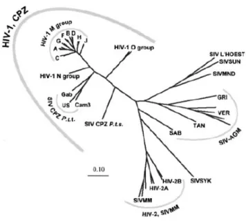
Characterization of the HIV-2 virion showed a similar genomic organization to HIV-1, even though they only share 40% to 60% of nucleotide homology (36). Interestingly, HIV-2 is genetically closer to the Simian Immunodeficiency Virus (SIV), with over 75% nucleotide homology (37), a result of interspecies transmissions from the natural host sooty mangabey (Cercocebus atys) as illustrated in Figure 2 (27, 35, 38, 39).
SIV infects several nonhuman primates without causing disease or AIDS; such is the case of sootey mangabeys or african green monkeys, considered natural hosts for SIVsm or SIVagm, respectively (40-42). On the other hand, SIVsm can infect several species of Asian macaques, such as the rhesus macaque, causing severe immunodeficiency and AIDS–like syndrome (43, 44). Thus, the study of SIV infected macaques and comparative analyses of pathogenic and non pathogenic SIV infections have constituted important models for the study of HIV pathogenesis.
40
Figure 2 – Phylogenetic relationship of primate lentiviruses based on identity of pol gene sequences.
SIVCPZ corresponds to SIV from chimpanzee (Pan troglodytes); SIV L’HOEST from L'Hoest's Monkey
(Cercopithecus lhoesti); SIVSUN from Sun-tailed Monkey (Cercopithecus solatus); SIVMND from
Mandrill (Mandrillus sphinx); SIVAGM African Green Monkeys; SIVSYK from Sykes' Monkey
(Cercopithecus albogularis); SIVMM from Sooty Mangabey (Cercocebus atys). Adapted from Reeves, et al (39).
Figure 3 – Representation of the genomic organization of HIV-1 and HIV-2/SIV. Adapted from Marlink, R. (27).
41
Replication cycle and latency
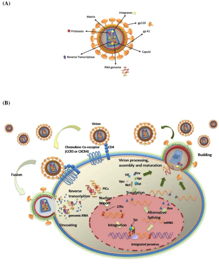
HIV-1, HIV-2 and SIV are members of the lentivirus genus included in the Retroviridae family, all characterized by the unique enzyme, reverse transcriptase which utilizes the viral genomic RNA as a template for synthesis of proviral cDNA (45).
The HIV genome is approximately 10kb, consisting of nine genes flanked by long terminal repeat sequences (LTRs) (46), depicted in Figure 3. The HIV-1’s genome, like HIV-2 and all other retroviruses, has three major coding regions: gag which encodes for the structural proteins of the viral capsid, pol encoding enzymes involved in both viral replication and integration and env which encode the viral envelope glycoproteins. The genes gag and pol are well conserved in HIV-1, HIV-2, and SIV, and account for most of the cross-reactivity seen in enzyme-linked immunosorbent assays (ELISA) for HIV-1 (47, 48). HIV-1 and HIV-2 env genes encode for different glycoproteins (gp) that seem to have similar functions during infection; the outer membrane gp120 or gp105 and the transmembrane gp41 or gp36, respectively in HIV-1 or HIV-2. The LTRs are required for the integration of the viral cDNA (provirus) into the host cell genome and subsequent initiation of transcription of the integrated provirus (49). HIV-1 and HIV-2 share five additional reading frames tat, rev, nef,
vif and vpr, coding for the gene products Tat, Rev, Nef, Vif and Vpr, accessory proteins with
different key regulatory roles during HIV infection. HIV-1 vpu and HIV-2 vpx, although similar in sequence encode for proteins, Vpu and Vpx, respectively, that appear to have different functions in the infected cell (48). Most of the viral genes have overlapping reading frames, which allow the virus to encode several proteins in a small genome, through alternative splicing (50).
The virion capsid has a particular cone shape, contained within an external lipid envelope. Embedded in this membrane are the transmembrane glycoproteins responsible for binding to the host cells, and for fusion of the viral particle with the cell membrane, as illustrated in
Figure 4A. Two copies of genomic RNA and several viral proteins such as reverse
transcriptases, proteases and integrases are packaged inside the viral capside (51).
The viral replication cell cycle is schematically represented in Figure 4B. Both HIV-1 and HIV-2 enter cells by a membrane fusion process that requires the interaction of their external envelope gp with the CD4 molecule and co-receptors from the chemokine receptor family
42
(mainly CCR5 or CXCR4). The first step in cellular infection involves high affinity binding of the outer membrane gp to the CD4 molecule of host cells (52). Then, the outer membrane gp undergoes a conformational change allowing a better binding to either one of the cellular co-receptors (53). The induced conformational shift exposes a transmembrane gp which mediates cell-virus fusion. Its fusion domain binds and penetrates the host cell membrane bringing the two membranes closer (54), allowing virion content to enter the cytoplasm (55). The limited exposure of the key surfaces that mediate virus-host interaction during the extracellular stages of the HIV infection is considered a main reason for the failure to generate neutralizing antibodies that can control this phase of infection (56, 57).
Once in the cytoplasm, the RNA genome is reverse transcribed into double-stranded DNA by the viral reverse transcriptase. This process is highly error prone, leading to mutations of the viral genome (49). During the process of trafficking and transport of the viral DNA to the nucleus, the double stranded DNA and all viral proteins in the cytoplasm of the host cell aggregate, forming preintegration complexes (PICs). At this stage, PICs recruit host cellular factors to aid in the cell trafficking to the nucleus (58).
In the nucleus, integration occurs through the interaction of the viral integrase and of host cell DNA repair enzymes (59). Furthermore, the sites of HIV integration are not randomly distributed in the genome, but occur preferentially in transcriptionally active regions, downstream of promoters (60). When the provirus first starts to be transcribed, the messenger RNA is fully spliced leading to the production of Tat, Rev and Nef. Tat ensures transcription of viral mRNA (59). With the accumulation of Rev in the nucleus mRNA is exported leading to the production of the other viral proteins – Env, Vif, Vpr and Vpu or Vpx – and later in infection, of Gag and Pol (61). In the cytoplasm, the proteins are assembled to produce the viral capsid and envelope. The unspliced mRNA is packaged in the viral particles as well as the viral enzymes reverse transcriptase, integrase and protease (62). The completely assembled viral particles bud out of the cells, ready to infect other cells. All these processes critically depend upon the state of cell activation and cell cycling.
43
(A)
(B)
Figure 4 – The life cycle of HIV-1.
(A) Organization of the HIV-1 virion. (B) Schematic description of the events occurring after HIV infection of a susceptible target cell.
44
A state of latency is reached when integrated provirus have no active transcription. With the development of effective therapies that reduce the plasma viral load by targeting cells that are actively replicating virus, the persistence of a latent viral reservoir is considered the most important factor precluding the discontinuation of antiretroviral therapy (63-65). The factors that determine whether a cell will be latently infected or actively transcribing virus remain elusive.
Cell targets: co-receptor usage and tropism
The primary receptor for the HIV-1 particle is the CD4 molecule and, therefore, its main targets are CD4 T cells (66) and cells of the monocyte-macrophage lineage (67). Other cells have been shown to be infected in vitro though the in vivo relevance is not clear (68). HIV-2 infection of target cells has also been shown to require CD4 as the primary receptor together with one of several chemokine receptors as a co-receptor (69). HIV-2 appears to be more flexible in co-receptor usage than HIV-1, that mainly uses CCR5 or CXCR4 (53, 70 , 71, 72). Moreover, several HIV-2 strains have been shown to infect certain CD4-negative cell lines, generally through the use of CXCR4 or CCR5 alone (73, 74). However, in spite of the promiscuity of co-receptor usage exhibited by HIV-2 in vitro, several lines of evidence suggest that CCR5 and CXCR4 are as the major co-receptors for HIV-2 infection in vivo (75-77).
The chemokine receptors, CCR5 and CXCR4, belong to the family of transmembrane chemokine receptors.
CCR5 is upregulated on the surface of effector/memory T cells upon T cell activation and is a receptor for CCL3 (macrophage inflammatory protein 1α: MIP-1α), CCL4 (MIP-1β) and CCL5 (regulated upon activation, normal T cell expressed and secreted: RANTES). Interactions between these molecules regulate homing of effector/memory cells to specific lymphoid tissue (78). The importance of CCR5 as an HIV co-receptor, particularly during HIV-1 acute infection, became apparent when it was discovered that individuals homozygous for a mutation in the CCR5 gene (CCR5∆32) were protected from HIV infection (79). CXCR4, the receptor for CXCL12 (stromal derived factor 1: SDF-1), is constitutively expressed on CD4 T cells, including naive T cells, and several other cell populations (78). These chemokines play diverse roles both during normal lymphocyte development and
45
during inflammation, varying from chemoattractant factors for monocytes and T cells, to activators of granulocytes or in homing of hematopoietic stem cell.
Tropism of the HIV particle is currently classified according to its binding to CCR5 (R5-Tropic) or CXCR4 (X4-(R5-Tropic). The R5 variants are associated with the primary infection, since they are the predominant phenotype in newly infected individuals. In around 50% of the HIV-1 infected patients in advanced disease stage, the viral phenotype switches to an X4 variant and this change is usually followed by a rapid decline in CD4 T cell numbers and progression to AIDS (80). An evolution from CCR5 to CXCR4 co-receptor usage is less clear in HIV-2-infected individuals. Many primary isolates have been shown to use a range of co-receptors including both CCR5 and CXCR4 and only a limited number of X4 viruses have been isolated from symptomatic patients (76, 81). In addition, it is not known whether primary infection is mainly associated with R5 2 strains as it has been described in HIV-1 infection (8HIV-1, 82).
46
Natural history of HIV-1 infection
HIV-1 is transmitted by contact with infected body fluids, mainly by sexual contact, blood transfusion and/or contact with blood products, and by vertical transmission (from mother to child during pregnancy, delivery or breast feeding). Despite the multiple modes of transmission, there are no obvious differences in disease manifestations in individuals infected by different routes. This suggests that even if the initial target cells or route of HIV-1 infection differ, the subsequent viral spread ultimately results in a similar outcome (83).
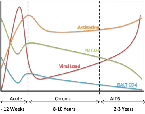
Typical course of untreated HIV-1 infection
The acute phase of HIV-1 infection is characterized by viral dissemination throughout the lymphoid tissue. The highest levels of viremia are typically achieved during the acute infection. A selective depletion of memory CD4 T cells occurs coincidently with the active viral replication. Following exposure, around 50% of individuals experience an acute phase syndrome that may persist for up to several weeks, with symptoms ranging from a mild cold to a flu-like or mononucleosis-like disease. At the end of the acute stage, viremia decreases, mainly as a result of the expansion of HIV-1 specific CD8 T cells (84), but also due to the response of other components of the immune system, such as specific antibodies. Concomitantly, the CD4 T cell counts increase, although to values usually below the pre-infection levels. The parallel expansion of CD8 T cells, that is only partially due to HIV specific responses, causes an inversion of the CD4:CD8 ratio, which persists throughout the infection. The viral set point, attained through a balance between the viral replication and the host immune response, is predictive of the rate of progression to AIDS in untreated individuals (85), such that the higher the viral set point, the faster the disease progresses (86).
Recent studies have provided evidence for the role of the infection of gut-associated lymphoid tissue (GALT) in the profound CD4 depletion during acute infection. In the gut of acutely HIV-1-infected individuals a massive mucosal CD4 T cell depletion is observed, 4–6 weeks post-infection, much more marked than the decrease in CD4 T cell counts in the peripheral blood (87). It is important to note that the majority of CD4 memory T cells in uninfected humans resides in the GALT (more than 95%), with the remaining cells distributed by the blood (less than 2%), spleen, peripheral lymph nodes and other lymphoid tissues. As a consequence, CD4 memory T cell depletion is more rapid and profound in the
47
GALT during HIV-1 primary infection (88). Although this depletion is observed both in pathogenic and non pathogenic SIV infection (89, 90), the extent of this initial depletion has been suggested to determine the outcome of the HIV-1 infection.
~ 12 Weeks 8-10 Years 2-3 Years
Figure 5 – Typical course of untreated HIV-1 infection.
Following resolution of the acute stage, a chronic persistent infection is established, that is associated with an asymptomatic period of clinical latency, prior to advanced immunodeficiency or AIDS. Numerous defects in immune function are evident in this phase of HIV-1 disease prior to substantial CD4 T cell loss (86). Despite the lack of clinical symptoms, ongoing virus replication can be detected consistently in lymphoid tissue attested by the usually significant levels of viremia. In vitro tests have shown that cell-mediated immune responses are progressively lost, first to microbial recall antigens, then to the more potent stimulation by alloantigens, and finally to mitogens. These in vitro functional T cell defects are also reflected in vivo by the loss of delayed type hypersensitivity skin testing (91).
48
Thus, despite a potent immune response and the marked downregulation of virus replication during the acute phase, HIV-1 succeeds in establishing a state of chronic infection with a variable degree of persistent viral replication.
The final stage of disease progression is characterized by an increased risk of opportunistic infections and tumors – AIDS, leading to death in 2 to 3 years in the absence of antiretroviral therapy (ART). Complex interactions between variable host responses and a highly variable pathogen determine the rate of the deterioration of the immune system. The median period from infection to AIDS is estimated to be 8-10 years, but AIDS can occur in months in the so called rapid progressors, whereas other infected individuals have remained asymptomatic for more than 10 years with relatively well preserved CD4 T cell counts, which are named long term non-progressors (91). A number of severe clinical manifestations, associated with reduced cell-mediated immunity in HIV-1 infected patients, are included in the Centers for Disease Control and Prevention (CDC) definition of AIDS. The AIDS-defining criteria also include a low CD4 T cell count (less than 200 CD4 T cells/µL blood). Since progression of HIV-1 disease is usually directly associated with a decrease in CD4 T cell counts and an increase in plasma HIV-1 viral load, measurement of these surrogate markers of infection has been used as a tool for assessing disease stage, prognosis and monitoring therapeutic response (86).
Antiretroviral therapy
Antiretroviral therapy or ART, using combinations of three or more drugs, is also termed highly active antiretroviral therapy in agreement with its major impact on slowing the progression of disease in a substantial proportion of adequately treated patients, by controlling viremia and allowing CD4 T cell counts to rise. It is the most important achievement in the battle against HIV-1 infection so far, with impressive impact on morbidity and mortality (92). Recent data from a comparative study of mortality before and after the introduction of ART in HIV-1 cohorts in Norway show that in the ART era the mortality was reduced by 80% (93). In another study, Walensky et al (94) demonstrated that since 1989, at least 3 million years of life have been saved in the United States as a direct result of ART.
49
Reservoirs
The number of available antiretroviral drugs has increased rapidly in the last decade, allowing for the use of several effective combinations of 3 or more drugs. However, strong evidence of the inability of ART to eradicate HIV-1 infection has come from studies of individuals who began antiretroviral therapy during the chronic stage of HIV-1 infection, showing that interruption of effective long-term antiretroviral therapy resulted in a rapid rebound of plasma viremia in almost all individuals (95, 96). Moreover, individuals whose ART was interrupted and remained off therapy for 4 to 6 weeks, experienced increases in the plasma HIV-1 RNA, to the same levels reached prior to the initiation of ART (97). These data suggest that ART does not eradicate HIV-1 infection and has no effect upon the post-therapy replication kinetics of the virus. HIV-1 proviral DNA and mRNA can be detected in CD4 T cells in individuals who have maintained "undetectable" plasma viremia (<50 copies of HIV-1 RNA/ml) for prolonged periods of time while receiving ART. Thus, viral reservoirs and some ongoing HIV-1 replication persist in the presence of effective antiretroviral therapy and it is currently consensual that complementary immune-based therapies will be required to eradicate or control the viral reservoirs.
50
HIV Immunopathogenesis
The hallmark of HIV infection is the progressive CD4 T cell depletion associated with active viral replication. There is considerable controversy regarding the relative contribution of the various putative mechanisms to the depletion of CD4 T cells during the course of HIV infection.
Tap and drain hypothesis
In 1995, Ho and colleagues (98) proposed that the virus was the major driving force behind the CD4 depletion, whether it was by direct infection and concomitant cytolytic effects, or by the removal of the infected cells by the immune response. This was termed the “tap and drain” hypothesis. In this analogy a sink represented the CD4 T cell compartment, an unplugged drain depicted the impact of HIV infection on CD4 numbers and water flowing from an open tap symbolized the production of new CD4 T cells. Over time the open tap, i.e., the production of new CD4 T cells would be unable to counteract the “flow of water going down the drain”, ultimately resulting in the depletion of the CD4 compartment. The major evidence that supported this hypothesis came from the study of the reduction of viremia and increase in CD4 T cells in the blood after treatment with a protease inhibitor (98, 99). Mathematical modeling applied to these data estimated that everyday there was a destruction of 2x10⁹ lymphocytes by the virus, which was compensated by the regenerative capacity of the immune system in the steady state (98-100). In this model, the ability to produce new cells progressively deteriorates resulting in AIDS. This mathematical model predicted that the half-life of the infected CD4 T cells was such that in 10 years the CD4 compartment would be empty and also that continuous therapy use would be able to eradicate infection in less than 2 years (98-100).
One of the first observations that directly challenged this simplistic view was that the initial rise in CD4 T cells did not signify the production of new cells as predicted. In fact, the increase in CD4 T cell counts that occurred immediately after initiation of therapy was subsequently shown to result mainly from the redistribution of CD4 T cells into the blood stream, that were trapped in the lymph nodes and other tissues, due to decrease in antigen load (101, 102). Also, the frequency of infected CD4 T cells was not sufficient to explain the CD4 T cell depletion by direct virus killing (103). Moreover, the major CD4 depletion could