Microbiological profile in Serra ewes’ cheese during ripening
A.C. Macedo, F.X. Malcata and T.A. HoggEscola Superior de Biotecnologia I Universidade Catolica Portuguesa , Porto, Portugal
5052/09/94: received 13 September 1994, revised 1 1 January 1995 and accepted 15 January 1995
A . C . M A C E D O , F . X . M A L C A T A AN D T . A . H O G G . 1995. T h e microflora of Serra cheese was monitored during a
35
d ripening period at three different periods within the ewe’s lactation season. After7
d ripening, the numbers of micro-organisms reached their maximum, and lactic acid bacteria (LAB) and coliforms were the predominant groups. Pseudomonads were not detected after1
week of ripening. At all stages of ripening, cheeses manufactured in spring exhibited the lowest numbers of L A B and yeasts, whereas cheeses manufactured in winter showed the lowest numbers of coliforms and staphylococci.faecium and Lactococcus lactis spp. lactis exhibited the highest decrease in percentage composition. Numbers of both Leuc. mesenteroides and Lactobacillus paracasei tended to increase throughout ripening. T h e most abundant coliform was Hafnia alvei. Klebsiella oxytoca was found in curd but declined in number during ripening. Staphylococcal flora of curd was mainly composed of Staphylococcus xylosus, Staph. aureus and Staph. epidermidis. Staphylococcus xylosus was the major species found at the end of ripening. Pseudomonas fltlorescens was the only Pseudomonas species isolated from the curd. Although a broad
spectrum of yeasts were found in Serra cheese, Sporobolomyces roseus was the most abundant yeast isolated.
Leuconostoc lactis was the most abundant
LAB
found in Serra cheese whereas EnterococcusINTRODUCTION
Serra da Estrela cheese (or simply Serra cheese) is a semi- soft cheese variety manufactured at the farm level from raw ewes’ milk using traditional methods in the mountainous centre region of Portugal. This cheese is made in batches twice daily (early morning and late afternoon) from October to May using the unpasteurized milk immediately after col- lection. Coagulation is catalysed by a crude vegetable rennet, which consists of a suspension of the dry flowers of Cynara cardunculus, prepared at room temperature and fil- tered through a piece of cotton cloth. Coagulation takes place at 27-30°C for 1-2 h. Once the right degree of con- sistency is attained, the curd is cut in irregularly shaped pieces, mainly by hand. After cutting, the curd is poured into perforated plastic moulds and lightly pressed by hand, and the cheese is salted by rubbing dry salt on the surface. Cheeses are ripened on wood shelves, namely in a basement for ca 40 d. The use of Bordaleira ewes’ milk which pos- sesses a high fat content (&20%) (Macedo et al. 1993), coupled with the use of the vegetable rennet, which pos- sesses a strong, unselective proteolytic activity (Barbosa et
Correspondence to : Dr F. Xavier Malcata, Escola Supertor de Bioternologia, Universidade Catilica Portuguesa, Rua Dr Antinio Bernardino de Almeida, 4200 Porto, Portugal.
0 1995 The Society for Applied Bacteriology
al. 1981), results in a cheese with unique bouquet and creamy texture.
Studies on the microbial flora of ripened Serra cheese have been reported by Hiscox et al. (194l), Cruz (1945) and Antunes and Santos (1943); however, very little was known until recently about the nature and the evolution of the main microbial groups during ripening. Studies focused on the changes in the numbers of the main groups of micro- organisms in the interior and on the surface of Serra cheese, throughout the lactation season, and during ripen- ing have been conducted by Macedo et al. (1995). The aim of the present study was to complement such previous information via the identification of the species of lactic acid bacteria (LAB), yeasts, coliforms, staphylococci and pseudomonads in Serra cheese during ripening and throughout the lactation season ; such work was developed hoping that a standardized starter suitable for Serra cheese manufacture will eventually be possible.
MATERIALS AND METHODS Manufacture and sampllng of cheese
Three batches of 12 0.5 kg Serra cheeses were manufac- tured and ripened according to the traditional practice
2 A . C . M A C E D O E T A L
(Macedo et al. 1993) in November, February and May in an attempt to adequately represent autumn, winter and spring manufactures. The 12 cheeses were randomly divided into four sets of three cheeses each; one set picked at random was sent to this laboratory on the day of cheese- making, and the others after 7 d, 21 d and 35 d of ripening, respectively. All samples were transported under refriger- ated conditions (ca 4°C). Sampling of the interior of the cheese and all microbiological analyses thereafter were per- formed immediately upon receipt of the cheeses ( < 1 h). Microbiological analyses
Ten g of each cheese sample were homogenized with 90 ml of a sterile solution of 2% (w/v) sodium citrate (Merck, Darmstadt, Germany) at 45°C for 1 min in a Stomacher Lab-Blender 400 (Seward Medical, London, UK). Decimal dilutions were prepared in sterile 0.1 O h peptone water (Sigma Chemical, St Louis, MO, USA) and plated in duplicate. Lactic acid bacteria (LAB) were grown anaero- bically (Gas-Pak anaerobic system BBL, Cockeysville, MD, USA) on M17 Agar (M17A; LabM, Bury, UK) and on Rogosa Agar (RA; Oxoid, Basingstoke, UK) at 30°C for 3 and 5 d. Cycloheximide (Sigma) was added (100 mg I-') to prevent the growth of yeasts (Kandler and Weiss 1986). Coliforms were determined on Violet Red Bile Agar (VRBA; Lab M) and staphylococci on Baird-Parker egg yolk tellurite medium (BPM; Lab M), at 37°C for 1 and 2 d, respectively. Pseudomonads were grown on Pseudo- monas Base Agar (PSDA; LabM) supplemented with cephaloridine, fucidin and cetrimide (Lab M) and yeasts on Potato Dextrose Agar (PDA; LabM) acidified with 10% lactic acid (Merck), at 25°C for 2 d and 5 d, respectively. The technique of surface viable count was used for all media except VRBA for which the pour-plate and overlay technique was used. Results were expressed as cfu g - ' of cheese.
For the curd and 35-d-old cheese, as well as from each of the aforementioned agars, colonies with different morphol- ogies were counted, picked (three colonies of each type), purified, and stored at 4°C as slope cultures until further characterization.
identification of mlcro-organisms
The API 50 C H L system (BioMCrieux, Marcy-I'Etoile, France) was used to identify to the species level the follow- ing genera : (i) Lactobacillus (Gram-positive, catalase- negative rods) ; (ii) Leuconostoc (Gram-positive, catalase-negative, non-producers of ammonia from arginine and heterofermentative cocci) ; (iii) Lactococcus (Gram- positive, catalase-negative and homofermentative cocci that grow at 10" but not at 45°C). Identification of enterococci (considered here as Gram-positive, catalase-negative,
homofermentative cocci that grow at both 10" and 45°C) was performed using the API STREP system (BioMCrieux). Coliforms (considered here as Gram- negative, catalase-positive, glucose-fermenters, nitrate- positive and oxidase-negative short rods) were identified by the API 20E system (BioMtrieux). Staphylococci (considered here as Gram-positive, glucose-fermenters under anaerobic conditions and catalase-positive cocci) were identified by the API STAPH system (BioMirieux). Staphylococcus aureus were confirmed by testing coagu- lation of lyophilized rabbit plasma (BioMirieux) at 37°C within 24 h. Results obtained from the different API systems were matched with the aid of a commercially avail- able software for the automatic identification of bacteria (Anon. 1993). Identification of pseudomonads (considered here as Gram-negative and oxidase-positive rods) was according to the method of Collins and Lyne (1984). Bio- chemical and physiological characterization of yeasts included: (i) assimilation of carbon compounds such as L-
arabinose, cellobiose, erythritol, galactose, gluconate, glu- cosamine, glucose, cr-methyl-mglucoside, glucuronate, glycerol, 2-keto-gluconate, DL-lactate, lactose, mannitol, maltose, melibiose, melezitose, raffinose, rhamnose, ribose, saccharose, sorbose, trehalose and D-XylOSe; (ii) assimilation of nitrogen compounds (nitrate) ; and (iii) temperature tol- erance (growth at 25", 30" and 37°C). I n addition, morpho- logical criteria such as budding/splitting of cells and colony pigmentation were taken into account. T h e computer program of Barnett et al. (1990) was used for automatic identification of yeasts.
Statistlcai analysis
ANOVA tables (not shown) were constructed with the rep- licated data pertaining to the numbers of micro-organisms us the factors ripening time and period within the lactation season.
RESULTS
The data statistically significant on the 5% level encom- passing the numbers of LAB on M17A, LAB on RA, coli- forms on VRBA, staphylococci on BPM, and yeasts on PDA are plotted in Figs 1, 3, 5, 7 and 9, respectively, as the average of the three replicates considered in ANOVA tables. The percentage composition in terms of microbial species of each of the aforementioned groups for both the curd and the 35-d-old cheese are depicted in Figs 2, 4, 6, 8 and 10.
D I S C U S S I O N
Inspection of Figs 1, 3, 5, 7 and 9 indicates that the growth patterns of the main groups of micro-organisms studied, as 0 1995 The Society for Applied Bacteriology, Journal of Applied Bacteriology 78, 1-11
M I C R O B I O L O G Y I N SERRA CHEESE 3
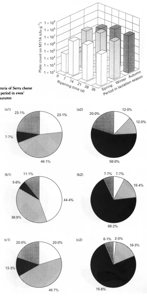
Fig. 1 Changes in numbers of lactic acid bacteria of Serra cheese (grown on MI7 agar) with ripening time and period in ewes’ lactation season. 0 , Spring; 0, winter;
.,
autumnFig. 2 Composition of LAB of Serra cheese (grown on M17A) in (1) curd and (2) 35-d-old cheese, produced in (a) autumn, (b) winter, and (c) spring, in terms of Enterococcus faecrum (O), Lactococcus lactrs ssp. lactis (N), Leuconostoc lactis ,).( Leuc. mesenterordes
ssp. mesenterordesldextranrcum
(a)
4 A . C . M A C E D O E T A L .
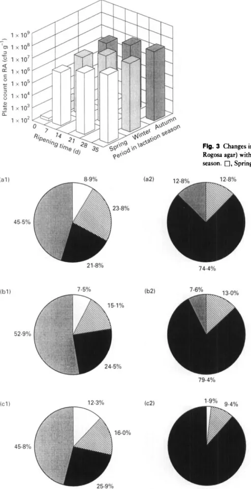
Fig. 3 Changes in numbers of LAB of Serra cheese (grown on Rogosa agar) with ripening time and period in ewes’ lactation season. 0, Spring; 0 , winter;
.,
autumnFig. 4 Composition of LAB of Serra cheese (grown on RA) in (1) curd and (2) 35-d-old cheese, produced in (a) autumn, (b) winter, and (c) spring, in terms of Lactobacrllus plantarum (O), Lact. paracaser ssp. paracase;
(m),
Leuconostoc lactis ).( and Leuc. rnesenterordes ssp. mesentero;desldextran;cum ( 0) 0 1995 The Society for Applied Bacteriology, Journal of Applied Bacterioiology 70, 1-11M I C R O B I O L O G Y I N S E R R A CHEESE 5
Fig. 5 Changes in numbers of coliforms of Serra cheese (grown on Violet Red Bile Agar) with ripening time and period in ewes’ lactation season. 0, Spring; 8 , winter;
.,
autumnFIg. 6 Composition of coliforms of Serra cheese (grown on VRBA) in ( 1 ) curd and (2) 35-d-old cheese, produced in (a) autumn, (b) winter, and (c) spring, in terms of Hujinra alvei ( O ) , Klebsrellu uxyroca
(a),
Escherrchra colt ).( andCrrrobucterfieundri ( 0 )
6 A.C. MACEDO E T A L .
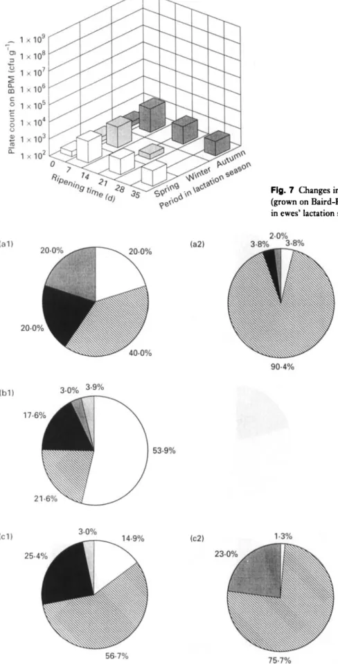
Fig. 7 Changes in numbers of staphylococci of Serra cheese (grown on Baird-Parker Medium) with ripening time and period in ewes’ lactation season. 0, Spring; 13, winter;
.,
autumnFig. 8 Composition of staphylococci of Serra cheese (grown on BPM) in ( 1 ) curd and (2) 35-d-old cheese, produced in (a) autumn, (b) winter, and (c) spring, in terms of Staphylococcus aureus (O), Staph. xylosus (H), Staph. eprdermidis
(m),
(U)) and Staph. homrnis 0 1995 The Society for Applied Bacteriology, Journal of Applied Bacteriology 79, 1-1 1
M I C R O B I O L O G Y IN SERRA CHEESE 7
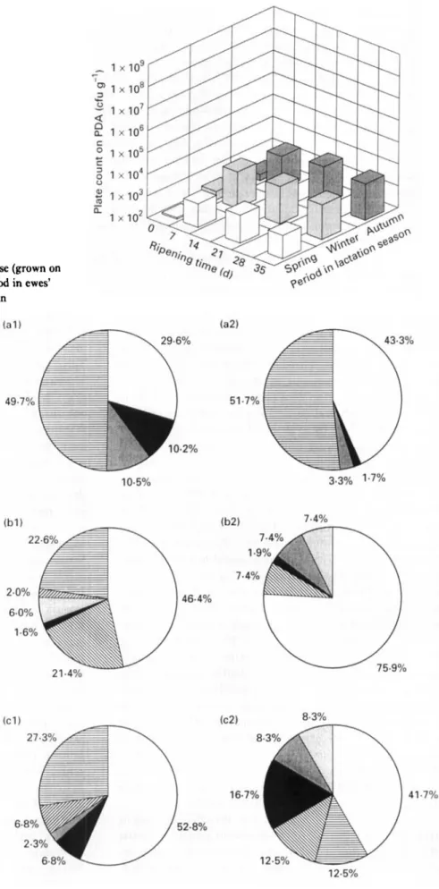
Fig. 9 Changes in numbers of yeasts of Serra cheese (grown on Potato Dextrose Agar) with ripening time and period in ewes’ lactation season. 0 , Spring; 0, winter;
.,
autumnFig. 10 Composition of yeasts of Serra cheese (grown on PDA) in ( 1 ) curd and (2) 35-d-old cheese, produced in (a) autumn, (b) winter, and (c) spring, in terms of
Sporobolomyces roseus
(n),
Kluyveromyces marxianus(m),
Rhodotorula aurantraca(.
), Yarromra lrpolytrca (
membranaefacrens
(a),
Trrchosporum bergelrr(m),
Leucosporrdrum scottrr/Debaryomyces hansenrt ( H)8 A . C . M A C E D O E T A L .
well as the growth rates of LAB, staphylococci and yeasts, with ripening time and period within the lactation season were similar to those reported previously by Macedo et al. (1995). However, the initial contamination of the curd by LAB enumerated on M17 (ca lo4), LAB enumerated on RA (ca lo2), staphylococci (ca 10’) and yeasts (ca 10’) were lower by 1000-fold, 100-fold, 1000-fold and 100-fold, respectively, whereas initial contamination of the curd by coliforms tended to be the same (ca lo6). This suggests that better standards of hygiene during milking and cheese- making were used in the present experimental study, but also that the inefficient refrigeration of raw milk is possibly still contributing to some extent to a fast growth of psych- rotrophic coliforms (such as Hafnia alvei which is the main coliform in curd).
LAB and coliforms were the major components of the microflora during ripening throughout the entire lactation season. LAB enumerated on M17 agar (Fig. I), LAB enu- merated on RA (Fig. 3) and coliforms (Fig. 5) increased their numbers during the first week of ripening by lo3,
lo4 and lo2, respectively, with respect to the initial numbers in the curd. Thereafter, the tendencies exhibited by these bacteria were: (i) to increase at a much lower growth rate from 7 d to 21 d (10-fold increase); and (ii) to stabilize for LAB and to decrease for coliforms (10-fold decrease) from 21 d to 35 d. Strict microbiological regula- tions concerning cheese made from raw milk do not for- mally exist in Portugal ; however, Spanish regulations pertaining to some cheese varieties manufactured from pas- teurized milk specify maximum counts of lo4 cfu g-’ for Enterobacteriaceae and lo3 cfu g - ’ for Escherichia coli (Gaya et al. 1983). Cheeses obtained from all batches uti- lized in the present work exceed those thresholds after 35 d of ripening (Fig. 5), figures which should be carefully con- sidered due to public health reasons. Numbers of staphylo- cocci (Fig. 7) reached their maximum at 7 d, and they tended to decrease thereafter at higher rates than LAB and coliforms. Numbers of pseudomonads in curd for autumn, winter and spring, were 1.2 x lo3, 2.6 x lo3 and 2.3 x lo3 cfu gi’, respectively. After 7 d of ripening, numbers of pseudomonads were negligible. I t is known that certain pseudomonads exhibit proteolytic and lipolytic extracellular activities that may cause undesirable effects in texture and flavour; however, the number of this group of micro-organisms present during the ripening process sug- gests that their role in flavour development is quite limited. Yeasts (Fig. 9) tended to increase during the first 2 weeks of ripening (10-fold increase), but much more slowly than I,AB and coliforms, and then tended to stabilize.
As shown in Figs 1, 3, 5, 7 and 9, the effect of ripening time on the numbers of micro-organisms present is greater than that of the period within the lactation season, an observation which is consistent with results reported pre-
viously by Macedo et al. (1995). LAB and yeasts were favoured by the environmental conditions prevailing in the maturation rooms during winter and autumn, i.e. lower temperatures and higher relative humidities than in spring; conversely, staphylococci and coliforms survived better in spring. Fernandez del Pozo et al. (1988) reported that lower counts of coliforms were obtained in La Serena cheese made in spring than in winter. Following the consideration of the inhibitory effect of LAB on the growth of coliforms in Manchego cheese (Gaya et al. 1983), the higher numbers of LAB recorded in winter might explain the apparent accelerated decrease of coliforms during this period. T h e initial contaminations by coliforms and staphylococci in Serra cheese are also in agreement with seasonal variations of coliform counts in raw ewes’ milk which apparently decrease from autumn to spring (Gaya et al. 1987), and with seasonal variations of staphylococci counts in raw ewes’ milk which have been reported as highest in spring and lowest in winter (Bautista et al. 1986).
Among LAB, L. lactis ssp. lactis and Ent. faecium were the most abundant bacteria found in curd (Figs 2 and 4). These bacteria showed the largest decrease in their percent- age composition from the curd to the fully ripened cheese (Fig. 2). However, the presence of lactococci and entero- cocci during the late stages of maturation (Fig. 2) suggests that these bacteria may play an important role in the ripen- ing of this type of cheese. It has been reported that L . lactis is the most abundant Lactococcus species in La Serena cheese (Fernandez del Pozo et al. 1988), Hrudkovy and Bryndza cheeses (Prekoppova 1990), and L . lactis ssp. lactis is the most abundant subspecies in Roncal cheese (Arizcun et al. 1992). Enterococcus faecium is commonly found in Manchego cheese (Nuiiez and Martinez-Moreno 1976), La Serena cheese (Fernandez del Pozo et al. 1988) and Teleme cheese (Tzanetakis and Liptopoulou-Tzanetaki 1992). During ripening, however, the bacterium that grows faster appears to be Leuc. lactis (Figs 2 and 4) because it domi- nates in the fully ripened cheese. Leuconostoc mesenteroides ssp. mesenteroidesldextranicum was also found in both curd and 35-d-old cheese. Leuconostoc species can play an impor- tant role in aroma development and eye formation through their ability to metabolize citrate and ferment carbo- hydrates, respectively. T h e predominance of the Leuco- nostoc genus in Serra cheese is consistent with results presented elsewhere : Nuiiez et al. (1984) reported that this genus is predominant in the Gram-positive psychrotrophic genera obtained from refrigerated raw ewes’ milk, and Arizcun et al. (1992) reported high numbers of Leuconostoc in Roncal cheese. (In this latter case, ca 20% of the Leuco- nostoc species found were Leuc. mesenteroides ssp. mesen- teroides and ca 24% were Leuc. mesenteroides ssp. dextranicum.) The main Leuconostoc species found in La Serena cheese was Leuc. mesenteroides (Fernandez del Pozo 0 1995 The Society for Applied Bacteriology, Journal of Applied Bacteriology 79, 1-11
M I C R O B I O L O G Y IN S E R R A CHEESE 9
et al. 1988). Leuconostoc species have been also deliberately added to Manchego cheese as a part of the microbial starter with good results (Ramos et al. 1990). Among the genus Lactobacillus, Lact. plantarum and Lact. paracasei ssp. para- casei could be identified in the curd (Fig. 4). During ripen- ing, Lact. paracasei ssp. paracasei tended to grow while Lact. plantarum tended to disappear, indicating a possible role for Lact. paracasei ssp. paracasei in Serra cheese matu- ration. It has been asserted that Lact. plantarum and Lact. casei were the dominant lactobacilli in La Serena cheese (Fernandez del Pozo et al. 1988), Hrudkovy and Bryndza cheeses (Prekoppova 1990), and Roncal cheese (Arizcun et al. 1992), and that Lact. plantarum was the predominant lactobacillus in Feta and Teleme cheeses (Tzanetakis and Liptopoulou-Tzanetaki 1992). T h e data presented in the present study also indicate that lactation season has an influence on the initial contamination of the curd and the growth of each group of LAB throughout ripening. The level of enterococci has been correlated with the level of hygiene during milk processing (Thompson and Marth 1986); higher values of the percentage composition of Ent. faecium in the curd (Fig. 2) in winter than in autumn or spring might be explained by the poor housing conditions in winter, when the animals spend most of their time in stables due to adverse weather. The ripening conditions in spring contributed preferentially to the growth of Leuc. lactis (Figs 2 and 4) with respect to such other lactic cocci as L e u . mesenteroides ssp. mesenteroidesldextranicum and Ent. faecium. T h e lowest percentage compositions of L . lactis ssp. lactis (Fig. 2) and Lact. paracasei ssp. paracasei (Fig.
4)
were obtained in autumn and spring, respectively.As a result of the absence of good sanitary conditions during milking and cheesemaking, coliforms are, like enterococci, important elements of the bacterial inventory of raw ewes’ milk. The levels of coliforms found in the present work (which are of the same order of magnitude as those of LAB) are of considerable public health concern and technological relevance for Serra cheese since this cheese variety is made directly from raw ewes’ milk without any type of thermal processing. Hafnia alvei was the most abundant and proliferating coliform found both in the curd and the 35-d-old cheese (Fig. 6). Gaya et al. (1983) also isolated H. alvei in very high proportions in 60-d-old Man- chego cheese, and Kleeberger et al. (1980) stated that H . alvei is the typical coliform isolated from milk and dairy products. Nuiiez et al. (1984) concluded that refrigeration of raw ewes’ milk had effects of decreasing or increasing the counts of psychrotrophs depending on whether the refriger- ation was made at
4“
or 7”C, respectively. Additionally, Juven et al. (1981) reported that psychrotrophic Enterobac- teriaceae exhibited proteolytic and lipolytic activities during refrigeration. Considering that the raw ewes’ milk used for Serra cheesemaking is lightly refrigerated below room tem-perature, one concludes that the predominance of H. alvei (as a psychrotrophic bacterium) may play an important role in ripening of Serra cheese. Gaya et al. (1987) reported that H. alvei and Klebsiella oxytoca were the predominant species in dairy farm samples. Klebsiella oxytoca was found in the curd but not in the 35-d-old cheese (Fig. 6). This fact may be partly accounted for by the weak resistance of this species to low p H ; Tornadijo et al. (1993) also found KI. oxytoca in the curd but not in 1- or 2-week-old cheeses made from raw goats’ milk which undergo pH variations similar to those in Serra cheese (Macedo et al. 1995). Escherichia cola was isolated from all ripened cheeses but only from the curd during the winter period; since it is able to ferment lactose, it may be responsible for the develop- ment of pin eyeholes in cheese. Failure to isolate E . coli until virtual completion of ripening may be probably attrib- uted to the fact that the initial contamination of milk by these bacteria is very low when compared with contami- nation by other micro-organisms. Since E . coli is particu- larly resistant to such adverse conditions as the low p H which tends to develop towards the end of the ripening period, this bacterium is likely to increase its percentage composition importance as time elapses. Gaya et al. (1983) reported that Enterobacter aerogenes, E . coli and H . alvei were the most resistant species against the acidic environ- ment generated by lactic acid bacteria. Citrobacter freundii was found in both curd and 35-d-old cheese, and its per- centage composition was lowest in spring and highest in winter (Fig. 6).
Due to their low numbers during ripening (ca lo2), staphylococci are not likely to play as important a role in the maturation process as LAB (Fig. 7). T h e predominant Staphylococcus species found in curd were Staph. xylosus, Staph. aureus and Staph. epidermidis, and at lower propor- tions, Staph. simulans and Staph. hominis (Fig. 8). Similar staphylococci species have been reported by Bautista et al. (1986) and Nuiiez et al. (1989) for ewes’ milk, and by Fern- andez del Pozo et al. (1988) for ewes’ cheese. Staphylococcus aureus showed a tendency to disappear as ripening time elapses (Fig. 8), so it is less likely that it causes health hazards by the normal time of cheese consumption (i.e. 45 d). For ewes’ milk, Bautista et al. (1986) reported higher values for the percentage composition of coagulase-positive staphylococci in spring than in the rest of the year; coagulase-positive staphylococci were not detected in La Serena cheese after 45 d (Fernandez del Pozo et al. 1988). The highest percentage composition of coagulase-positive strains among the staphylococci in curd was observed in winter. However, these species were less resistant in this period than in either spring or autumn since they could not be found in the 35-d-old cheese. This observation probably can be explained by the fact that more acidic cheese matrices, which are unfavourable to the growth of 0 1995 The Society for Applied Bacteriology, Journal of Applied Bacteriology 79, 1-11
10 A . C . M A C E D O E T A L .
coagulase-positive strains, are observed in winter (Macedo et al. 1995). Staphylococcus xylosus is the most abundant of the staphylococci in curd in the autumn and spring, whereas Staph. aureus is the most abundant species in winter; Staph. xylosus is also the most resistant and fastest growing bacteria during ripening (Fig. 8). Staphylococcus epidermidis was found in the curd and tended to disappear as ripening progressed ; the percentual compositions associ- ated with this strain in the curd and in the three periods within the lactation season were similar (Fig. 8). Initial contamination of the curd by Staph. simulans was highest in the autumn and lowest in the spring. However, this bacte- rium proved to be more resistant in spring than in autumn (Fig. 8). This fact probably can be correlated with the higher p H in cheese and higher ripening temperatures observed in spring (Macedo et al. 1995). Staphylococcus hominis was found in the curd at very low proportions and could not be detected in the 35-d-old cheese.
Among the Pseudomonas species, Ps. Juorescens was the only one found in Serra cheese. Nuiiez et al. (1984) have stated that Pseudomonas are the predominant psychrotro- phic genus in refrigerated raw ewes’ milk, although H. alvei was also found. For Serra cheese, H. alvei is more abun- dant than Ps. Juorescens.
Although the numbers of yeasts are 104-fold lower than those of LAB, they deserve special attention because some yeasts are able to synthesize lipolytic and proteolytic enzymes which may eventually contribute to the develop- ment of aroma and flavour during ripening. I t is, however, well established that their major contribution to the ripen- ing process is the utilization of lactic acid, which, by increasing the pH, encourages growth of the bacteria sensi- tive to acidic environments, and thus helps initiating the second stage of maturation. Since yeasts are ubiquitous in agricultural environments, a broad spectrum of yeasts was, as expected, found in the cheese samples analysed (Fig. 10). Sporobolomyces roseus and Leucosporidium scottail Debaryomyces hansenii (lactose-utilizing yeasts) were the predominant yeasts in both the curd and the 35-d-old cheese, but their proportions were dependent on lactation season (Fig. 10); the highest proportions occurred in the spring and the autumn, respectively. Predominance of lactic acid-utilizing yeasts over lactose-utilizing yeasts in La Serena cheese were reported by Fernandez del Pozo et al. (1988). Sporobolomyces roseus was, on the other hand, more resistant and more actively growing in winter than in autumn and spring. This observation is in agreement with previous data (Macedo et al. 1995) indicating that the lowest pH values in cheese occur in winter; therefore, higher growth rates of lactic acid-utilizing yeasts possibly can be expected in winter because the p H variations in Serra cheese are due mainly to the formation of lactic acid. Rhodotorula aurantiaca was present in both the curd and
the 35-d-old cheese although at low proportions, and was more resistant and proliferating in spring than in autumn. Yarrowia lipolytica was isolated in all curds and ripened cheeses except for the curd obtained in the autumn. T h e proportions of Kluyveromyces marxianus (a lactose-utilizing yeast) and Pichia membranaefaciens both in the curd and the 35-d-old cheese exhibited variations within the lactation season; these yeasts could be isolated in the 35-d-old cheeses made in the winter and spring but not in curds made in the autumn and spring. Trichosporum beigelii was found only in the curd, and at very low proportions (Fig. 10).
As final remarks, considering that Leuc. lactis, L . lactis ssp. lactis and Lact. paracasei ssp. paracasei were the most resistant and proliferating LAB found in this study, further research on the possibility of including these bacteria as part of a starter and on their effects on the organoleptic characteristics of Serra cheese seems very promising. Refrigeration of ewes’ raw milk on the farm level should also be optimized in order to control the growth of H. alvei. The temperature and relative humidity conditions of ripen- ing must also be set and controlled because they have an influence on the growthldeath rates of various micro- organisms which may impart relevant organoleptic charac- teristics to the final Serra cheese.
ACKNOWLEDGEMENTS
The authors are grateful to Professor Edmund Zottola (University of Minnesota, USA) for critical discussions, and to the members of the technical board of ANCOSE (AssociaGio Nacional de Criadores de Ovinos Serra da Estrela) for their cooperation encompassing the local manu- facture and transport of the cheeses. Financial support for ACM was provided by a PhD fellowship (BD-1741/91-IF) granted by J N I C T (Junta Nacional de Investigacio Cientifica e Tecnologica) within the framework of the CIENCIA program. This R&D work was partially funded by Agincia de InovaGio via project MAQUETTE (MelhorAmento de QUEijos Tradicionais e sua TEcnologia).
REFERENCES
Anon. (1993) APILAB PLUS Software. Version 3.2.2. Version B. BioMkrieux, France.
Antunes, T.M. and Santos, I. (1943) Elementos para o estudo do
queijo da Serra. Boletim Peculirio 11, 101-156.
Arizcun, C., Barcina, Y. and Torre, P. (1992) Identification of lactic acid bacteria isolated from Roncal cheese. In Identification of Bacteria : Present Trends-Future Prospects ed. Federation of
European Microbiology Societies. pp. 58, Granada, Spain : ICE University of Granada.
M I C R O B I O L O G Y I N S E R R A C H E E S E 11
Barbosa, M., Corradini, C. and Battistotti, B. (1981) Cheese- making experiments carried out on some Italian cheese with vegetable rennet from cardo (Cynara cardunculus, L). Scienzia Tecnica Lattiero Caserio 32, 204-221.
Barnett, J. A., Payne, R.W. and Yarrow, D. (1990) Yeasts identi- fication PC program. Version 2.
Bautista, L., Bermejo, M.P. and Nuiiez, M. (1986) Seasonal varia- tion and characterization of Micrococcaceae present in raw ewe’s milk. Journal of Dairy Science 53, 1-5.
Collins, C.H. and Lyne, P.M. (1984) Pseudomonads, acinetobac- ter, alkaligenes, achromobacter, flavobacterium, chromo- bacterium and acetobacter. In Microbiological Methods ed. Collins, C.H. and Lyne, P.M. pp. 261-267. London: Butter- worths.
Cruz, A.A. (1945) Lacticinios da Beira Baixa. Queijo i ovelheira e queijo d cabreira. Boletim Pecuario 12, 105-125.
Fernandez del Pozo, B., Gaya, P., Medina, M., Rodriguez-Marin,
M.A. and Nuiiez, M. (1988) Changes in the microflora of La Serena ewe’s milk cheese during ripening. Journal of Dairy Research 55, 449-455.
Fontecha, J., Peliez, C., Juirez, M., Requena, T. and Gbmez, C.
( 1990) Biochemical and microbiological characteristics of artesa- nal hard goat’s cheese. Journal of Dairy Science 7 3 , 115CL1157. Gaya, P., Medina, M. and Nuiiez, M. (1983) Accelerated decrease
of Enterobacteriaceae counts during ripening of raw milk Man- chego cheese by lactic culture inoculation. Journal of Food Pro- tection 46, 305-309.
Gaya, P., Medina, M. and Nuiiez, M. (1987) Enterobacteriaceae, coliforms, faecal coliforms and salmonellas in raw ewes’ milk.
Journal of Applied Bacteriology 62, 321-326.
Gomez, R., Peliez, C. and De La Torre, E. (1989) Microbiologi- cal study of semi-hard goat’s milk cheese (Majorero). Interna-
tional Journal of Food Science and Technology 24, 147-151. Hiscox, E.R., Rowland, S.J., Wolf, J. and Jacob, M. (1941) Nota
acerca da bacteriologia e quimica do queijo portuguts de leite de ovelha (Serra). Boletim Pecuurio 9(4), 163-165.
Juven, B.J., Gordin, S., Rosenthal, I. and Laufer, A. (1981) Changes in refrigerated milk caused by Enterobacteriaceae.
Journal of Dairy Science 64, 1781-1784.
Kandler, 0. and Weiss, N. (1986) Regular nonsporing Gram- positive rods. In Bergey’s Manual of Systematic Bacteriology ed.
Krieg, N.R. and Holt, J.G. pp. 1209-1234. Baltimore: Williams
& Wilkins.
Kleeberger, A., Braatz, R. and Busse, M. (1980) Zur Taxonomie und Okologie der Enterobakterien in Milch. Milchwissenschaji 35,457-460.
Macedo, A., Malcata, F.X. and Oliveira, J.C. (1993) The tech- nology, chemistry, and microbiology of Serra cheese: a review.
Journal of Dairy Science 7 6 , 1725- 1739.
Macedo, A.C., Costa, M.L. and Malcata, F.X. (1995) Character- ization of the microflora of Serra cheese: Evolution throughout ripening time, lactation period, and axial location. International
Dairy Journal (in press).
Nuiiez, M. and Martinez-Moreno, J.L. (1976) Flora microbiana del queso Manchego I. Evolucion de la flora microbiana de quesos Manchegos artesanales. Anales del Instituto Nacional de
Investigaciones Agrarias 4, 33-40.
Nuiiez, J.A., Chavarri, F.J. and Nuiiez, M. (1984) Psychrotrophic bacterial flora of raw ewes’ milk, with particular reference to Gram-negative rods. Journal of Applied Bacteriology 57, 23-29. Nuiiez, M., Medina, M. and Gaya, P. (1989) Ewes’ milk cheese:
technology, microbiology and chemistry. Review article.
Journal of Dairy Research 56,303-321.
Prekoppova, J. (1974) Microflora of ewe’s milk Hrudkovy and Bryndza cheese. XIX International Dairy Congress. Conference proceedings, IE, pp. 705.
Ramos, M., Barneto, J. and Ordbnez, J.A. (1981) Evaluation of a
specific starter for Manchego cheese production. Milchwissens-
chaji 36, 52S534.
Thompson, T.L. and Marth, E.H. (1986) Changes in Parmesan cheese during ripening: Microflora-oliforms, enterococci, anaerobes, propionobacteria and staphylococci. Mikhwissens-
chaji 41, 201-204.
Tornadijo, E., Fresno, J.M., Carballo, J. and Martin-Sarmiento, R. (1993) Study of Enterobacteriaceae throughout the manufac- turing and ripening of hard goats’ cheese. Journal of Applied Bacteriology 7 5 , 24&246.
Tzanetakis, N. and Litopoulou-Tzanetaki, E. (1992) Changes in numbers and kinds of lactic acid bacteria in Feta and Teleme, two greek cheeses from ewes’ milk. Journal of Dairy Science 7 5 ,
138%1393.