Effects of Large Pressure Amplitude Low
Frequency Noise in the Parotid Gland
Perivasculo-Ductal Connective Tissue
Efeitos do Ruído de Baixa Frequência de Alta Amplitude no Tecido Conjuntivo
Perivasculo-Ductal da Glândula Parótida
1. Centro de Investigação Interdisciplinar Egas Moniz (CiiEM). Instituto Superior de Ciências da Saúde Egas Moniz. Monte de Caparica. Portugal. 2. Instituto de Ciências Biomédicas Abel Salazar. Porto. Portugal.
Recebido: 30 de Novembro de 2012 - Aceite: 24 de Janeiro de 2013 | Copyright © Ordem dos Médicos 2013
Pedro OLIVEIRA1, José BRITO1, João MENDES1, Jorge DA FONSECA1, Artur ÁGUAS2, José MARTINS dos SANTOS1
Acta Med Port 2013 May-Jun;26(3):237-242
RESUMO
Introdução: Em tecidos e órgãos expostos a ruído de baixa frequência de alta amplitude ocorre fibrose na ausência de sinais
infla-matórios, que se pensa ser uma resposta protetora. No tecido conjuntivo perivasculo-ductal da glândula parótida seguem artérias, ve-ias e a árvore ductal. Crê-se que o tecido conjuntivo perivasculo-ductal funcione como um estabilizador mecânico do tecido glandular.
Material e Métodos: Para quantificar a proliferação de tecido conjuntivo perivasculo-ductal em ratos expostos a ruído de baixa
frequência de alta amplitude foram utilizados 60 ratos Wistar igualmente divididos em seis grupos. Um grupo mantido em silêncio, e os restantes 5 expostos a ruído de baixa frequência de alta amplitude continuamente: g1-168h (1 semana); g2-504h (3 semanas); g3-840h (5semanas); g4-1512h (9 semanas) e g5-2184h (13 semanas). Após a exposição, as parótidas foram removidas e o tecido conjuntivo perivasculo-ductal foi medido em todos os grupos. Foi efectuada análise estatística com ANOVA por SPSS 13.0.
Resultados: A tendência é um aumento global das áreas do tecido conjuntivo perivasculo-ductal, que se desenvolve de forma linear
e significativa com o tempo de exposição (p < 0,001).
Discussão: Tem sido sugerido que a resposta biológica à exposição ao ruído de baixa frequência de alta amplitude está associada
à necessidade de manter a integridade estrutural. O reforço estrutural seria conseguido através do aumento do tecido conjuntivo perivasculo-ductal.
Conclusões: Assim, estes resultados mostram que o tecido conjuntivo perivasculo-ductal aumenta em resposta à exposição ao ruído
de baixa frequência de alta amplitude.
Palavras-chave: Glândula Parótida; Ratos; Tecido Conjuntivo.
ABSTRACT
Introduction: In tissues and organs exposed to large pressure amplitude low frequency noise fibrosis occurs in the absence of
inflam-matory signs, which is thought to be a protective response. In the parotid gland the perivasculo-ductal connective tissue surrounds arteries, veins and the ductal tree. Perivasculo-ductal connective tissue is believed to function as a mechanical stabilizer of the glan-dular tissue.
Material and Methods: In order to quantify the proliferation of perivasculo-ductal connective tissue in large pressure amplitude low
frequency noise-exposed rats we used sixty Wistar rats which were equally divided into 6 groups. One group kept in silence, and the remaining five exposed to continuous large pressure amplitude low frequency noise: g1-168h (1 week); g2-504h (3 weeks); g3-840h (5 weeks); g4-1512h (9 weeks); and g5-2184h (13 weeks). After exposure, parotid glands were removed and the perivasculo-ductal connective tissue area was measured in all groups. We applied ANOVA statistical analysis, using SPSS 13.0.
Results: The global trend is an increase in the average perivasculo-ductal connective tissue areas, that develops linearly and
signifi-cantly with large pressure amplitude low frequency noise exposure time (p < 0.001).
Discussion: It has been suggested that the biological response to large pressure amplitude low frequency noise exposure is
associ-ated with the need to maintain structural integrity. The structural reinforcement would be achieved by increased perivasculo-ductal connective tissue.
Conclusions: Hence, these results show that in response to large pressure amplitude low frequency noise exposure, rat parotid glands
increase their perivasculo-ductal connective tissue.
Keywords: Fibrillar Collagens; Connective Tissue; Parotid Gland; Rats.
ARTIGO ORIGINAL
INTRODUCTION
Mechanical stressors have long been known to cause morphologic cellular changes1-3 and organic alterations with
functional repercussion4-6 Large pressure amplitude low
fre-quency noise (LPALFN) – sound pressure >90dBL band-width <500Hz – which is present in professional, residential and leisure environments, is a powerful mechanical stress-or7 that causes degenerative cellular changes and organic
alterations,8,9 both in humans and animals.
The abnormal proliferation of connective tissue has
been pointed out as one of the main consequences of the exposure to LPALFN in the biologic tissues.9,10 The lesions,
which correspond to fibrosis, occur with surprisingly ab-sence of inflammatory signs.8,10
The respiratory system12-17; the cardiovascular
sys-tem18-20;or the digestive system,21-24 have been shown to
develop fibrosis when exposed to LPALFN.
The proliferation of components of the extracellular ma-trix fibrosis is thought to be not only a degenerative sign
– associated to the immunological25-27 and genotypical
al-terations28,29 – but also a protective response of the tissues
and organs, by increasing the production of elements with structural role and viscoelastic properties, to the impact of such a strong mechanical stress.17
In previous studies on the parotid gland we showed mi-croscopical degenerative cellular alterations, proliferation of perivasculo-ductal connective tissue (PVDCT) and vascular lesions with qualitative and quantitative repercussions on the salivary gland function.24
In the parotid gland, the PVDCT surrounds the arteries, the veins and the ductal tree and is thought to have func-tions as a mechanical stabilizer of the glandular tissue.30
Our aim is to quantify the proliferation of PVDCT in-duced by different exposure times to LPALFN.
MATERIALS AND METHODS Animals
The Wistar rats used in this study were acquired from Charles River Laboratories, Barcelona Spain, males and females, ages ranging between 8 and 12 months, average weights 300g (SD ± 91g), caged in groups of 2 or 3 with the same gender, with no limits to their movements, and exposed to cycles of 12 hours light/dark. The animals were randomly divided in 6 groups of 10 animals and treated differently. Rats assigned to groups 1 to 5 were submit-ted to continuous LPALFN for: g1–168h (1week); g2–504h (3 weeks); g3–840h (5 weeks); g4–1512h (9 weeks); g5– 2184h (13 weeks). Group 6 was kept in similar laboratory conditions but in silence for control purposes.
All the animals (control and exposed) were fed with the same standard rat food, had unrestrained access to water, and were treated according to the EU directive on Animal Protection for Experimental and Scientific Purposes (86/609/CE) and also according to the Portuguese laws in that concern.
The animals were sacrificed at the end of the exposure pe-riods by a blow to the head followed by cervical dislocation; at the same time two animals from the control group were also sacrificed. Then an incision was made in the upper part of the animal neck and parotid glands were surgically re-moved, according to the protocol for dissection of cervical structures.31
After surgical removal, one of the parotid glands was fix-ated in formalin, embebed in paraffin and sectioned for light microscopy. Sections were stained with hematoxylin-eosin (HE).
Large Pressure Amplitude Low Frequency Noise expo-sure
The cages were placed in an isolated compartment, measuring 217×211×195 cm, in front of a noise genera-tor, consisting of a subwoofer Magnat xtc 1200 (Magnat, Germany) that reproduced, continuously, a sound signal, previously recorded, consisting of white noise, amplified and frequency filtered, by a computer Compaq (Pentium 133 MHz) (Compaq, USA) and a QSC amplifier (QSC Au-dio Products, Inc., USA), creating an acoustic environment rich in low frequency components The noise produced was measured with a digital spectral analyzer, B&K 2144. The spectral analysis was similar in the entire compartment and levels were above 90 dB in the frequencies ranging from 50 to 500 Hz as shown in previous studies by Oliveira et al.24
PVDCT analysis/measurement
We undertook an analysis of variance with an ordinal factor in order to determine whether the time of exposure to LPALFN (hours of exposure) had a significant effect on the dependent variable defined by us - the area of PVDCT. For that purpose we’ve made blinded measures, as many as possible for each HE section, for an ordinal absolute value, the area of PVDCT in all groups. The measurements were made with image analysis software (Optimas, MediaCyber-netics) on images captured with a NIKON D100 coupled to an optical microscope (Leica) with magnification ×100. The areas were chosen regardless of their form and taking into consideration the following criteria: a) the presence of vessels and tubules within their boundaries; b) being sur-rounded by glandular parenchyma.
Statistical data analysis
For the purpose of multiple comparisons between groups we used ANOVA analysis. Data analysis was per-formed with SPSS 13.0.
ARTIGO ORIGINAL
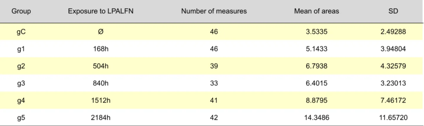
Table 1 – Mean areas of PVDCT with exposure time and SD within groups
Group Exposure to LPALFN Number of measures Mean of areas SD
gC Ø 46 3.5335 2.49288 g1 168h 46 5.1433 3.94804 g2 504h 39 6.7938 4.32579 g3 840h 33 6.4015 3.23013 g4 1512h 41 8.8795 7.46172 g5 2184h 42 14.3486 11.65720
ARTIGO ORIGINAL
RESULTS
Morphometric analysis
Table 1 and its graphic representation (Fig. 1) show that the areas of PVDCT have a growing change to the g2, 3 weeks, decreasing slightly in the g3 (5 weeks), to grow again until the g5 (13 weeks) (Fig. 1).
As is well known, the use of One-Way ANOVA for group comparisons requires that the data have certain properties, namely normal distribution within groups and homogeneity of variances between groups; therefore, the validity of these assumptions was checked prior to the data analysis, using the Shappiro-Wilk test of normality and the Levene test for variances. These procedures showed that the two assump-tions were violated, in which case the Welch ANOVA is pref-erable for the purpose of group comparisons. The results revealed the existence of significant differences in the mean PVDCT between groups, with p < 0.001 (Table 2).
In these conditions, multiple comparisons between
groups were conducted using the Tamhane test, which is an appropriate post-hoc test in the case of unequal vari-ances. It was found that the control group shows significant differences when compared to the other groups. We’ve also found that there are significant differences in the groups exposed to the LPALFN, with the exception of the g4 (9 weeks of exposure to LPALFN) which only shows a signifi-cant difference towards the control group. The g5 (13 weeks of exposure to LPALFN) is the one that presents the larg-est differences relative to the other groups exposed to the LPALFN and seems to indicate a trend for higher growth ar-eas of PVDCT with growing exposure to the LPALFN. Com-paring groups g2 (3 weeks) and g3 (5 weeks of exposure to the LPALFN) we can see that there is a stabilization of the average areas of PVDCT (p = 1.000). Finally, a contrast analysis was performed to see if there was an increasing linear trend in areas across the groups (Table 3).
The results presented on the last table show that the global trend for areas of PVDCT is increasing, and that the average area of PVDCT increases linearly and significantly with time of exposure to LPALFN (p < 0.001).
DISCUSSION
Alterations attributed to LPALFN are secondary to the degenerative cellular changes; to fibrosis8,10;
immunologi-cal25,26 and genotypical alterations28,29 and vascular lesions
that occurs in small, medium and large caliber vessels.19,20
The fibrotic processes are characterized by an abnormal and abundant deposition of extracellular matrix compo-nents.32 They have a multifactorial nature and result of a
long-term activation of fibroblasts in the affected organs33
in response to chronic or inflammatory stimuli,34 occurring
also in senescence, in response to ischemia or in neuro-endocrine changes.35 In the case of fibrotic proliferation
at-tributed to LPALFN, the described alterations occur in the absence of inflammatory signs.10 In our study we did not Table 2– Welch test
Statistic df1 df2 Sig.
Welch 12.308 6 116.544 < 0.001
Asymptotically F distributed.
Robust Tests of Equality of Means - Area
Table 3 – ANOVA (contrast analysis) Area
Sum of Squares df Mean Square F Sig. Between Groups Within Groups Total (Combined) Linear Term Weighted Deviation 4542.024 4320.384 221.640 20408.408 24950.432 6 1 5 276 282 757.004 4320.384 44.328 73.944 10.238 58.428 0.599 0.000 0.000 0.700 ANOVA - Area
Figure 1 – Evolution of mean PVDCT with time of exposure to LPALFN (p < 0.001).
ARTIGO ORIGINAL identify cells or signs of any inflammatory process. The absence of inflammatory signs makes the situation
unusual from the point of view of conventional medical con-cepts, since processes of fibrosis are usually accompanied by inflammation.10 However there are descriptions of clinical
fibrotic processes in the absence of inflammatory signs. The idiopathic pulmonary fibrosis is an example where there is fibroblastic proliferation and remodeling of the extracellular matrix without inflammation that leads to an irreversible dis-tortion of the architecture of the lung.36
In the parotid gland there are two populations of colla-gen fibrils: those associated with fibroblasts and those pres-ent in the epithelium of excretory ducts and blood vessels. There is no significant difference in the thickness of the two types of collagen fibrils, which are separated only by a small cluster of elastic fibers.30 According to Hosoyamada et al.
the location of the collagen fibrils in laminae densae of the basement membrane indicates that the epithelial cells syn-thesize and secrete collagen fibrils,30 as is the case of the
fibrous layer of the liver37 - leaflet of Glisson -, or the
glomer-ular capsule of Bowman.38 The production of fiber-forming
collagen fibrils type I, III and V, was also detected in several types of epithelia in experimental models of fibrosis.30
In intestinal fibrosis associated with radiation, the ex-pression of connective tissue growth factor (CTGF) is in-creased,39 and in vitro isolation of intestinal muscle cells
also exposed to radiation40 showed a high concentration of
constituent CTGF and a collagen increased production ca-pacity. Bourgier et al. were able to verify that the path Rho / Rho kinase is related to the activation of CTGF, and that its blockade reduced the fibrogenic differentiation.40
We have to call up the attention to the references found in relation to the regulation of gene over-expression of mol-ecules such as collagen, in particular by growth factors, in diseases related to mechanical stress, resulting from in-creased external forces applied to tissues, and mechani-cal changes in the actin cytoskeleton of the cells. These forces are significantly increased in several conditions such as hypertension, the organic obstruction or hemodynamic overload.6 In these conditions the cellular components of
or-gans, particularly fibroblasts, endothelial cells and smooth muscle cells are subjected to a mechanical stress that goes beyond what happens under normal conditions.41 The
transmission of such forces to cells in organs such as blood vessels causes a production of growth factors, cytokines or hormones that leads to hypertrophic, hyperproliferative or fibrotic responses.41 Somehow the impact caused by
mechanical vibration or by direct LPALFN may have com-mon paths on a physical point of view with what happens in some of the diseases in which abnormal mechanical stimuli are applied cyclically to the tissues. Consistent with these considerations is that in vitro studies demonstrated that mechanical forces applied to cells, for example fibroblasts, result in profound histomorphometric, phenotypic and func-tional changes.2,4,5 In parotid gland exposed to LPALFN we
have seen morphometric changes. Oliveira et al proved the existence of functional changes, with reduction in saliva
se-cretion and its qualitative change.24
The apparent stabilization of fibrosis caused by LPALFN on g2 to g3 may indicate an adaptive mechanism in which compensatory signaling pathways are activated to enable the gene transcription to go back to normal levels.42 In the
same line is the statement of Reis Ferreira et al that, un-like the fibrotic proliferation in response to an inflammatory stimulus, the tissue exposed to LPALFN, particularly in the airways, seems to reflect a structural reinforcement in or-der to assimilate the abnormal vibration stress.17 This
struc-tural reinforcement would be achieved by mass production of collagen.17 In the parotid gland structural reinforcement
would be achieved by increased PVDCT. The PVDCT at-tached to the arteries, veins and the ductal tree is thought to function as mechanical stabilizer of the glandular tissue.30
Verrecchia et al reported that in fibrosis caused by chronic stimulation, the tissue injury and the attempt of regeneration process are implicated.32 Thus, in the parotid gland, the
pe-riod from the 3rd to the 5th weeks of exposure to LPALFN, could be a recovery response in the stroma, which tends to imbalance with the continued application of mechanical stimulation. Also Kisseleva et al. reported that despite the fibrogenesis is a repair mechanism it tends to imbalance with extensive deposition of the matrix proteins and fibrosis with the chronicity of the stimulus on the tissue.43
It seems important at this stage to discuss the mecha-nisms that lead to fibrosis found in the gland and caused by LPALFN, trying to understand the transmission of mechani-cal energy to the cell, the concept of mechanotransduction; this was associated to the vibroacoustic disease and biolog-ical effects of LPALFN as the pathway by which the sound pressure damages the cells and tissues.10 A starting point
for the interpretation of this recent concept in the cell biol-ogy that may have an application in the area of biological effects of LPALFN is the interesting experience of Naruse et al. that used low-intensity pulsed ultrasound to acceler-ate the repair of fractures and distraction osteogenesis.44
The authors attributed the osteoblastic differentiation to mechanical stimuli, noticing that many physical forces may have consequences in the micro-environment of each cell. The authors also suggested the involvement of integrins in this process at the level of focal adhesions, which are a connection between the matrix and the cells.3 The integrins
seem to be the primary route for the transmission of forces into the cell and are seen as candidates for the starting of mechano-sensitive events.45 Munger et al demonstrated
that bleomycin, a known fibrotic agent,46 didn’t cause
pul-monary fibrosis in mice deficient in integrin αvβ6, despite the intense inflammatory reaction.47
Mechanotransduction is the mechanism that converts physical stimuli into biochemical signs and integrates these signs into cellular responses at the molecular level.3 We
assume that in intact tissues the cells react to mechanical stimuli with molecular responses that aim to protect their integrity. These forces on the cells may be applied in many different ways provided they have a sufficient magnitude to cause a biological response in the cell.45 The response of
ARTIGO ORIGINAL endothelial cells to mechanical stress caused by fluid
pres-sure is the best studied.48 It is known the contribution of this
kind of mechanical stimulus in the induction of endothelial cell proliferation49;it is also known that the critical level of
force to initiate various biological responses in cells is ap-proximately 1 Pascal.50
Although we cannot make a direct comparison with the processes mentioned in the previous paragraph we should not forget the conclusion of Huang et al: despite the appar-ent complexity of the process of cell mechanotransduction is likely that cells stimulated in different ways can be acti-vated by similar molecular mechanisms.45
CONCLUSIONS
We can conclude that exposure to LPALFN causes PVDCT fibrosis. This fibrosis is significant from the first week of exposure and increases with the increase of expo-sure time. LFN may activate mechanisms of cellular mecha-notransduction that we intend to characterize in future stud-ies. CONFLICT OF INTERESTS None stated. FUNDING SOURCES None stated. REFERENCES
1. Wang N, Butler JP, Ingber DE. Mechanotransduction across the cell surface and through the cytoskeleton. Science. 1993;260:1124-7. 2. Ingber DE. Mechanical signalling and the cellular response to
extracellular matrix in angiogenesis and cardiovascular physiology. Circ Res. 2002;91:877–87.
3. Ingber DE. The mechanochemical basis of cell and tissue regulation. Mech Chem Biosyst. 2004;1:53-68.
4. Silver FH, Devore D, Siperko LM. Role of mechanophysiology in aging of ecm: effects of changes in mechanochemical transduction. J Appl Physiol. 2003;95:2134–41.
5. Janmey PA, Weitz DA. Dealing with mechanics: mechanisms of force transduction in cells. Trends Biochem Sci. 2004;29:364–70.
6. Chaqour B, Goppelt-Struebe M. Mechanical regulation of the Cyr61/ CCN1 and CTGF/CCN2 proteins. FEBS J. 2006;273:3639-49. 7. Alves-Pereira M. Noise-induced extra-aural pathology: a review and
commentary. Aviat Space Environ Med. 1999;70:A7-21.
8. Castelo Branco NA. The Vibroacoustic disease: an emerging pathology. Aviat Space Environ Med. 1999;70:A1-6.
9. Castelo Branco NA, Alves-Pereira M. Vibroacoustic disease. Noise Health. 2004;6:3-20.
10. Alves-Pereira M, Castelo Branco NA. Vibroacoustic Disease: biological effects of infrasound an low frequency noise explained by mechanotransduction cellular signaling. Prog Biophys Mol Biol. 2007;93:256-79.
11. Alves-Pereira M, Joanaz de Melo J, Marques MC, Castelo Branco NA. Vibroacoustic disease: the response of biological tissue to low frequency noise. Prog Biophys Mol Biol. 2004;295-308.
12. Grande NR, Águas AP, Sousa Pereira A, Monteiro E, Castelo Branco NA. Morphological changes in the rat lung parenchyma exposed to low frequency noise. Aviat Space Environ Med. 1999;70:A70-7.
13. de Sousa Pereira A, Grande NR, Monteiro E, Castelo Branco MS, Castelo Branco NA. Morphofunctional study of rat pleural mesothelial cells exposed to low frequency noise. Aviat Space Environ Med. 1999;70:A78-85.
14. Oliveira MJR, Pereira AS, Castelo Branco NA, Grande NR, Águas AP. In utero and postnatal exposure of wistar rats to low frequency/high intensity noise deplets the tracheal epithelium of ciliated cells. Lung. 2002;179:225-32.
15. Oliveira MJR, de Sousa Pereira A, Ferreira PG, Grande NR, Águas AP, Guimarães L, et al. Reduction of rat pleural microvilli caused by noise pollution. Exp Lung Res. 2003;29:445-54.
16. Castelo Branco NA, Monteiro E, Costa e Silva A, Reis Ferreira J, Alves-Pereira M. Respiratory epithelia in Wistar rats. Rev Port Pneumol. 2003;IX-5:381-8.
17. Reis Ferreira J, Sousa JA, Foreid P, Antunes M, Cardoso S, Alves-Pereira M, et al. Drive respiratório anormal na doença vibroacústica. Rev Port Pneumol. 2006;12:369-74.
18. Castelo Branco NA, Águas AP, Sousa Pereira A, Monteiro E, Fragata FIG, Tavares F, et al. The human pericardium in vibroacoustic disease. Aviat Space Environ Med. 1999;70:A54-62.
19. Martins dos Santos J, Grande NR, Castelo Branco NA, Zagalo C, Oliveira P. Vascular lesions and vibroacoustic disease. Eur J Anat. 2002;6:17-21.
20. Martins dos Santos J, Grande NR, Castelo Branco NA, Zagalo C,
Oliveira P, Alves-Pereira M. Lymphatic lesions and vibroacoustic disease. Eur J Lymphol. 2004;12:17-20.
21. da Fonseca J, Martins dos Santos J, Castelo Branco N, Alves-Pereira M, Grande NR, Oliveira P. Noise-induced duodenal lesions. Eur J Anat. 2006;9:29-33.
22. da Fonseca J, dos Santos JM, Branco NC, Alves-Pereira M, Grande NR, Oliveira P, et al. Noise-induced gastric lesions: a light and scanning electron microscopy study of the alterations of the rat gastric mucosa induced by low frequency noise. Cent Eur J Public Health. 2006;14:35-8. 23. Mendes J, Martins dos Santos J, Oliveira P, Castelo Branco NA. Low
frequency noise effects on the periodontium of the wistar rat: a light microscopy study. Eur J Anat. 2007;11:27-30.
24. Oliveira PM, Pereira da Mata AD, Martins dos Santos JA, da Silva Marques DN, Branco NC, Silveira JM, et al. Low-frequency noise effects on the parotid gland of the Wistar rat. Oral Dis. 2007;13:468-73. 25. Águas AP, Esaguy N, Grande NR, Castro AP, Castelo Branco NA. Effect
of low frequency noise exposure on BALB/c mice splenic lymphocytes. Aviat Space Environ Med. 1999;70:A128-31.
26. Águas AP, Esaguy N, Grande NR, Castro AP, Castelo Branco NA. Acceleration of lupus erythematosus-like processes by low frequency noise in the hybrid NZB/W mouse model. Aviat Space Environ Med. 1999;70:A132-6.
27. Oliveira MJ, De Sousa Pereira A, Águas AP, Grande NR, Monteiro E, Serrano I, et al. Effects of low frequency noise upon the reaction of pleural milky spots to mycobacterial infection. Aviat Space Environ Med. 1999;70:A137-40.
28. Silva MJ, Carothers A, Castelo Branco N, Dias A, Boavida MG. Increased levels of sister chromatid exchanges in military aircraft pilots. Mutat Res. 1999;441:129-34.
29. Silva MJ, Dias A, Barreta A, Nogueira PJ, Castelo-Branco NA, Boavida MG. Low frequency noise and whole-body vibration cause increased levels of sister chromatid exchange in splenocytes of exposed mice. Teratog Carcinog Mutagen. 2002;22:195-203.
30. Hosoyamada Y, Sakai T. The ultrastructure of periductal connective tissue and distinctive populations of collagen fibrils associated with ductal epithelia of exocrine glands. Arch Histol Cytol. 2003;66:407-18. 31. Krinke, GJ. The handbook of experimental animals. The laboratory rat.
Salt Lake City: Academic Press; 2000.
32. Verrecchia F, Mauivel A. TGF-Beta and TNF-alpha: antagonistic cytokines controlling type I collagen gene expression. Cell Signal. 2004;16:873-80.
33. Verrecchia F, Mauviel A. Control of connective tissue gene expression by TGF beta: role of smad proteins in fibrosis. Curr Rheumatol Rep. 2002;4:143-9.
34. Samuel CS. Relaxin: antifibrotic properties and effects in models of disease. Clin Med Res. 2005;3:241-9.
35. Swynghedauw B. Molecular mechanisms of myocardial remodelling. Physiol Rev. 1999;79:215-62.
36. Selman M, King TE, Pardo A, American Thoracic Society, European Respiratory Society, American College of Chest Physicians. Idiopathic pulmonary fibrosis: prevailing and evolving hypotheses about its pathogenesis and implications for therapy. Ann Intern Med. 2001;134:136-51.
ARTIGO ORIGINAL
distribution of collagen fibrils in Glisson’s sheath of rat liver: implications for mechanical environment and possible producing cells. J Anat. 2000;196:327-40.
38. Mbassa G, Elger M, Kriz W. The ultrastructural organization of the basement membrane of Bowman’s capsule in the rat renal corpuscle. Cell Tissue Res. 1988;253:151-63.
39. Vozenin-Brotons MC, Milliat F, Sabourin JC, de Gouville AC, Francois A, Lasser P, et al. Fibrogenic signals in patients with radiation enteritis are associated with increased connective tissue growth factor expression. Int J Radiat Oncol Biol Phys. 2003;56:561–72.
40. Bourgier C, Haydont V, Milliat F, François A, Holler V, Lasser P, et al. Inhibition of Rho kinase modulates radiation induced fibrogenic phenotype in intestinal smooth muscle cells through alteration of the cytoskeleton and connective tissue growth factor expression. Gut. 2005;54:336-43.
41. Hill MA, Davis MJ, Meininger GA, Potocnik SJ, Murphy TV. Arteriolar myogenic signalling mechanisms: implications for local vascular function. Clin Hemorheol Microcirc. 2006;34:67–79.
42. Abdel WN, Mason RM. Connective tissue growth factor and renal diseases: some answers, more questions. Curr Opin Nephrol Hypertens. 2004;13:53–8.
43. Kisseleva T, Brenner DA. Mechanisms of fibrogenesis. Exp Biol Med.
2008;233:109-22.
44. Naruse K, Miyauchi A, Itoman M, Mikuni-Takagaki Y. Distinct anabolic response of osteoblast to low-intensity pulsed ultrasound. J Bone Miner Res. 2003;18:360-9.
45. Huang H, Kamm, RD, Lee RT. Cell mechanics and mechanotransduction: pathways, probes, and physiology. Am J Physiol Cell Physiol. 2004;287:C1-11.
46. Thrall RS, McCormick JR, Jack RM, McReynolds RA, Ward PA. Bleomycin-induced pulmonary fibrosis in the rat: inhibition by indomethacin. Am J Pathol. 1979;95:117-30.
47. Munger JS, Huang X, Kawakatsu H, Griffiths MJ, Dalton SL, Wu J, et al. The integrin αVβ6 binds and activates latent TGF beta 1: a mechanism for regulating pulmonary inflammation and fibrosis. Cell. 1999;96:319-328.
48. Malek AM, Izumo S. Control of endothelial cell gene expression by flow. J Biomech. 1995;28:1515–28.
49. White CR, Haidekker M, Bao X, Frangos JA. Temporal gradients in shear, but not spatial gradients, stimulate endothelial cell proliferation. Circulation. 2001;103:2508–13.
50. Helmke BP, Thakker DB, Goldman RD, Davies PF. Spatiotemporal analysis of flow-induced intermediate filament displacement in living endothelial cells. Biophys J. 2001;80:184–94.