ASSESSMENT OF CHROMOPHORES IN CHEMICALLY TREATED AND
AGED WOOD BY UV-VIS DIFFUSE REFLECTANCE SPECTROSCOPY
TEREZA TRIBULOVÁ,* FRANTIŠEK KAČÍK,* DMITRY EVTUGUIN** and IVETA ČABALOVÁ*
*
Department of Chemistry and Chemical Technologies, Technical University in Zvolen, 24, T. G. Masaryka, 960 53 Zvolen, Slovak Republic
**
CICECO, Department of Chemistry, University of Aveiro, Campus Universitário de Santiago, 3810-193 Aveiro, Portugal
✉Corresponding author: František Kačík, [email protected]
Dedicated to the 50th anniversary of Cellulose Chemistry and Technology and to the memory of its founder Acad. Prof. Cr. I. Simionescu
The optical properties and degradation of fir wood treated by different inorganic salts, which are components of waterborne wood preservatives, were studied before and after wet-thermal accelerated ageing (T=85 °C, RH=65%, for 30 days) using UV-Vis diffuse reflectance spectroscopy. The colour of all impregnated samples was due to the same chromophoric structures, thus revealing that the primary reactions leading to colour changes are essentially very similar. These chemically treated as well as standard (non-treated, unaged) and reference (treated only with distilled water, unaged/aged) samples have a significant absorption band at 358 nm, along with a shoulder near 265 nm of various intensity. The broad band at higher wavelength is attributed to the chromophoric structures derived especially from lignin, i.e. quinones, stilbenes, charge transfer complexes causing a bathochromic shift and other conjugated carbonyl structures. Coniferaldehyde is also a major contributor to this absorption. The band at 265 nm belongs to chromophores arising predominantly from carbohydrate degradation products, i.e. furan derivatives, quinone-type derivatives, catechol structures. Catechol structures (colourless) form coloured complexes with transition metal cations (Cu2+, Fe3+) or are oxidized to form highly coloured benzoquinone structures. Alkaline solutions of borax cause the reduction of conjugated and non-conjugated carbonyls and the degradation of coniferaldehyde. Generally, after ageing, the samples show higher absorptions in the visible region of the spectra in comparison with the unaged ones, which explains their yellowing and loss of brightness after the action of elevated temperature and moisture.
Keywords: accelerated ageing, chemically treated wood, chromophores, diffuse reflectance spectroscopy, inorganic salts
INTRODUCTION
Wood is a fairly good absorber of light and is capable of interacting with a range of wavelengths of electromagnetic radiation, owing to the wide range of chromophoric groups associated with its basic components.1,2 Cellulose and hemicellulose have been shown to have a relatively low absorption in both the visible and the ultraviolet regions. Lignin, on the other hand, has an absorption that is low in the visible wavelength region but increases rapidly with decreasing wavelength into the ultraviolet regions.3,4 Lignin contributes by 80-95%, the carbohydrates by 5-20%, and the extractives by about 2% to the absorption coefficient.5 According to Falkehad et
al.,6 the colour of natural light-yellow wood is most likely caused by chromophores in lignin and extractives, and also possibly sometimes by organometallic complexes in extractives.
Oxidative and hydrolytic reactions, including depolymerization, which change both the appearance and chemical properties of wood, are primarily considered to be the cause for the production of chromophores during thermal treatment of wood, where hydrolytic reactions are generally the dominant process when moisture is present.7
Considering the colour changes in wood and other lignocellulosics due to modification and
ageing, many investigations have been done and there is a variety of explanations for colour formation and the degradation mechanism resulting from weathering, heating and drying,8-11 but no literature sources were found to thoroughly describe the chemical reasons for the colour changes in solid chemically treated wood by inorganic compounds used as components of wood preservatives after wet-thermal accelerated ageing. However, after a literature overview, it can be concluded that these changes cannot be solely explained by a wood component. These colour changes seem to originate from complex changes and degradation of hemicelluloses, lignin and certain extractives.
The aim of this paper is to evaluate the method of UV-Vis diffuse reflectance (UV-Vis DR) spectroscopy in studies of optical properties of chemically treated and wet-thermally aged wood. This paper describes the changes in the UV-Vis DR spectra of the thus treated fir wood samples with the objective of characterizing their UV-active components and the chemical changes occurring after impregnation by certain chemicals and after accelerated ageing.
EXPERIMENTAL Materials
Fir wood (Abies alba) samples were first conditioned to constant weight at a temperature of 23 ± 1 °C and relative air humidity of 50 ± 2%, and then chemically treated. For the treatment of 10 sample series, aqueous solutions of selected chemicals at a 5 wt% concentration level were used, i.e. Na2B4O7.10H2O, H3BO3,CuSO4.5H2O, ZnSO4.7H2O, Fe2(SO4)3, NaCl, NH4Cl, (NH4)2SO4, (NH4)2HPO4, NH4H2PO4. Moreover, standard non-treated unaged samples and reference unaged and aged samples impregnated by distilled water were also included into this experiment.
Impregnation of individual series of samples was performed in the laboratory desiccator under reduced pressure for 60 minutes with subsequent immersion into a salt solution under atmospheric pressure for another 60 minutes. This double-step process was repeated three times, each time with a freshly prepared solution.
One half of the treated wood samples were kept in a climatic chamber (Memmert HCP 108, Germany) at 80 °C and 65% RH for 30 days. (The samples that released ammonia during accelerated ageing were subjected to elevated temperature and humidity separately in another climatic chamber with the intention to prevent contamination of other samples with ammonia.) Prior to testing, all specimens were
re-equilibrated to constant weight at 23 ± 1 °C and 50 ± 2% RH, according to standard ISO 187.12
Methods
The contribution of chromophores to chemically treated and aged wood colour was analyzed by UV-Vis diffuse reflectance spectroscopy (UV-Vis DR) in the range of wavelengths 200-600 nm. UV-Vis DR spectra were recorded for early wood on the tangential cross section of the plywood (10 × 10 mm and thickness about 1mm) samples, using a JASCO V-560 spectrophotometer (scanning speed of 200 nm/min, bandwidth of 5 nm) equipped with a JASCO ISV-469 integrating sphere, and with BaSO4 standard as a background reference. Two replicates were analysed for each sample. The amount and type of light absorbing materials or chromophores were characterized by the Kubelka-Munk light absorption coefficient (k/s) at different wavelengths.
Brightness values (R∞, %) were taken from the k/s absorption spectra at a wavelength of 457 nm and are not identical with ISO or TAPPI brightness values, for which a specific geometry is required.13
RESULTS AND DISCUSSION
UV-Vis DR spectroscopy provides a tool for characterizing wood chromophores and changes in their concentration correlated with the discolouration of wood during chemical and wet-thermal treatment.14 Chemical treatment with selected inorganic chemicals and wet-thermal ageing of wood result in an increase of the absorption band at around 358 nm and its shoulder near 265 nm, in comparison with the standard sample. The only exception is the wood treated by Na2B4O7.10H2O after ageing with lower intensity of both absorption bands, compared to the standard. The close similarity observed in the spectra of the samples impregnated by different chemical compounds indicates that the changes in the optical properties of wood after treatment and after ageing may be assigned in all these cases to the same group of chromophores.
Moreover, the colour of wood is modified, turning to a yellowish-brown, upon ageing caused by an elevated temperature and the presence of moisture. According to Chen et al.,1 5 the increase in chromophores upon heating is enhanced with an increase in the initial moisture content. This can be explained as being due to the formation of coloured degradation products, which absorb the light in the visible region. The absorptions at >300 nm are indicative of conjugated structures.16 The absorbance of blue and violet colour (380-
520 nm) is recognized as a yellow colour. The absorption coefficient of all samples (except the sample treated by ZnSO4.7H2O) after ageing, in the visible region, is always higher than that for the same samples before ageing, i.e. the brightness of treated wood after ageing is always lower in comparison with that of treated unaged wood (note the k/s values at 457 nm).
Contribution of lignin
The absorption maximum of lignin structures in wood in the UV region of the measured spectra is detected at around 358 nm and is caused by various conjugated structures, containing both polyunsaturated aromatic structures and conjugated carbonyl groups.
Schmidt et al.17 and Paulsson et al.18 reported that near-UV irradiation causes increases in the absorption coefficient due to the formation of aromatic carbonyl groups at approx. 330 nm and as yet unidentified chromophores at approx. 360 nm (possibly methoxylated para- or ortho-quinones), and that these two peaks coalesce into a single broad peak with maximal intensity at approx. 360 nm. According to Agarwal,19 the formation of carbonyl groups can be explained by the photo-reduction of p-quinones in the presence of primary or secondary alcohols originated from either carbohydrates or lignin, which act as hydrogen donors. Imsgard et al.20 found that a polymeric quinone made by oxidizing absorbed at 380 nm. This band can have a broad tail into the visible region and would certainly be a major contributor to the wood colour. Simple stilbenes have an absorption maximum at about 330 nm in solution, but it is possible that this maximum can be shifted to higher wavelengths when incorporated in the lignin, respectively a wood matrix. A bathochromic shift of the UV-Vis absorption maxima of quinone and stilbene model compounds in the solid state (to a wavelengthof about 350 nm), compared to the absorption in solution, has been reported.21,22 Charge transfer complexes (quinon-phenol) and some auxochromes might also contribute to this intensification and to the shift to longer wavelengths.23 It is also possible that coniferaldehyde, the major contributor to the colour of native lignin in wood with an absorption maximum at 350 nm,24 generated through oxidation of coniferyl alcohol end-groups, contributes to this lignin characteristic UV-absorption peak of chemically treated and aged wood.
Simple prototypes for the chromophoric structures in lignin are given in Fig. 1. Structures referred to as leucochromophores are potential chromophores that can be converted into chromophores upon oxidation.25
Contribution of polysaccharides
The absorption band, respectively a shoulder of broad band corresponding to lignin chromophore structures, at about 265 nm belongs to chromophores arising predominantly from carbohydrate degradation products. In this case, sugar degradation products, the least stable wood components against chemical treatment and wet-thermal accelerated ageing process, could be intermediates in the colour formation. However, the cellulose may contribute to the formation of chromophores during some oxidation reactions due to free radicals, too.26
Sugar degradation is linked to the presence of furanic aldehydes in wood. Hodge27proposed a mechanism explaining that 5-hydroxymethyl-furfural and 5-methyl5-hydroxymethyl-furfural came from hexoses existing in cellulose, and how furfural comes from pentoses, the principal constituents of hemicelluloses. Several authors note that untreated and non-aged wood already contains small amounts of furfural, 5-methylfurfural28 and 5-hydroxymethylfurfural.29 For instance, 2-furancarboxylic acid can be formed under acidic conditions. Soares et al.30 suggest that furfural is the most abundant in thermally degraded wood, due to hemicelluloses being highly instable at increasing temperature. Chemical treatment plays an important role in hexoses and pentoses degradation coupled with the creation of chromophores, as indicated by the loss of brightness,26 but the effect of wet-thermal ageing seems to be stronger.
In particular, certain aromatic products (Fig. 1) absorbing at this wavelength (e.g. reduced structures of quinones) can also contribute to this band. Oxidised sugar residues absorbing at ≤ 230 nm may be degraded during wet-thermal accelerated ageing to quinone-type derivatives, respectively catechols (Theander products). Therefore, after ageing, new unsaturated structures with absorption above 250 nm are formed because of the degradation of cellulose components absorbing below 240 nm.31 Ziobro32 reported that the degraded model sample of glucose has a high absorptivity at 254 nm and at around 280 nm. This author also stated that the termination of the peeling reactions of cellulose in
alkaline media could result in terminal keto-enols and that these enols could be responsible for colour. Figure 2 shows some examples of the
family of chromophores originating from carbohydrates.
Figure 1: Lignin derived chromophoric and leucochromophoric structures and their associated absorbance25
Figure 2: Some proposed chromophoric structures as carbohydrate degradation products, a) furan derivatives;27 b) chromophores proposed by Ziobro along with their absorbance maxima32
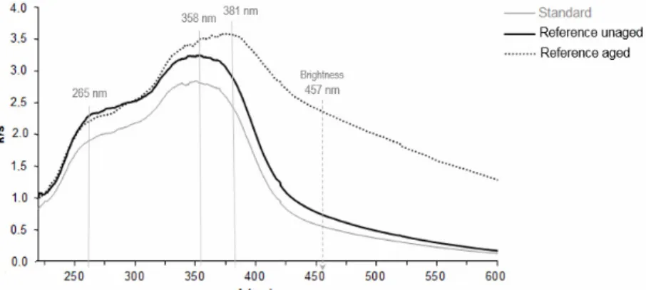
Spectra of reference sample and bathochromic shift of its aged form
The reference sample (treated only with distilled water) provides a very similar spectral curve to that of the standard sample, but with slightly higher absorption coefficients at 265 nm and 358 nm. However, the absorption coefficient of the aged reference sample at 265 nm is lower and at 358 nm much higher in comparison with the values for the same sample before ageing. In the spectrum of the aged reference sample, a bathochromic shift from approximately 23 nm to 381 nm is observed for the absorption band corresponding to lignin chromophoric structures (Fig. 3). A similar red-shift is also characteristic of the aged samples impregnated by NaCl (from about 19 nm to 377 nm) and sulphates containing transition metal cations, i.e. CuSO4.5H2O and Fe2(SO4)3 (from about 34 nm to 392 nm).
Comparison across all spectra of treated and aged samples
In general, the intensity of two main absorption bands (265 nm and 358 nm, respectively) of impregnated and of aged wood samples can be lower or higher in comparison with that of the reference sample before and after ageing (Fig 4). The group of chemicals, which cause an increase of the absorption coefficient at 358 nm with respect to the unaged reference wood, includes: (NH4)2SO4, (NH4)2HPO4, ZnSO4.7H2O, whose impact is recognized in treated wood before ageing, NaCl and Fe2(SO4)3, which cause increasing intensity before as well as after ageing, and CuSO4.5H2O, which causes this change after wood ageing. It is worth noting that the course of spectral curves of the samples treated by the last two listed chemicals, i.e. NaCl (pH ≈ 5.4) and CuSO4.5H2O (pH ≈ 2.8), is very similar despite their different pH values, which may result in different intensity of the degradation reaction.33The highest absorption intensity in all parts of the visible and UV regions of the spectra (two absorption bands at 265 nm and 392 nm) was recorded for the sample treated by Fe2(SO4)3 after wet-thermal ageing. This is also the only sample with a higher absorption coefficient related to chromophores generated from lignin, in comparison with the aged reference sample.
On the other hand, the group of chemicals leading to a reduction of the absorption coefficient of impregnated wood, compared to the reference sample, before ageing at the wavelength
of 358 nm and after ageing at 265 nm, consists of: Na2B4O7.10H2O, H3BO3, ZnSO4.7H2O, NH4H2PO4, (NH4)2HPO4, (NH4)2SO4, NH4Cl. All these chemicals cause a drop of intensity for the analyzed wood samples after ageing, only Na2B4O7.10H2Ohas the same effect both after and before ageing. Aged wood treated by Na2B4O7.10H2O shows extremely low absorption coefficients in the UV region, compared to the other analyzed samples, but in the visible region the absorption intensity is even higher than that for the standard and reference unaged samples. Likewise, the above mentioned inorganic salts with lower absorption intensity in the UV region have an increased absorption in the visible region of the measured spectra, which is in each case higher than that of the reference unaged sample, and additionally, for phosphates and NH4Cl, it is very close to that of the reference sample after ageing.
The highest absorption coefficients, even higher than for the aged reference sample, were recorded in the visible region for wood samples treated by Fe2(SO4)3, CuSO4.5H2O, NaCl (at 428 nm) and partially NH4Cl (between 421 nm and 468 nm). For these samples, due to their strong absorption at the wavelength of 457 nm, the lowest brightness values of all analysed wood samples can be observed. Conversely, the standard sample shows the highest brightness (note the k/s values at 457 nm).
Role of borates and reduction of chromophores due to alkaline environment
The highest intensity in the UV region of the spectra measured for the borate-treated sample series has been recorded for unaged wood impregnated by H3BO3 and in the visible region for the same sample but after ageing (Fig. 4a). Before ageing, the wood impregnated by Na2B4O7.10H2O shows at the wavelength of 265 nm a similar absorption coefficient to those of the unaged and aged samples treated by H3BO3. The absorption coefficient of this borax-treated sample is at the wavelength of 358 nm, close to that of the aged sample treated by H3BO3, and its spectral intensity in the visible region is slightly higher than that of the unaged wood treated by H3BO3.Also, it is nearly the same as the absorption coefficient for the same sample after ageing. The extreme decline in the intensity of two absorption bands in the UV region for the aged sample impregnatedbyNa BO.10HO is
explained by the influence of the alkaline environment (the pH of the cold water extract of this sample was 9.4), which significantly reduced
the early formation of chromophores in modified wood.34
Figure 3: k/s absorbance spectra of standard (untreated non-aged fir wood sample) and reference samples (treated by distilled water) before and after wet-thermal accelerated ageing process (T = 85 °C, RH = 65%, for 28 days)
Figure 4: k/s absorbance spectra of chemically treated fir wood samples with selected inorganic chemicals; a) Na2B4O7.10H2O, H3BO3; b) CuSO4.5H2O, ZnSO4.7H2O, Fe2(SO4)3; c) NaCl, NH4Cl d) (NH4)2SO4, (NH4)2HPO4, NH4H2PO4,before and after wet-thermal accelerated ageing process (T =85 °C, RH=65%, for 28 days) compared with the standard (untreated non-aged)fir wood sample
Alkaline solutions of borates also cause the reduction of conjugated and non-conjugated carbonyls to the corresponding alcohols.35The decrease of the absorption band at 358 nm can be attributed to the degradation of coniferaldehyde.36
Role of transition metal sulphates and Fenton reactions
From the series of unaged samples treated by sulphates containing transition metal cations, the lowest spectral intensity was noted for the sample
impregnatedbyCuSO4.5H2O and the highest spectral intensity – for the sample impregnated by ZnSO4.7H2O (except the visible region from the wavelength of 420 nm, where the wood treated by Fe2(SO4)3 provided stronger absorption) (Fig. 4b). Conversely, the lowest spectral intensity of the aged wood was recorded for the sample treated by ZnSO4.7H2O, a significantly higher one – for the sample treated by CuSO4.5H2O, and the highest – for the one treated with Fe2(SO4)3. The sample impregnated by CuSO4.5H2O shows the greatest difference in spectral intensity before and after ageing. Sulphates with Cu2+ and Fe3+ cations cause a bathochromic shift of the absorption band from 358 nm to the wavelength of 392 nm, which may result from a relatively higher energy of electronic transitions in the ultraviolet region. However, in some cases, such as the ferric ion and catechol complex, the energy of the electronic transitions related with the charge transfer is reduced significantly due to an increase in the resonance system afforded by the aromatic ring, thus increasing the overall absorbance in the visible region. Also, an increase in the resonating system of the conjugated ligand would tend to release the restraint on the electrons causing the absorption band.37
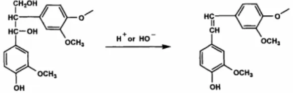
In the presence of transition metal sulphates (with Fe3+ or Cu2+ cations), the β-1 phenylic lignin structures are easily converted into the corresponding stilbene structure, according to the reaction shown in Fig. 5.38 The stilbene, in turn, is
easily oxidized to form deeply coloured quinones and stilbenequinones of various types (Fig. 1). Catechol structures (colourless) can either form coloured complexes with transition metals or be oxidized to form highly coloured benzoquinone structures (Fig. 6).The colour of metal to ligand complexes may be due to either the d-d interactions, the charge transfer between the metal ions and the ligand, the conjugated ligand, or all of the above. The d-d transitions depend largely on the transition metal ion and the donor atom of the ligand.38,39
Role of chlorides
The highest absorption coefficient at the band of 265nm was noted for the unaged sample treated by NaCl and the lowest – for the aged samples treated by both chlorides (Fig. 4c). The wood samples impregnated by NH4Cl before and after ageing show the same absorption at the wavelength of 358 nm and only slightly higher intensity is remarked for the unaged wood impregnated by NaCl. The highest absorption coefficient of this band corresponding to lignin chromophoric structures is observed for the aged sample treated by NaCl, whose maximum is shifted to 377 nm. The lowest spectral intensity in the visible region is noted for the unaged wood treated by NaCl and the highest one – for the same sample but after the treatment.
Figure 5: Formation of stilbene from β-1 structures in wood38 Role of ammonium phosphates and ammonium
sulphate
The unaged wood impregnated by (NH4)2HPO4 from this series shows the highest intensity in the UV and visible regions of the spectra (Fig. 4d). From the aged samples, the lowest spectral intensity in the UV region was exhibited by the wood treated with NH4H2PO4, which is shifted in the visible region from the wavelength of 420 nm to the highest absorption
maxima for all the samples in this series. The aged sample impregnated by (NH4)2SO4 had the highest absorption coefficients in the UV region from all the aged samples in this group. Conversely, it showed the lowest absorption intensity in the visible region. The spectrum of the aged (NH4)2SO4-treated sample is very similar to that of the unaged sample treated by (NH4)2HPO4, which, on the other hand, has the highest spectral intensity in the visible region from all unaged
samples. The wood treated by (NH4)2HPO4, in unaged and aged form, shows even higher intensity of the measured spectra in comparison with those of the unaged and, respectively, aged wood treated by NH4H2PO4. The only exception
is in the visible region from 420 nm, where the spectral intensity of the wood impregnated by NH4H2PO4 is slightly higher than that of the samples subjected to the (NH4)2HPO4 treatment.
Figure 6: Degradation of phenol structures by Fenton reaction through Fe3+(a)37and Cu2+(b)40
CONCLUSION
Upon oxidation, in lignin, there appears a cleavage of β-O-4 linkages and a reduction of methoxyl content, leading to more condensed structures. The acidic oxidation of cellulose involves both a progressive stepwise reaction, commencing with the oxidation of the methylol groups of glucose units, and a direct hydrolytic cleavage of the glycosidic bonds, followed by its destruction.
Comparing all chemically treated and aged fir wood samples, we can conclude that the main chromophoric compounds are common to all of them, namely some carbonyls, aromatic rings and aliphatic double bonds. The absorption band at 358 nm (in some cases shifted to higher wavelengths) comes from the lignin chromophoric structures, containing phenolic aldehydes (coniferaldehyde), quinones, stilbenes and charge transfer complexes. The second absorption band in the UV region of the spectra at 265 nm belongs to the chromophores arising from carbohydrate degradation. This band may be assigned to furanic derivatives (furfural, 5-hydroxymethylfurfural, 5-methylfurfural) and
some aromatic compounds (quinones, benzoquinones, catechols). Catechol structures are oxidized to form highly coloured benzoquinone structures or form coloured complexes with transition metal cations (Cu2+, Fe3+) through Fenton type reactions. These metal sulphates cause a bathochromic shift of the absorption band at 358 nm to the wavelength of 392 nm, which results from a relatively higher energy of the electronic transitions in the UV region and the band can overlap into the visible region. An alkaline solution ofNa2B4O7.10H2Ocauses the reduction of conjugated and non-conjugated carbonyls and the degradation of coniferaldehyde. The greatest difference between the spectral intensity of the unaged and aged samples is observed for the wood impregnated by CuSO4.5H2O and, on the other hand, the wood impregnated by H3BO3 has very similar spectra before and after ageing. After ageing, all the samples show higher absorption coefficients (except the sample treated withZnSO4.7H2O) in the visible region in comparison with the unaged ones, which means that after wet-thermal accelerated ageing the samples have lower values
of brightness than the same samples before ageing. Controversially, unaged samples impregnated by chemicals provide higher absorption coefficients in the UV region in comparison with their aged equivalents.
ACKNOWLEDGEMENT: This work was
supported by funding from the Slovak Scientific Grant Agency VEGA (1/0521/15).
REFERENCES
1 D. N. S. Hon, in “Developments in Polymer Degradation“, edited by N. Grassie, Applied Science Publishers, 1981, vol. 3(8), pp. 229-281.
2 H. F. Launer and W. K. Wilson, Pap. Trade J., 116, 28 (1943).
3 N. Hartler and H. Norrström, Tappi J., 52,1712 (1969).
4 H. Norrström, Sven. Papperstidn., 75, 891 (1972). 5 H. Norrström, Sven. Papperstidn., 72, 25 (1969). 6 I. S. Falkehag, J. Marton and A. Adler, in “Lignin Structure and Reactions“, edited by J. Marton, J. Am. Chem. Soc., Washington D.C., USA, 1966, pp.75-89. 7 D. Fengel and G. Wegener, “Wood: Chemistry, Ultrastructure, Reactions”, Mníchov,Veglag Kessel, 2003, p. 613.
8 W. Sandermann and F. Schlumbom,
HolzRoh-Werkstoff, 20, 285 (1962).
9
W. C. Feist and D. N. S. Hon, in “The Chemistry of Solid Wood“, edited by R. M. Rowell, J. Am. Chem. Soc., Washington D.C., 1984, vol. 11, pp. 401-451. 10 M. Polovka, J. Polovková, K. Vizárová, S. Kirschnerová, L. Bieliková et al., Vib. Spectrosc., 41,112 (2006).
11 E. O. dos Santos, A. M. S. Silva, W. D. Fragoso, C. Pasquini and M. F. Pimente, Vib. Spectrosc., 52, 154 (2010).
12 ISO 187, Paper, board and pulps – Standard atmosphere for conditioning and testing and procedure for monitoring the atmosphere and conditioning of samples, 1990, p. 8.
13 P. E. G. Loureiro, A. J. S. Fernandes, M. G. V. S. Carvalho and D. V. Evtuguin, Carbohyd. Res., 345, 1442 (2010).
14 Y. Chen, Y. Fan, M. A. Tshabalala, N. M. Stark, J. Gao et al., BioResources, 7, 1474 (2012).
15 Y. Chen, Y. Fan, J. Gaoand N. M. Stark,
BioResources, 7, 1157 (2012).
16 A. Yamamoto, A. Rohumaa, E. Kontturi, M. Hughes and T. Vuorinen, BioResources, 10, 6610 (2015).
17 J. Schmidt and C. Heitner, in “Advances in Lignocellulosic Characterization“, edited by D. Argyropoulos, TAPPI Press, Atlanta, 1999, pp. 187-209.
18
M. Paulsson and A. J. Ragauskas, Nord. Pulp
Pap. Res. J., 13,191 (1998).
19 U.P. Agarwal, J. Wood Chem. Technol., 18, 381 (1998).
20 F. Imsgard, S. I. Falkehag and K. P. Kringstad,
Tappi J., 54,1680 (1971).
21 A. Castellan, A. Nourmamode, N. Colombo, C. Jaeger, C. Noutary et al., in Procs. 6th
International Symposium of Wood and Pulp Chemistry, Melbourne,
Australia, 1991, pp. 151-158.
22 L. Zhang and G. Gellerstedt, in Procs.3rd
European Workshop on Lignocellulosics and Pulp, Stockholm,
Sweden, 1994, pp. 293-295.
23 G. S. Furman and W. F. W. Lonsky, IPC Technical
Paper Series, Number 198, The Institute of Paper
Chemistry, Appleton, WI, USA, 1986, p. 6.
24 J. C. Pew and W. J. Connors, Tappi J., 54, 245 (1971).
25
J. Polcin and W. H. Rapson, Pulp Pap. Mag. Can., 72, 80(1971).
26 C. Mateo, H. Furstoss, A. Jeunet, D. Lachenal and C. Chirat, Pulp Pap. Mag. Can., 103, 28 (2002). 27 J. E. Hodge, in “Symposium on Foods”, edited by W. Schultz, E. A. Day and L. M. Libbey, Westport, USA, AVI Publishing, 1967, pp. 465-491.
28 J. Gétaz, J. Canetti, J. Lebedeff and D. Horisberger, Rev. Suisse Viticult. Arboricult. Horticult., 28,183(1996).
29
E. Masson, R. Baumes, M. Moutounet and J. L. Puech, Am. J. Enol. Viticult., 51,201(2000).
30 B. Soares, R. Garcia, A. M. C. Freitas and M. J. Cabrita, Ciência Téc. Vitiv., 27,17 (2012).
31
T. Rosenau, A. Potthast, K. Krainz, Y. Yoneda, T. Dietz et al., Cellulose, 18, 1623 (2008).
32 G. C. Ziobro, J. Wood Chem. Technol., 10, 133(1990).
33 M. Milichovský, B. Češek and J. Gojný, Cellulose
Chem. Technol., 49, 854 (2015).
34 J. L. Minor and M. A. Smith, in Procs. The
International Pulp Bleaching Conference, Vancouver,
British Columbia, 1994, pp. 193-197.
35 F. A. Carey and R. J. Sundberg, “Advanced Organic Chemistry”, Part B: Reaction and Synthesis. Plenum, 1977, p. 1203.
36 A. S. Jääskeläinen, A. M. Saariaho, J. Vyorykka, T. Vuorinen, P. Matousek et al., Holzforschung, 60, 231 (2006).
37 A. Ghosh and Y. Ni, in Procs. The 9th
International Symposium on Wood and Pulping Chemistry,
Montreal, 1997, M6-1–M6-6.
38 D. N. S. Hon and N. Shiraishi, “Wood and Cellulosic Chemistry”, 2nd ed., Marcel Dekker, Inc., New York, Basel, 2001, pp. 513-546.
39 J. Gratzl, in Procs. The TAPPI Oxygen
Delignification Symposium, Atlanta, Georgia, 1990 pp.
1-21. 40
A. Aguiar, A. Ferraz, D. Contreras and J. Rodríguez, Quím. Nova, 30,623 (2007).