UNIVERSIDADE DE LISBOA
Faculdade de Medicina de Lisboa
GLIAL CELL-DERIVED NEUROREGULATORS CONTROL
TYPE 3 INNATE LYMPHOID CELLS AND GUT DEFENCE
Bethania García Cassani
Orientador: Doutor José Henrique Veiga Fernandes
Tese especialmente elaborada para obtenção do grau de
Doutor em Ciências Biomédicas e especialidade em
Imunologia
UNIVERSIDADE DE LISBOA
Faculdade de Medicina de Lisboa
GLIAL CELL-DERIVED NEUROREGULATORS CONTROL TYPE 3
INNATE LYMPHOID CELLS AND GUT DEFENCE
Bethania García Cassani
Orientador: José Henrique Veiga Fernandes
Tese especialmente elaborada para obtenção do grau de Doutor
em Ciências Biomédicas e especialidade em Imunologia
Presidente: Doutor José Augusto Gamito Melo Cristino, Professor
Catedrático e Presidente do Conselho Científico da Faculdade de Medicina
da Universidade de Lisboa
Vogais: Doutor Nuno Miguel de Oliveira Lages Alves, Especialista de Reconhecido Mérito, Investigador, Group Leader do Instituto de Biologia Molecular e Celular, Porto;
Doutora Ana Isabel Dias Neto Domingos, Investigadora Principal do Instituto Gulbenkian de Ciência;
Doutor Luís Filipe Ferreira Moita, Investigador Principal do Instituto Gulbenkian de Ciência;
Doutor Bruno Miguel de Carvalho e Silva Santos, Professor Associado com Agregação da Faculdade de Medicina da Universidade de Lisboa;
Doutora Luisa Maria Vaqueiro Lopes, Especialista de Reconhecido Mérito, Investigadora, Group Leader do Instituto de Medicina Molecular da Faculdade de Medicina da Universidade de Lisboa;
Doutor Cláudio Areias Franco, Especialista de Reconhecido Mérito, Investigador, Group Leader do Instituto de Medicina Molecular da Faculdade de Medicina da Universidade de Lisboa;
Doutor José Henrique Veiga Fernandes, Especialista de Reconhecido Mérito, Investigador Group Leader do Instituto de Medicina Molecular da Faculdade de Medicina da Universidade de Lisboa (Orientador);
“ A impressão desta tese foi aprovada pelo Conselho Científico
da Faculdade de Medicina de Lisboa em reunião de dia
i
TABLE OF CONTENTS
ACKNOWLEDGEMENTS ... 1
RESUMO... 5
SUMMARY ... 9
INTRODUCTION ... 11
The intestinal barrier ... 12
The enteric immune system ... 18
Pattern recognition receptors ... 19
Innate lymphoid cells ... 21
Development of Innate Lymphoid cells ... 22
Group 3 Innate lymphoid cells ... 24
Environmental sensing by ILC3s ... 25
Dual effects of Interleukin 22 ... 27
Inflammatory bowel pathologies ... 29
Faecal microbiota transplantation studies ... 30
Enteric nervous system ... 33
Enteric glial cells ... 34
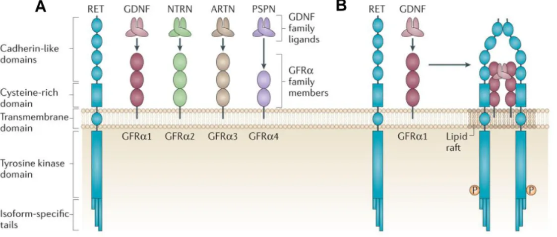
RET tyrosine kinase receptor ... 36
RET associated pathologies ... 38
Neuro-immune interactions at barrier sites ... 40
AIMS OF THIS THESIS ... 41
GLIAL CELL-DERIVED NEUROREGULATORS CONTROL TYPE 3 INNATE
LYMPHOID CELLS AND GUT DEFENCE ... 43
Abstract ... 44
Methods ... 45
ii
Discussion ... 60
SUPPLEMENTARY FIGURES ... 61
GENERAL DISCUSSION AND PERSPECTIVES ... 71
REFERENCES ... 75
1 ACKNOWLEDGEMENTS
First of all, I would really like to apologize for writing the acknowledgements in three different languages .
Henrique, quero-te agradecer por ter-me dado a oportunidade de participar neste fantástico projecto e de ter-me permitido crescer em muitos aspectos tanto da minha vida profissional como pessoal. Obrigada!
Sales, no podemos decir que el camino fue fácil pero llegamos y juntas. Creo que estos 4 años de días, noches, madrugadas, charlas, malos pero también buenos momentos van a formar parte de nuestras memorias para siempre. Prueba superada. Sobrevivimos! Hélder, obrigada por todo o trabalho, mas sobretudo e mais importante obrigada pela amizade, pela cumplicidade, pela paciência, pelas conversas, pelos choros de desespero mas também pelas horas de chorar a rir. Acho que isto foi mesmo uma grande prova que claramente, embora que não sei como, conseguimos superar.
Sílvia, são tantas as coisas que é impossível dizer tudo. Fico com as horas e horas de conversas e como não, as horas e horas de queixas. Sempre que pensava que me estava a queixar a mais aparecias tu com as tuas próprias histórias. Foste um apoio em todos os momentos bons e maus dentro e fora do laboratório. Obrigada amiga pela paciência, pelos ouvidos, pelos abraços e por fazer deste um caminho muito mais fácil. Rita, essa pessoa que sem saber quem eu era ensinou-me a identificar “plugs”. Passaste de ser essa colega de secretaria maluquinha a ser a minha amiga (maluquinha também). Obrigada pelas conversas sem sentido nenhum dessas que só tu sabes fazer e pelas tantas e tantas outras. Obrigada pelos risos que tu sem perceber provocas e que fizeram me tanto bem.
Sem vocês os três, sem o Concerto, sem os cafés, lanches, jantares, brunches, cervejas/gin (Síl) e tanto mais…isto não tivesse corrido nada bem ♥ ♥ ♥ Obrigada!
Luís, o meu mais querido towe ara, ainda tenho que pedir desculpas por não ter conseguido o towe ara 2.0 mas achei que o teu era tão bom que não podia haver melhora possível. Obrigada por partilhar todas as pérolas que o nosso “projecto” nos ofereceu só para poder rir um bocadinho.
2
Carlos, obrigada pelas tuas maravilhosas laminas próprias e pelo tempo que dedicavas a explicar, pela paciência (mutua claro) pelas conversas, pelos jantares no meio das activações por tudo e mais que não posso escrever aqui (só porque não há espaço), mas sobretudo pela amizade e por ser o meu PT que é isso que importa agora em momentos de stress.
I also want to thank (now in English because of you, of course, ma chéri) to all the people in the lab for their help throughout the years. Every one of you has a part in this project. Thank you for every conversation (in particular to Bruno), every coffee and every moment that we shared. Of course, thanks to the padel HVFs team! Ao Bruno (a pessoa mais organizada que eu já conheci na vida), à Manuela (a cara da felicidade), to Julie (the crazy one), ao Diogo (o comentador politico), ao Afonso (o dos tantos conselhos e o protector dos anticorpos que fica muito serio nas fotos), à Vânia (a pessoa sempre disposta a fazer e organizar todas as actividades de equipa), à Filipa (a pessoa que roubou a minha bancada e secretaria ) à Cristina (a fit super angel com muito jeito para o Photoshop de “personalidades”) e ao Telmo (o herdeiro, tiveste uma professora tão boa que o teu doutoramento só pode ser maravilhoso).
Tenho de agradecer especialmente ao serviço de Bioimaging do IMM, ao António, a Ana e ao Rino, pela dedicação constante, pelo profissionalismo, pela ajuda incondicional, paciência e por me aturarem em tantas e tantas conversas.
Também quero agradecer a unidade de citometria de fluxo à Maria, à Ana, à Sílvia, à Andreia, à Mariana e à Isabel pelo trabalho sempre bem feito e por tanta ajuda. Especialmente a Ana “de FACS” pelas conversas e pela paciência com as nossas células com RFP que as vezes sim e as vezes não, as misturas estranhas de anticorpos e as milhares de tentativas de sorting, tu conhecias mais do que nós aquelas células acredita Obrigada!
Á unidade de histologia, Tânia, Andreia, Ana, Bruna por toda a ajuda, a boa disposição e pela simpatia. Andreia, obrigada especialmente por tanta ajuda, pela disponibilidade incondicional e as longas conversas.
Ao biotério pela ajuda sempre disponível, pela paciência e compreensão. Obrigada Joana, Dolores, Iolanda, Carlos, Olena, Pedro e Felícia.
André Faustino, obrigada pelas dicas durante a escrita e pelas conversas e o apoio. Ao IFA , Antónia e Ema por toda a ajuda durante a submissão mesmo que eu aparecesse lá sempre à última hora.
3 Of course I would like to thank to all the authors in this wonderful paper that I did not mention yet. Thanks to Rute Marques, Ana M. Misic, Casey Bartow-McKenney, Denise M. Larson, William J. Pavan, Gérard Eberl and to Elizabeth A. Grice. Great job!
I want to thank Stroma ITN for funding and for giving me the opportunity to join a fantastic group of scientists to perform my PhD thesis. Thanks to all stroma people and also thanks to those that we met throughout the different courses and secondments, for all the advices and the time that we spent together, was very nice to meet you all! Marion, my brownie, I have to give you a special and big thank you for all the advices, the conversations, the good moments in Köln and traveling around. Thank you for your friendship.
Aos vizinhos do P2C, ao laboratório do Bruno Silva-Santos e do Luís Graça, por tudo, pelas conversas e pelos emprestamos como os anticorpos sobretudo em horas da noite desesperadas. Especialmente agradecer aos primos: ao Tiago (o primo original), ao Pedro, ao Sérgio e ao Miguel pelas conversas todas, pela companhia, pelos desabafos, pelos risos, pelos jantares, viagens e momentos. Á Julie pelas aventuras no Sh´bam, Pilates e bedbalances que foram fundamentais para relaxar durante a escrita .
A Turminha do Magritte, o lugar aonde qualquer queixa ia ser bem-vinda e aonde mesmo sem ir ao seminário, dava para perceber ao detalhe tudo o que se estava a passar lá “em baixo”.
Martita, mi amiguita, gracias por la paciencia, por escucharme, por preocuparte por mí, por estar incondicionalmente y por aguantarme tanto. Tantas cosas vividas en estos años, tantas aventuras, discusiones, llantos, risas, no imaginas cuánto más fácil me hicieron ésta loca travesía hasta aquí.
Borjita, sin ti mi vida hubiese sido bastante más aburrida estos años . Tantas conversaciones, tantos satélites, tantos secretos y entremeadas que nunca me dejaste probar, tantas cervezas, tantos print screens y mundos todays, tantas caminadas Lisboetas, tantos desayunos y tantos días sin azúcar. Gracias por acompañarme tanto amiguito.
Caetano, filho, sobrinho ou primo, eu diria amigo de coração, obrigada por tudo, por me cuidares, por sempre ter uma palavra quando eu necessitava, por estar, obrigada por aguentar desabafos e por me ouvires.
Dani, gracias por acompañarme durante esta última y nada fácil etapa, fuiste la razón por la cual escribir una tesis de repente se transformó en una tarea infinita. Estuviste presente en la mayor parte de los tantos lugares improbables en los que escribí alguna
4
línea. Por escucharme tanto y por formar parte de mi vida en esta pequeña parte del camino.
Às minhas barbies Ângela e Luciana obrigada por partilharem esta parte da minha tese e da minha vida, obrigada por estar sempre para tudo o que for preciso mas sobretudo para jogarrr, beberrr, comerrr e fumarrr (sim isto é por ti Angy)!
A todos mis amigos que aunque desde muy lejos estuvieron muy presentes. A Vir y Dani, a Ivana, a Andrea, a Jose, a los pajaritos Ale y Gere, También a los no tan lejanos que fueron parte de estos años, Paulita y Chris mis Gandienses (¿?) preferidos y a mis bioloquitos Lara, Silvia, Faus y Moni porque esas horas de micro y cito en la biblio valieron la pena. A todos por estar siempre presentes.
Gustavo gracias por preocuparte por mis cristales allá por el 2011, por venir conmigo a aquel 25 de abril y por siempre estar interesado en mis historias raras. Edu, Pablito (si tú, el de la mazorca) Javi, Ferrán, Pedro Pinto (proprietario), Pedro (el guapo), Manu y María gracias por formar parte de mi vida, en alguno de todos estos años, con algunos altos y con otros bajos muchas gracias por estar presentes.
A mis HH, a los latinos desaparecidos Javi y Moni, a Annina y a mis especialmente presentes Alvarito y Lu, gracias a todos por siempre estar, por ser más que amigos, unos más cerca que otros, por escuchar, por saber, por aguantar, por reírnos y llorar, por los encuentros espontáneos que me alegran la vida, por todo. Obrigada irmãos.
A mi tío Santiago, a mi primo Javier (el de la epigenética) y a Diego, gracias por estar ahí siempre y por interesarse siempre por lo que hago. A mi padre, gracias por preocuparte por mí aunque sea a la distancia, por preguntarme dónde voy, a qué y por qué. A los Cassani-Cuyunyian y Muiños, a Anita, Carmen, Romeo, Santi y Guz porque aún sin saber nada de lo que hago están ahí.
A mi familia. Mamá, Pablo y Daniel gracias por apoyarme siempre, de una forma o de la otra, sin entender muy bien lo que hacía, ni a dónde iba, ni por qué iba, ni por qué no tenía suficientes vacaciones, por qué trabajaba tantas horas, gracias por aguantarme sobre todo, por escucharme, por no matarme (sobre todo esto) y tenerme paciencia. A vos, por bancarme, por escucharme, por tantas horas y horas de conversaciones, de aguantes de cabeza, por frenar mis locuras, por enojarte porque siempre hablaba del mismo “tema”, por quererme y hacerme bien, por contarle a la gente lo que era una cryptopatche, por interesarte al detalle de lo que hacía y creer que lo hacía bien aunque te tuviese engañado. Felipe (ese nombre horrible sí) sin vos esto ni hubiese empezado.
5 RESUMO
No intestino, três tipos celulares são fundamentais na defensa do organismo perante as agressões externas: as células linfóides inatas, as células epiteliais e as células da glia. Esta relação funcional entre tecidos sugere a existência de um mecanismo de regulação entre o sistema imunitário, o epitélio e o sistema nervoso.
As células linfóides inatas (ou innate lymphoid cells-ILCs) são os novos membros na família dos linfócitos. Como tais, apresentam morfologia linfóide mas por outro lado as ILCs não possuem os genes activadores da recombinação (ou recombination activating
genes-RAG) nem marcadores específicos de outras linhagens celulares. As ILCs
integram sinais do ambiente e respondem a citoquinas, alarminas e moléculas produzidas pelas células epiteliais, as células do estroma e outras células do sistema imunológico. Podem ser divididas em três grupos dependendo dos factores de transcrição que regulam o seu desenvolvimento e a sua função.
O grupo 3 das ILCs têm como função regular os tecidos revestidos por mucosas sendo de grande importância na preservação da homeostasia. Este grupo compreende células indutoras de tecidos linfóides (ou lymphoid tissue inducer cells- LTis) e as células inatas linfóides do grupo 3 (ou innate lymphoid cells group 3 – ILC3s e possuem RORγt como factor de transcrição fundamental no seu desenvolvimento e função. No intestino, as ILC3s encontram-se principalmente agrupadas, junto com outras células do sistema imunológico e células do estroma, em estruturas chamadas cryptopatches. As ILC3s são de grande importância na formação de tecidos linfóides (LTis), na protecção frente a bactérias e são cruciais durante doenças inflamatórias devido ao seu grande potencial para produzir citoquinas importantes na protecção da barreira epitelial. Citoquinas como a IL-22 vão activar as células epiteliais que são as responsáveis de produzir proteínas antimicrobianas e mucus.
No nosso laboratório, foi descoberto que as ILC3s expressam elevados níveis da proteína tirosina cinase RET, o receptor dos factores neurotróficos da família do GDNF (glial cell
line-dereived neurotrophic factor) (GDNF family ligands – GFLs). Isto foi confirmado tanto
ao nível de RNA como de proteína utilizando um modelo animal GFP (green fluorescent protein) para RET.
Ret gene - em inglês “rearranged during transfection”- foi descrito em 1985 como um
6
desenvolvimento, maturação e manutenção de diferentes tipos celulares e tecidos. RET é expresso em tecidos embrionários, está envolvido na formação dos rins, manutenção das células estaminais e no desenvolvimento do sistema nervoso. Para além disso, foi também demonstrado que possui um papel crucial na formação de tecidos linfóides, em particular nas células hematopoiéticas envolvidas na organogénese das placas de Peyer no intestino durante a vida embrionária.
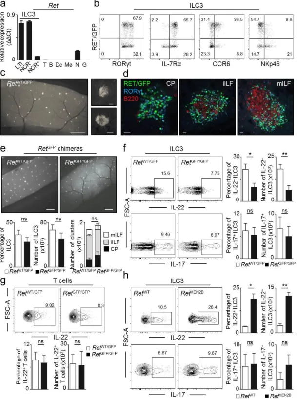
Dado que a deficiência em RET é embrionariamente letal, para poder perceber o papel do RET nas ILC3s foram desenvolvidas quimeras com o fígado fetal de ratinhos deficientes nesta proteína. Curiosamente, apesar de não terem sido encontradas diferenças no número de células nem de cryptopatches foi observada uma deficiência na função das ILC3s, em particular uma diminuição na produção da citoquina IL-22. Em concordância, no modelo animal em que há uma activação constitutiva de RET (RetMEN2B) encontramos um aumento na citoquina IL-22 procedente das ILC3s, o que se traduz numa maior protecção da barreira epitelial.
Para conseguir estudar o papel de RET nas ILC3s foi desenvolvido no laboratório um modelo de ratinho com uma ablação específica de RET nas células com expressão de Rorγt. No modelo RorγtCreRetfl/fl (ou RetΔ) encontramos uma diminuição da IL-22 e uma redução dos genes envolvidos na reactividade epitelial: mucinas e defensinas.
Com o fim de perceber o papel de RET não só em condições basais mas também em estados de inflamação, os animais RetΔ foram tratados com DSS (dextran sodium sulfate) para induzir colite. Nestes animais observamos uma redução na produção de IL-22 e consequentemente uma maior ruptura da barreira epitelial o que ao mesmo tempo produz maior translocação bacteriana desde o intestino para outros órgãos, tais como, nódulos linfáticos e o fígado. Do mesmo modo, os animais RetMEN2B foram tratados com DSS e após tratamento, mostraram uma maior produção de IL-22 junto com uma maior protecção da barreira epitelial e menor translocação bacteriana para órgãos periféricos. Estes resultados mostram o papel protector das RET+ILC3s em modelos de inflamação. De forma a estudar o processo inflamatório num contexto mais fisiológico, os animais RetΔ foram infectados com a bactéria Citrobacter rodentium que adere às células epiteliais e causa colite. Nestes animais, tanto a produção de IL-22 como a expressão dos genes de defesa aparecem diminuídos. Além disso observamos um aumento da inflamação intestinal, infecção e mortalidade.
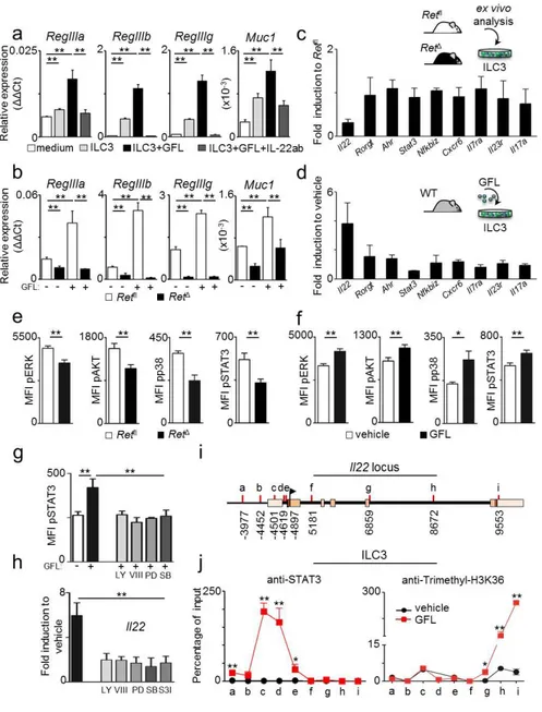
Com o objectivo de perceber a relação de RET e de GFLs na produção de IL-22 por parte das ILC3s foi usado um sistema in vitro de organóides epiteliais ou “mini intestinos”.
7 Observamos que quando são adicionados GFLs aumenta a expressão de genes de reactividade epitelial de forma dependente de IL-22. Para além disso a presença de GFLs nas ILC3s deficientes em RET (RetΔ) não afecta a expressão destes genes em relação aos controlos. Estes resultados mostram o papel fundamental de RET na activação das ILC3s através de factores neurotróficos.
Por outro lado, descobrimos que a regulação molecular da produção de IL-22 tem lugar após a activação das RET+ILC3s por GFLs e da fosforilação da cascata de sinalização p38 MAPK/ERK-AKT. Isto leva a activação de STAT3 que se liga ao promotor da Il22 iniciando a sua transcrição.
O sistema nervoso entérico ou também chamado “segundo cérebro” contem uma enorme rede de células nervosas. Em particular, nas células da glia do intestino, foi descrita a presença de receptores de reconhecimento de patógenos tais como “toll like receptors ou TLRs”. Isto juntamente com o facto das células da glia produzirem GDNF – necessários para a sinalização de RET- levou-nos a pensar numa regulação nervosa das células do sistema imunitário, neste caso, das RET+ILC3s.
As células da glia exprimem a proteína GFAP (glial fibrillary acidic protein) e para identificar a sua localização em relação às ILC3s foi preciso desenvolver um modelo de ratinho duplo fluorescente GFP/RFP para as proteínas RET e GFAP. Com este modelo, conseguimos perceber que as projecções das células da glia estão localizadas muito próximas as cryptopatches, na lâmina própria do intestino.
Por outro lado, foram desenvolvidas co-culturas de ILC3s e células da glia que mostraram a capacidade para produzir GFLs em resposta à activação por TLR2 e TLR4 e também por IL-1β e IL-33 promovendo a produção de IL-22. Estudos in vivo com ratinhos deficientes em Myd88 (molécula adaptadora para a sinalização por TLRs) nas células da glia e tratados com DSS, manifestaram um aumento da inflamação e uma redução na expressão de GFLs e IL-22 para além de sofrer uma maior perda de peso em comparação com os controlos. Quando infectados com Citrobacter Rodentium os animais deficientes em Myd88 foram mais susceptíveis a inflamação intestinal e a infecção. Resumindo, as células de glia processam os sinais do ambiente via MYD88 para produzir GFLs que ao mesmo tempo activam as RET+ILC3s e pela cascata das MAP cinases e STAT3 induzem a transcrição de Il22. A produção de IL-22 por sua vez promove a produção de péptidos antimicrobianos e genes de defesa e reparação do epitélio. A unidade glia-ILC3-epitélio é crítica na manutenção da homeostasia intestinal sendo fundamental na protecção barreira epitelial. Esta descoberta abre mais uma porta na
8
compreensão e nos possíveis tratamentos de doenças inflamatórias intestinais tais como doença de Crohn ou cancro.
9 SUMMARY
In this thesis we demonstrate that three distinct players form a novel multi-tissue defence unit in the intestinal wall: group 3 of innate lymphoid cells (ILC3s), intestinal epithelial cells and enteric glial cells (EGCs). This interplay reveals a neuro-immune interaction unit that regulates epithelial homeostasis and mucosal defense.
ILC3s are major regulators at mucosal surfaces being critical in tissue repair and in the maintenance of gut homeostasis. Intestinal ILC3 – that mainly aggregate into cryptopatches - integrate environmental signals leading to the production of the pro-inflammatory cytokines IL-22 and IL-17. IL-22 in turn induces intestinal epithelial cells to produce antimicrobial peptides and mucus.
We found that ILC3s express high levels of RET, a neuroregulatory receptor for GDNF family ligands (GFLs). In order to address the effect of RET in ILC3s development and function RET-deficient mice foetal liver chimeras were analyzed. Interestingly a decrease of IL-22 expressing ILC3s was observed when compared to WT controls. In addition a RET gain of function model (RetMEN2B) resulted in increased IL-22 expressing ILC3s. In line with these experiments, cell-autonomous ablation of RET in Rorγt expressing cells was performed. RorγtCreRetfl/fl, (RetΔ) mice had decrease IL-22 expressing ILC3s and a reduction of epithelial reactivity genes such as mucins and defensins comparing with their littermate controls. Upon infection with the attaching and effacing bacteria Citrobacter
rodentium, RetΔ mice had marked gut inflammation, reduced IL-22 producing ILC3, increased C. rodentium infection and translocation, reduced epithelial reactivity genes, increased weight loss and reduced survival. All these data together, suggest that RET cell autonomous ILC3 signals regulate IL-22 production.
Signals downstream of Ret were regulated via GFLs which directly controled rapid phosphorylation of the p38 MAPK/ERK-AKT cascade and STAT3 activation in ILC3s. In turn, STAT3 bound to the Il22 promoter to induce transcription.
Finally, we found that enteric glial cells integrated commensal and environmental signals to produce GFLs that control IL-22 production. Physical localization of glial cells in the vicinity of ILC3 was observed taking advantage of double reporter mice for GFAP (glial fibrillary acidic protein) and RET. Enteric glial cells had a stellate shape morphology, projecting into cryptopatches.
10
In vitro co-culture studies showed EGCs capacity to produce GFLs in response to TLR2
and TLR4 activation and IL-1β and IL-33 stimulation, promoting IL-22 production by ILC3s. In vivo studies with DSS induced colitis in glial specific Myd88 deficient mice (Gfap-Cre.Myd88Δ) showed an increase of gut inflammation and weight loss along with a reduced expression of intestinal GFLs and ILC3-derived IL-22 levels compared with their littermate controls. When infected with Citrobacter Rodentium Myd88 deficient mice exhibited a pronounced susceptibility to bowel inflammation and infection.
In summary, we were able to show that the enteric glial cells sense environmental cues through MYD88 to produce GFLs that in turn activate RET expressing ILC3s and via MAP kinase and STAT3 induce the transcription of Il22. The production of IL-22 promotes the expression of defence and repair genes. Thus, this novel glial-ILC3-epithelial unit is critical in the maintenance of intestinal homeostasis providing protection and repairing the epithelial barrier after injury.
11 INTRODUCTION
The intestinal mucosal barrier isolates the internal body from the external environment and is necessary for nutrient absorption and waste secretion1,2. Epithelial cells in the mucosa are in contact with the commensal gut microbiota and through recognition of multiple receptor molecules are able to identify danger signals from the environment, thus avoiding pathogen penetration into the gut wall. Furthermore, the gut mucosa contains a vast number of immune cells responsible for the maintenance of tissue homeostasis. Importantly, disruptions of the intestinal mucosal barrier and dysfunction of any of its components can produce disease3.
Malfunction of immune cells and their secreted molecules can increase gut permeability, altering nutrient absorption and allowing microbial product translocation that further increases inflammation4. However, many other factors are involved in the progression of intestinal pathologies. As such, we need to take into consideration not only immune alterations, but also environmental changes, genetic backgrounds, alterations in the gut microbiota and the extensive cell network that constitutes the enteric nervous system, which was shown to be altered in inflammatory bowel pathologies3,5. Interactions between the central and the enteric nervous system with the intestinal microbiota, diet products and immune responses are currently being investigated6. Nevertheless, the mechanisms by which the nervous system contribute to inflammatory intestinal pathologies remains unclear7–9.
In Europe approximately 3 million people suffer from inflammatory bowel diseases (IBD) costing to the health-care system billions of euros annually10. Bowel inflammation pathologies are a worldwide group of diseases that cause patients significant suffering due to gastrointestinal tract inflammation and tissue damage. There are two major clinical manifestations: Ulcerative colitis (UC) and Crohn´s disease (CD). Despite all efforts made so far trying to eradicate these pathologies, there is still no cure for IBD3.
Since IBD is a chronic inflammation pathology actual treatments are focused on immune suppressive drugs that target cytokines and their receptors3. To improve therapeutic design and implement new effective treatments to IBD, a better understanding of the mechanisms that regulate intestinal barrier homeostasis is needed.
12
The intestinal barrier
After birth, new-borns are exposed to millions of different microorganisms through contact, inhalation and ingestion11. Body surfaces such as the skin, the respiratory tract and the intestine start to be colonised. From then on, the intestine and its epithelia will act as barriers and as a reservoir for trillions of microorganisms from hundreds of different species: the gut microbiota12–16. Different organs, cells and molecules are involved in the maintenance of a healthy tissue microenvironment and to ensure that, not only the immune system but also the nervous system is intimately connected to provide protection against infection17,18,6.
The intestinal epithelial barrier (IEB) is the largest exchange surface between the body and the external environment. The IEB consists of a monolayer of epithelial cells organized into invaginations, or Lieberkühn crypts (proliferation compartment), and finger-like projections or villi (differentiation compartment)19. The brush border of epithelial cells constitutes an essential interface during infection with adhering bacterial pathogens20,21. Importantly, epithelial cells have the ability to repair wounds after an insult22,23, proliferating and migrating into the injured tissue while integrating microbial signals through pattern recognition receptors. These receptors can sense a wide range of microbial products such as peptidoglycans, flagellins, amino acids, microbiota derived short-chain fatty acids and butyrate, among others57.
The IEB allows for absorption of nutrients, but it also controls the passage of pathogens and toxins; therefore, changes in paracellular permeability can lead to multiple inflammatory and digestive diseases. The IEB plays a critical role in the protection against aggressions by deploying different mechanisms: i. the production of antimicrobial peptides by enterocytes and Paneth cells; ii. electrolyte production by enterocytes; iii. hormone secretion by enteroendocrine cells; and iv. mucus production by goblet cells (Figure 1). Epithelial cell renewal in the small intestine relies on pluripotent intestinal stem cells located in the base of the crypts with a high rate of cell turnover. Differentiation occurs while they migrate upwards into the villi, acquiring their specific absorption and secretion functions24. In contrast, Paneth cells migrate downwards to the base of the crypt where they become majors elements of the epithelial stem cell niche21. In conclusion, the intestinal epithelial barrier is the first line of defence against enteric pathogens and the maintenance of an intact IEB function is crucial to ensure health17.
13
Goblet cells and mucus production
The intestinal mucus is produced by specialized mucin secreting cells, or Goblet cells, derived from the secretory epithelial cell lineage25. Mucins are synthesized in the endoplasmic reticulum and final glycosylations occurs in the Golgi26. Mature mucins are then packed into secretory granules to be secreted into the lumen27–30.
Mucins are proteins with serines, prolines, threonines and carbohydrate chains attached by glycoside bonds31. There is a wide range of different mucins classified into three groups: i. gel forming; ii. soluble; and iii. membrane-bound mucins21. Changes in the composition of mucus and goblet cell function was reported in response to changes in the microbiota or in the presence of pathogens, but the mechanism behind these changes remains poorly understood26.
The intestinal mucus allows for protection and lubrication of the epithelium but it is also known to be involved in the regulation of foetal development, epithelial renewal and carcinogenesis32. Moreover, some enteric bacteria can use mucus for nutrition purposes, which facilitates their growth and colonization of the gut33,34. As the mucus layer acts as a physical barrier, enteric pathogens have developed mechanisms to interfere with mucin production and to cross the mucus barrier34. However, resident commensal bacteria have also developed mechanisms to inhibit the adherence of pathogens by means of increasing the production of mucins35.
Paneth cells and antimicrobial peptides
Antimicrobial peptides (AMPs) are mostly produced by Paneth cells at the base of the crypts of Lieberkühn and are part of the innate immune response being the front line of enteric defence36. This antimicrobial system is also present in the airways, gingival epithelium, cornea, reproductive tract, urinary and gastrointestinal tract where they can develop a rapid response before the adaptive immune system become active37,38.
Paneth cells are pyramid shaped exocrine cells containing secretory granules with AMPs and other antimicrobial molecules such as lysozyme and phospholipase A21,39. Antimicrobial peptides are able to control the bacterial composition and, in conjunction with the innate immune system, contribute to maintain the crypt microenvironment free from pathogens39–42. AMPs are small hydrophobic peptides of 20 to 40 amino acids and
14
are subdivided in two groups: the defensins and the cathelicidins43–45. The antimicrobial peptides have also been subdivided according to their secondary molecular structure as α-defensins (α helical structure) and β-defensins (β -sheet structure)45. AMPs have a wide spectrum of microbicide activity against gram negative and gram positive bacteria, fungi, protozoa and viruses21. AMPs can induce damage to microorganisms, binding to their surface and forming transmembrane pores46,47. Nevertheless, certain enteric pathogens have developed AMPs resistance48.
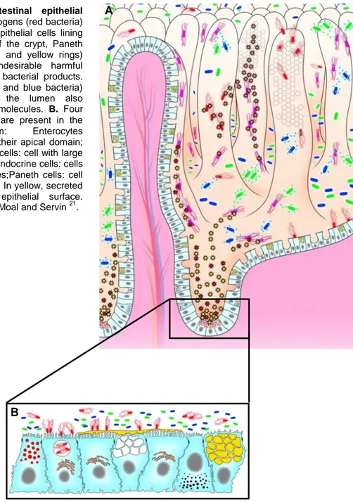
B
A
Figure 1 - The intestinal epithelial barrier. A. Enteric pathogens (red bacteria) interact with the host epithelial cells lining the villi. At the base of the crypt, Paneth cells releas AMPs (red and yellow rings) upon exposure to undesirable harmful pathogens and/or their bacterial products. The commensal (green and blue bacteria) intestinal bacteria in the lumen also produced antibacterial molecules. B. Four epithelial cell lineages are present in the
intestinal epithelium: Enterocytes
harbouring microvilli at their apical domain; mucus-secreting goblet cells: cell with large yellow granules; enteroendocrine cells: cells with small, dark granules;Paneth cells: cell with small, red granules. In yellow, secreted mucins coating the epithelial surface. Adapted from Liévin-Le Moal and Servin 21.
15
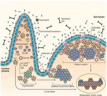
Gut associated lymphoid tissue
The gastrointestinal tract is considered the largest immune organ due to its well-organized immune system: the gut associated lymphoid tissue (GALT). The GALT (Figure 2) is composed by different lymphoid organs, such as mesenteric Lymph Nodes (mLN), Peyer’s Patches (PP), Cryptopatches (CP) and Isolate Lymphoid Follicles (ILF) where T and B cells can interact and become activated49. Development of secondary lymphoid organs is a well characterized50 process that starts between day 12.5 and 15.5 during embryogenesis where foetal liver hematopoietic progenitors start to colonize the gut51,52. Enteric secondary lymphoid organ development depends on lymphoid tissue inducer (LTi) cells53,54 and it was shown that LTi deficient animals fail to develop LNs, PPs, CPs and ILFs55–59. Peyer´s patches formation occurs due to interactions between RET+ lymphoid tissue initiator (LTin) and LTi cells with VCAM+ICAM+ lymphoid tissue organiser (LTo) stromal cells60. RET and LTo-derived ARTN signalling is crucial in the formation or PPs60. Nevertheless, it was shown that RET is not required for the development of LTin or LTi cells, but Ret deficient LTin and LTi cells fail to aggregate into PPs51,52. Different cytokines such as TRANCE or IL-7 induce LTi differentiation into LTαβ expressing cells which are attracted by chemokines such as CXCL13, CCL19 and CCL21 into the emergent anlagen structures60
.
Similar to Peyer´s patches, during the formation of lymphoid nodes LTi cells aggregate close to endothelial cells regulating the differentiation of LTo cells61. In this process, CD11c+ LTin cells are observed in the primitive lymph node but these cells and RET-ARTN interactions are dispensable for their development61.
In the lamina propria of the murine intestine we can also found clusters of group 3 (ILC3s) surrounded by dendritic cells (DC) named as cryptopatches. Although CPs start to aggregate around day 14 after birth49,62–65, they do not rely on microbiota, thus, it was observed that in germ free mice the number of CPs is unperturbed66,67. After contact with bacteria or during an inflammation, B cells are recruited to CP where critical IgA mediated-responses develop49,68,69. As occurs in PP, follicular B cells become plasma cells controlling microbiota in the lumen of the gut68,70,71. These mature follicles are named isolate lymphoid follicles (ILFs) and are large lymphoid structures composed by a single B cell follicle surrounded by ILC369. In contrast to LNs and PPs, ILFs maturation depends on microbiota62,72, notably in germ free mice that CPs fail to recruit B cells66,67. Thus, ILFs only form around weaning time and continuously throughout life when solid food intake increase bacterial load67. Pathogen associated molecular patterns induce ILFs
16
aggregation via NOD-1-dependent recognition by epithelial cells67. In addition, expression of LTα1β2 and CXCL13 by ILCs regulates ILFs development. LTα1β2 binds to LTβR on
stromal cells73 up-regulating the expression of the chemokine CXCL13 and adhesion molecules in order to recruit and retain lymphocytes72,74. In agreement, LTα and LTβ-deficient mice do not have CPs and ILFs 14,75–77.
Intestinal microbiota
Presumably, when thinking of a human body the first thing that comes into mind is the idea of a separate and unique entity. Far from this, we are the result of millions of years of co-evolution with prokaryotic entities which resulted into a balanced host-microbial symbiotic relationship. In the last decade the intestinal microbiota got our attention due to its capacity to affect the host´s metabolism, immune system and its response to infection. Recent studies also showed that the microbiota can affect and be affected by factors such as the diet, genetics and immune signals, thus, microbiota sensing by the host may have a critical impact in organismic homeostasis, in health and disease78–80. As an example,
Figure 2 - Associated lymphoid tissue or GALT. The lymphoid structures of the gastrointestinal tract include mesenteric lymphoid nodes, Peyer´s patches, cryptopatches and ILFs. Development of PPs and MLNs occurs during embryogenesis. After birth, ILC3s induce the formation of cryptopatches that, upon challenging with microbiota and recruitment of B cells may develop into ILFs66.
17 chronic infectious, inflammatory and metabolic diseases in humans have been associated with alterations in the composition or localization of commensal bacteria81.
The intestinal bacterial load is 10 times more abundant than the total number of cells in the body. The microbiome - the collective genome of the microbiota - contains at least 100 times more genes than the human genome12, therefore, the microbial composition and its metabolites can significantly shape gut microenvironment and dysbiosis can relate to disease82. Importantly, the gut microbiota, the epithelial cells and the immune system in the gut act together in order to provide protection against deviating pathobionts and pathogenic microbes, ensuring the maintenance of a balanced commensal bacterial population49,83,84.
18
The enteric immune system
It is well-established that a healthy immune system is necessary to prevent disease and immune cells are fundamental to limit pathogen invasion. For that purpose, the immune system deploys two major arms of response: the innate and the adaptive immune system. The innate immune system first appeared 750 million years ago at almost every level of the evolutionary tree of life, that is, vertebrates, invertebrates and plants. It has been remarkably conserved throughout the evolution. From a Darwinian point of view innate immunity has been crucial for surviving in the so-called “struggle for existence” 85–87. The innate immune system is composed by different cell types such as phagocytic cells, antigen presenting cells, killer cells and we should also take in consideration epithelial cells that are physical barriers, cytokine and chemokines producers, and express a broad repertoire of innate immune receptors88. This arm of the immune system also includes soluble recognition molecules and the complement system89. Innate immune sensing mechanisms include pattern recognition molecules that are able to integrate and perceive danger signals from the microenvironment.
The adaptive immune system arose 500 million years ago and is restricted to vertebrates. Its response is mediated by T and B lymphocytes that evolved to express a high number of recombinant receptors: the T-cell receptor and the immunoglobulins, respectively, and which are capable to recognize defined antigens. B and T lymphocytes have the ability to recall previous antigens encounters and mount memory responses that are stronger and faster that previous primary immune responses11,86,87,90.
Although adaptive immune responses are highly specific, they are slow as a consequence of the requirement for specific B and T cell clones to be activated and expanded after the first contact with a pathogen. The orchestration of immune responses and the production of cytokines is crucial for adaptive immunity to infection91. Since single bacteria can produce a 20 million progeny in an hour, the adaptive immune system can take a week before the response is effective. Accordingly, during the first critical hours and days after exposure to a new pathogen we mostly rely on our innate immune system to protect us from infection. As such, the innate immune system acts instantaneously as a first line of defence and is important until the adaptive immune system become efective11,89.
Adaptive T cells are activated after binding of their T cell receptor (TCR) with an antigen presenting cell. However, in order to control their response many different signals are integrated by the T cell; notably, diet derived products, cytokines, microbial products and
19 oxygen levels92. As an example, recent work showed T cell oxygen sensing by prolyl-4-hydroxylase domain (PHD) proteins as being critical for tumour metastasis into the lung93. Interestingly, B cells can also sense environmental cues, such as calcium. Notably, B cells can sense calcium levels fluctuations shaping B cell function94.
Pattern recognition receptors
The expression of microbe-associated molecular patterns or MAMPs in endogenous bacterial populations and pathogen-associated molecular patterns or PAMPs in enteric pathogens can be distinguish through pattern recognition receptors (PRRs)95. Sensing is one of the capacities that the organism implements to protect itself from aggressions. PAMPS can be recognized by different types of PRRs depending on their localization. They can be sub-divided in 3 groups: secreted, cytosolic and transmembrane receptors96.
The secreted PRRs are the collectins, ficolins and pentraxins and are involved in virus, bacteria and fungi infections, clearance of apoptotic and necrotic cells, resolution of allergic processes and inflammation96. These proteins can bind to the pathogen cell surface, activate the complement or opsonize the pathogen for phagocytosis by neutrophils and macrophages96.
The cytosolic PRRs include the retinoic acid inducible gene I (RIG-I) like receptors (RLRs) and the nucleotide-binding domain and leucine-rich repeat containing receptors (NLRs). These receptors can detect viral nucleic acids, microbial products and a variety of different stress signals95,97.
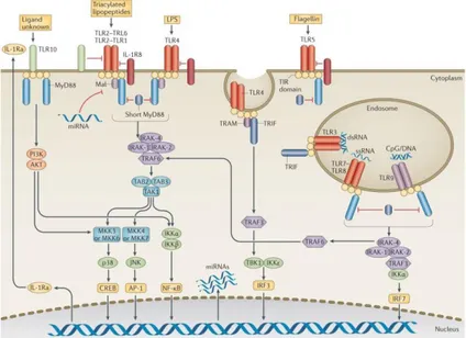
The transmembrane PRRs are the family of toll-like receptors (TLR) (Figure 3) and C-type lectin receptors that are express either in endosomal organelles or in the plasma membrane. Mammals possess 10–13 types of TLRs and each of them depends on the PAMP that can recognize. Thus, TLR4 recognizes lipopolysaccharide (LPS) of gram- bacteria; TLR1/TLR2 and TLR2/TLR6 lipoteichoic acids of gram+ bacteria and bacterial lipoproteins respectively and TLR5 recognizes flagellin. In addition, endosomal TLRs can sense microbial nucleic acids. For instance, dsRNA can be recognized by TLR3, ssRNA by TLR7 and dsDNA by TLR9. The C-type lectin family of receptors include Dectin-1 that recognizes β-glucans and Dectin-2 which senses fungi98.
20
TLRs are membrane-bound glycoproteins with leucine-rich repeats and cysteine-rich repeats ligand-binding motifs and with a Toll/IL-1 receptor [TIR] as cytoplasmic signalling domain99. TIR domains are involved in inflammation100. After activation, TIR domains use the adaptor molecule myeloid differentiation primary response gene 88 (MyD88) or the TIR domain–containing adapter-inducing INF- β (TRIF). Essentially, TLR responses are subdivided in two types depending on the molecular pathway that is used: Myd88 or TRIF100. For instance, TLR3 exclusively signal through TRIF but other TLRs signal primarily through MyD88. As an exception, TLR4 can activate both signalling pathways95,97,99. Interestingly was shown that TLRs are express not only in immune cells but also in non-immune cells as the enteric nervous cells in the intestine101–103
Figure 3 - Localization, signaling pathways and regulation of
Toll-like receptors (TLRs). TLR1, TLR2, TLR4, TLR5 and TLR6
localize in the cell surface, TLR3, TLR7, TLR8 and TLR9 localize in the endosomes and sense microbial and host-derived nucleic acids.
TLRs are membrane-bound glycoproteins with a Toll/IL-1 receptor [TIR] as cytoplasmic signalling domain. TIR domains use the adaptor molecule myeloid differentiation primary response gene 88 (MyD88) or the TIR domain–containing adapter-inducing INF- β (TRIF). TLR responses are subdivided depending on the molecular pathway that
is used: Myd88 or TRIF. dsRNA, double-stranded RNA; LPS,
lipopolysaccharide; miRNA, microRNA; ssRNA, single-stranded RNA. Adapted from O'Neill, L. A. et al.403.
21 Innate lymphoid cells
In addition to adaptive T lymphocytes, the immune system also harbours the recently identified family of innate lymphoid cells (ILCs). ILCs are members of the lymphoid lineage and are characterized by their lymphoid morphology, the absence of recombination activating genes (RAG) and the lack of cell lineage phenotypical markers83,104. ILC responses are important to resolve inflammation and infection53,91. Contrary to T cells, ILCs mainly reside in non-lymphoid tissues105. Importantly, ILCs can also express MHC class II and present antigens to adaptive lymphocytes106,107. Thus, ILCs can interact with other immune cells, such as T cells being important in the coordination of adaptive immune responses108–111. Deregulated ILC functions were also shown to contribute to chronic inflammatory diseases, metabolic disorders and cancer83. Several studies have investigated the role of ILCs in humans112, but little is known on their role in inflammatory bowel pathologies.
Different nomenclatures have been proposed for innate lymphoid cells53, nevertheless, there is a consensus in which ILCs can be sub-divided in three main groups based on their cytokine production and the expression of transcription factors that regulate their development and function (Figure 4). Innate lymphoid cell subsets mirror the T helper cell compartments104. As such, ILCs are the innate counterparts of the Th1, Th2 and Th17 subsets.
Group 1 ILCs (ILC1) comprises the cytotoxic NK cells113 - INFγ, TNF, perforin and granzyme producers- and helper ILC1s – INFγ and TNF producers - that express the transcription factor T-bet. Group 1 ILCs participate in immunity to viruses, intracellular pathogens and in tumour surveillance being involved in inflammatory diseases114,115.
Group 2 ILCs (ILC2) comprises IL-5, IL-9, IL-13 and amphiregulin producing cells. ILC2 dependent on GATA3, RORα and Notch for their development and function. ILC2s are involved in helminthic infections, wound healing and are associated with allergy and asthma116–118.
Group 3 ILCs (ILC3) comprises the foetal Lymphoid Tissue inducer cells (LTis) and helper ILC3 - IL-17 and/or IL-22, GM-CSF, TNF and lymphotoxin producers - that require RORγt, AHR and Notch for their development and function. Group 3 ILCs are involved in lymphoid tissue development, intestinal homeostasis, immunity to
22
extracellular bacteria and are associated with inflammation bowel disease91,119–122. Crohn´s disease patients were shown to have altered innate IL-17 119 and reduced ILC3 numbers together with increased ILC1s, possibly suggesting that ILC1s may derived from the ILC3s counterparts115.
Development of Innate Lymphoid cells
Innate lymphoid cells derive from a Flt3+IL7Rα+ Common Lymphoid Progenitor (CLP)123 (Figure 5). Therefore, CLPs in either adult bone marrow or foetal liver can give rise of all ILC subsets124. Downstream of CLPs, a common lymphoid precursor (αLP) and the early innate lymphoid progenitor (EILPs) are further committed to the helper ILC and NK cell lineages125,126. αLP was described as IL7Rα+CXCR6+NFIL3+TOX+α4β7+ 127,128 and EILPs as NFIL3+IL7Rα-TCF-1+129. EILPs express Id2 which is not required for their development129. NFIL3 regulates the expression of TOX, that is required for the development of NK cells and ILC subsets130 and also promotes the expression of the transcriptional repressor inhibitor of DNA binding 2 (Id2)131. Differentiation of ILC subsets requires ID2 mediated suppression of lymphoid cell fates (as B or T cells) 132. Restriction of αLPs into the specific ILC lineages occurs after losing NK-cell and LTi-cell potential131.
Figure 4 – Innate lymphoid cells. ILCs can be subdivided in cytotoxic ILCs or NK cells and in
non-cytotoxic or helper ILCs which include ILC1s, ILC2s and ILC3s. These different subsets were grouped depending on their transcription factors and cytokines that regulate their development and function. ILCs respond to a diverse range of signals such as neuropeptides, hormones or cytokines contributing to immunity, inflammation and homeostasis. However, dysregulated ILC responses can also contribute to chronic inflammatory diseases, metabolic disorders and cancer. AHR, aryl hydrocarbon receptor; Areg, amphiregulin; GM-CSF, granulocyte macrophage colony-stimulating factor; IFNγ, interferon-γ; IL, interleukin; LTi, lymphoid tissue inducer; NCR, natural cytotoxicity receptor; NK, natural killer; TNF, tumour necrosis factor; TSLP, thymic stromal lymphopoietin83
23 ILC1s, ILC2s and ILC3s subsets differentiate via a common helper innate cell precursor (ChILP) present in adult bone marrow and foetal which expresses IL-7Rα, α4β7 and ID2. A subset of ChILP that express promyelocytic leukemia zinc finger (PLZF) and GATA3 was designated as helper ILC progenitors (ILCP). ILCP give rise to all helper ILC subsets. In contrast, NK cells differentiate from a NK progenitor (NKP) whereas LTi cells from a PLZF-ChiLP83,124,127,133. Recently, it was demonstrated the role of PD-1 in ILC progenitors. A committed ILC progenitor identical to ILCP was described as PD-1hiIL-25Rhi and is critical in ILC2 development134,135.
ILC progenitors can be found in both in the embryo and adult sharing similar phenotypes and developmental programs136. The vast majority of peripheral ILCs are tissue resident cells and it was established that these cells have a slow cell turnover in steady state, thus suggesting that the majority of ILCs may arise during foetal life137. Nevertheless, it is believed that ILC replacement occurs in inflammation or stress situations136. In contrast, NK cells continuously move from the blood to tissues regenerating their peripheral pool137. The exact mechanism of how resident ILCs and
NK cells are regenerated and the pathways leading to cytotoxic or helper potential are not fully understood. It was proposed that ILCs may share the same factors that regulate T cell differentiation programs138. However, there are at least some differences between both systems such as the requirement for NFIL3 and ID2136. It remains unknown how different cell progenitors differentiate into adaptive lymphocytes, NK cells or LTis and how they acquired or lose their transcription factors. As an example, ILC3 were shown to loose RORt expression and give rise to IFN producing ILC1 (ex-ILC3s) in certain conditions136.
Figure 5 - Development of Innate lymphoid cells. Innate lymphoid cells derived from a common lymphoid progenitor (CLP). The common ILC/NK precursor also express the α4β7 integrin. Downstream is an
Id2-expressing precursor and a Id2+PLZF+
precursor for non-cytotoxic ILCs. However, LTi cells are not derived from that Id2+PLZF+.
24
Group 3 Innate lymphoid cells
ILC3s are key regulators of mucosal barrier defence, notably for the maintenance of a healthy gut microbial environment contributing for tissue repair and homeostasis83,53,132. ILC3s are required for the proliferation of intestinal stem cells and replenishing the epithelial barrier after tissue damage during inflammation139. Interestingly, during chemotherapy ILC3s were shown to be radio-resistant mediating interleukin 22 (IL-22) production required for tissue protection139. ILC3s have been most studied in the intestinal mucosa where they aggregate into cryptopatches and isolate lymphoid follicles134.
ILC3s rely on the transcription factor RORt140 which is necessary to regulate critical genes such as Il17 or Il22. ILC3 are also regulated by the transcription factor AHR, that is essential for Th17 and ILC3 survival and function in particular the production of IL-22 by NCR+ILC3 cells141 . Noteworthy, AHR is activated by environmental and endogenous signals such as diet-derived ligands and microbial products141,142. Another master regulator required for ILC3 development is ID2, which acts upstream of RORt and it was shown to affect the susceptibility to C. rodentium colonization143. RUNX3 is also required for the development of ILC1s and ILC3s144. RUNX3 binds to Rorc (which encodes RORt) and it is necessary for optimal RORt expression and also for Ahr transcription144. Likewise GATA3 regulates ILC3 development through regulation of CD127 (which encodes IL-7Rα) and controls RORt and T-bet in ILC3s145–147.
ILC3s can be further subdivided according to the expression of the chemokine receptor CCR6: i. CCR6+ ILC3 includeLTi cells (CD4+ and CD4-); ii. CCR6- ILC3s. CCR6+ LTi cells were discover in 1997148 and are crucial in the formation of secondary lymphoid organs during embryogenesis60,149. In the embryo, they are known for their Lin- CD45+ RORt+ CD4+LTα+LTβ+CD127+CD117+ phenotype150. In adulthood they mature into LTi-like cells and are capable of producing IL-22 and IL-17104. LTi-like cells may be fundamental in the reconstruction of lymph nodes after acute viral infection151. However, their exact immune function in adulthood is still unknown53,152,153.
CCR6- ILC3s can express the expression of the natural cytotoxicity receptor (NCR) NKp46. NCR+ ILC3 are specific IL-22 producers while NCR- can express 22 and IL-17154. Nevertheless, NCR- IL-17 producers can also produce GM-CSF having an important role in colitis154. NCR+ cells reside predominantly in mucosal tissues like the skin, lungs and intestinal tract mediating the balance between the symbiotic microbiota and the immune system91. These ILC populations also rely on signalling through IL-7Rα 53. Innate
25 IL-22- producers have also been shown to play a critical role against bacterial infections such as Citrobacter rodentium83,156. C.rodentium is an attaching and effacing bacteria that induce colitis in mice and mirrors the effect of E.coli in humans157. ILC3s were shown to have an essential function in C. Rodentium infection model being critical for IL-22 production121,158. In this model, ILC3s are the predominant population of IL-22 producers in the first weeks of infection121,159. Nevertheless, whether innate and adaptive lymphocyte functions are redundant is not fully understood. A specific mouse model for ILC3 deletion will be extremely useful to understand the exact contributions of T cells and ILC3s. Nevertheless, recent work of ILC3 deficiency in humans demonstrated that the functions of this subset may be redundant with T cells112, while in long-term-treated HIV-1-infected individuals with reduced T cell-derived IL-22 but preserved innate IL-22 displayed intact sigmoid mucosa integrity 160.
Environmental sensing by ILC3s
ILCs lack pattern recognition receptors but instead, they can sense cytokines, alarmins, lipids, hormones, dietary and microbial products and other molecules released by epithelial cells, stromal cells and other immune cells161. In contrast to mouse, human ILC3s can also be activated by recognition of pathogen-associated molecular patterns via the expression of Toll-like receptors162. As such, ILC3 have the capacity to sense their environment and integrate these signals in their functions. For instance, ILC1s can sense IL-12161; ILC2s, IL-33 or IL-25163–165 and ILC3s can sense IL-23 and IL-1β among others162,165. Innate lymphoid cells were also shown to work as hubs in the crosstalk between different systems. As an example, ILC2s can be activated by enteric neurons through the neuropeptide VIP possibly regulated by the circadian clock166 and ILC3s were shown to express molecules that might allow them to interact with the nervous system167.
ILC3s can respond to a sort of environmental signals, such as diet-derived molecules, microbial products and cytokines165. Several sensing mechanisms were shown to affect the development and function of different immune cells and organs.
Vitamin A-derived retinoic acid (RA), can bind to RAR or RXR nuclear receptors in the ILCs. Vitamin A is crucial for LTi cell differentiation and for the development of lymphoid organs in the embryo168 acting upstream of RORt169. As was previously mention, in the formation of lymphoid nodes, LTis are attracted by the chemokine CXCL13 from mesenchymal lymphoid organizer cells through LTβR61,170. For this to happen, stimulation
26
by TRANCE, IL-7 and RA is required72,165. This interaction promote the production of chemokines which will attract T cells and B cells into the follicle increasing the expression adhesion molecules72. In adult mice, RA signals promote ILC3 responses contrary to ILC2s that are supressed171. Hence, vitamin A deficient mice fail to resolve Citrobacter
rodentium infection168,171 although they can effectively resolve helminth infections.
Another dietary product sense by ILCs is the indole-3-carbinol (I3C) derived from cruciferous vegetables such as broccoli172. I3C ligands bind to the nuclear receptor aryl hydro-carbon receptor or AHR expressed in the ILC3s172. CCR6−ILC3s are dependent on
Ahr while CCR6+ILC3s can develop in its absence although their function is affected producing less IL-22173. Accordingly, Ahr deficientmice failed to develop cryptopatches and to control C. Rodentium infection142,174 .
ILC3s can sense IL-23 produced by dendritic cells (DCs) or mononuclear phagocytes inducing the production of IL-22 and IL-17, the master pro-inflammatory cytokines of the ILC3 lineage175. Stimulation of DCs is initiated by the LTβR-LTαβ binding on the ILC3s within the cryptopatches and is crucial for IL-23 production176. DCs-derived IL-23 is dependent of Notch2 and is required for ILC3 activation. Accordingly IL-23 deficient mice were shown to fail in resolving C. Rodentium infection165,175.
Another important environmental factor is represented by the microbiota. Commensals can induce IL-25 production in intestinal epithelial cells and IL-25 acts on DCs to limit ILC3s derived IL-22159. In addition, microbial products can be presented by CCR6+ILC3s to CD4+T cells through MHC class II inducing apoptotic cell death of activated commensal bacteria-specific T cells in the mesenteric lymph node111. Furthermore, in Crohn´s disease patients it was observed a reduction of ILC3s MHCII+ cells suggesting a role of this pathway in inflammatory bowel pathologies111.
In conclusion, a better understanding of ILC biology will certainly encompass the discovery of metabolites, microbial products and neuron-derived factors that are sensed by ILCs and can affect their function.
27 Dual effects of Interleukin 22
Interleukin (IL) -22 belongs to the 10 cytokine family (10, 19, 20, 24 and IL-26)177 and its expressed by innate (DCs, natural killer (NK) cells and ILCs) and adaptive (T cells) immune cells178. 22 can be regulated by many cytokines such as 23, 6 or IL-1β and transcription factors such as STAT3, RORγt or AHR. In this way, ILCs can respond to cytokines and produce IL-22 due to their expression of IL-1R and IL-23R104. IL-22 receptor is an heterodimer composed by IL-22R1 and IL10R2179. In contrast to IL10R2, which is expressed by most cells types, IL-22R1 is restricted to non-hematopoietic cells such as epithelial cells, stromal cells, keratinocytes or hepatocytes179,180. In enteric epithelial cells, IL22-IL-22R1-IL10R2 complex induces the activation of the STAT3 cascade that regulates many repairing genes, notably MYC, cyclin D1 and other proteins involved in proliferation and cell cycle as well as the expression of anti-apoptotic genes such as Bcl2, Bcl2l1, and Mcl1181–183. IL-22-derived ILC3 is necessary and sufficient for induction of epithelial cell fucosylation, a type of protein glycosylation which protects against enteric pathogens, possibly inducing epithelial cells to produce antimicrobial molecules158,184–188. The expression of IL22R1 in non-hematopoietic cells makes IL-22 an ideal therapeutic candidate specifically affecting tissue responses without targeting immune cells. Since Interleukin 22 is associated with IBD, the usage of IL-22 recombinant protein was proposed as an anti-inflammatory treatment189.
A dual “beneficial vs pathogenic” role was assigned to IL-22 due its ability to induce pro-inflammatory (IL-8 or IL-6)190,191 and anti-inflammatory or regulatory (antibacterial peptides or IL-10) molecules. IL-22 promotes epithelial cell proliferation to restore the epithelial barrier after an insult. IL-22 function is tissue specific contributing to the regulation of diseases like hepatitis or IBD187,192 and IL-22 is critical for mucosal homeostasis and tissue repair. Nevertheless, exacerbated production of IL-22 can also lead to inflammation188,190,193–195. Thus, IL-22 can be beneficial for the host in many diseases but in some others it can also be pathogenic showing a strong pro-inflammatory effect that can be enhanced when acting together with other pro-inflammatory cytokines such as IL-17.
In the C. rodentium colitis model, IL-22 deficient mice fail to resolve infection and to induce the expression of antimicrobial proteins, such as epithelial cell-derived defensins156, and mucus192. During colitis, IL-22 also induces wound healing through the STAT3 pathway196. 22 often acts together with 17; nevertheless the main role of
IL-28
17 is to recruit neutrophils and to induce the expression of pro-inflammatory cytokines197,198. In the DSS model, overexpression of IL-22 in T-cell receptor (TCRα)-deficient mice reduces disease192. Accordingly, blocking of the IL-22 pathway increases DSS induced colitis score183,192. Interestingly, IL-23R was shown to be a susceptible gene during Crohn’s disease and elevated levels of IL-22 were found in IBD patients, correlating levels of cytokine with disease severity199.
Constitutive expression of IL-22 in the small intestine contributes to the anatomical containment of commensal bacteria. While IL-22 is produced constitutively by ILC3s in the small intestine, depletion of ILC3s resulted in peripheral dissemination of commensal bacteria and systemic inflammation, which could be prevented by administration of IL-22 recombinant protein200.
Additionally, IL-22 can also be beneficial in allergy airway inflammation, graft-versus-host disease (GFHD) and autoimmune diseases201–203. In liver diseases, IL-22 stimulates the proliferation of hepatocytes and regenerates the tissue204. However, in many situations, IL-22 acts as a pro-inflammatory cytokine and the dysregulation of its expression can lead to pathology. During Toxoplasma gondii infection, IL22-deficient mice show decreased signs of inflammation in the small intestine205 while wild type IL-22 sufficient mice die of necrosis in the ileum.
A clear example of the dual effect of IL-22 is found in the skin. IL-22 induces tissue defence and wound healing; nevertheless, overexpression of IL-22 can cause skin inflammation. In fact, this overexpression produces psoriasis; an hyper proliferation and abnormal differentiation of keratinocytes (thickness of the skin) and infiltration of leukocytes206. This beneficial or pathogenic role of IL-22 depends on different factors such as the concentration and duration of local cytokine, the target tissues and the cytokine microenvironment. For instance IFN-α and TNF-α increase IL-22R and IL-10R2 expression on keratinocytes, thus increasing the capacity to respond to IL-22194,207. In addition IFN-α induce differentiation of monocytes into DCs that via IL-23 leads to IL-22 production208.
IL-17 has a pro-inflammatory role, but both IL22 and IL-17 can act together to determine the protective or pathogenic effect of IL-22198. Acute airway inflammation is lethal in wild type mice but is ameliorated in Il22 deficient mice, indicating a pro-inflammatory role of IL-22. In contrast, Il17 deficient mice are more protected from airway inflammation despite higher IL-22 production, thus indicating that IL-17 regulates the pro-inflammatory function of IL-22181.