UNIVERSIDADE FEDERAL DO PARÁ INSTITUTO DE ESTUDOS COSTEIROS (IECOS)
CAMPUS UNIVERSITÁRIO DE BRAGANÇA
PROGRAMA DE PÓS-GRADUAÇÃO EM BIOLOGIA AMBIENTAL
ADELSON SILVA DE SOUZA
DESCRIÇÃO LARVAL DE CARANGUEJOS BRACHYURA (CRUSTACEA: DECAPODA) DA REGIÃO AMAZÔNICA, OBTIDO EM LABORATÓRIO
ADELSON SILVA DE SOUZA
DESCRIÇÃO LARVAL DE CARANGUEJOS BRACHYURA (CRUSTACEA: DECAPODA) DA REGIÃO AMAZÔNICA, OBTIDO EM LABORATÓRIO
Dissertação apresentada ao Programa de Pós-graduação em Biologia Ambiental, Mestrado em Ecologia de Ecossistemas Costeiros e Estuarinos da Universidade Federal do Pará, Campus de Bragança, como um dos requisitos necessários á obtenção do título de Mestre em Biologia Ambiental.
Orientador: Prof. Dr. Fernando Araujo Abrunhosa
Co-orientador: Prof. Dr. Rauquírio Marinho da Costa
ADELSON SILVA DE SOUZA
DESCRIÇÃO LARVAL DE CARANGUEJOS BRACHYURA (CRUSTACEA: DECAPODA) DA REGIÃO AMAZÔNICA, OBTIDO EM LABORATÓRIO
Dissertação apresentada ao Programa de Pós-graduação em Biologia Ambiental, Mestrado em Ecologia de Ecossistemas Costeiros e Estuarinos da Universidade Federal do Pará, Campus de Bragança, como um dos requisitos necessários á obtenção do título de Mestre em Biologia Ambiental.
BANCA EXAMINADORA
_______________________________________________
Prof. Dr. Fernando Araujo Abrunhosa (Orientador).
Universidade Federal do Pará – UFPA - Campus de Bragança
_______________________________________________
Prof. Dr. João Marcos de Góes (Titular)
Universidade Federal do Piauí – UFPI – Campus Ministro Reis Vello
_______________________________________________ Profa. Dr. Cristiana Ramalho Maciel (Titular)
Universidade Federal do Pará – UFPA - Campus de Bragança
_______________________________________________
Profa. Dr. Bianca Bentes da Silva (Titular)
Universidade Federal do Pará – UFPA - Campus de Bragança
_______________________________________________ Prof. Dr. Marcus Emanuel Fernandes (Suplente)
“Talvez meio caminho andado seja a gente acreditar no que faz.
Mas acima de tudo, o que mais nos incentiva, que mais nos
valoriza e também mais nos torna conscientes de nossa
responsabilidade, é saber que outros crêem em nós. E não há
palavras que descrevam o que sentimos ao saber dos sacrifícios a
que eles se impõem por crerem não apenas em nós, mas também no
que cremos.”
AGRADECIMENTOS
Agradeço antes de tudo a Deus, por caminhar sempre ao meu lado conduzindo-me e proporcionado momentos de conforto, reflexão e paciência diante de todas as etapas a serem vencidas.
À minha família ―minha origem‖, pelo incentivo e confiança ao longo de cada conquista acrescida em minha vida.
Ao Prof. Dr. Fernando Abrunhosa, por propor e orientar este trabalho enriquecendo-o com grandes contribuições... sou muito grato.
Ao Prof. Dr. Rauquirio M. da Costa, pela confiança e contribuições para a finalização deste estudo.
A FAPESPA ‗Fundação de Amparo à Pesquisa do Estado do Pará‘, pelo financiamento deste trabalho através de uma bolsa de mestrado.
Aos grandes amigos (a)s Fábio, Marilia, Welliton, Paulo baiá, Tião, Ádila e Suellen, pela grande ajuda nas coletas das espécies e da água, além dos inesquecíveis momentos de descontração... sou muito grato. E não poderia deixar de lembar da ‗my friend‘ ―peça rara‖, por tudo.
A Dane Brito pela grande ajuda na formatação das figuras e a Dane Arruda pelas discussões e sugestões no momento de conclusão deste trabalho.
Aos professores João Góes, Cristiana Maciel e Bianca Bentes, por aceitarem avaliar este trabalho.
Ao programa de Pós-Graduação em Biologia Ambiental, pela oportunidade de desenvolver este projeto.
SUMÁRIO
ESTRUTURA DA.DISSERTAÇAO... 5
RESUMO... 5
ABSTRACT... 6
CAPITULO I: INTRODUÇÃO GERAL... 6
REFERÊNCIAS BIBLIOGRÁFICAS... 8
CAPITULO II: THE COMPLETE LARVAL DEVELOPMENT OF PANOPEUS LACUSTRIS DESBONNE, 1867 (BRACHYURA: PANOPEIDAE), FROM THE AMAZON REGION, REARED IN THE LABORATORY………. 16 ABSTRACT... 16
INTRODUCTO... 16
MATERIALS AND METHODS... 18
RESULTS... 19
DISCUSSION... 38
ACKNOWLEDGEMENTS... LITERATURE CITED... 41 41 CAPITULO III: DESCRIÇÃO DO PRIMEIRO ESTÁGIO LARVAL DE TREZE ESPÉCIES DE CARANGUEJOS BRACHYURA (CRUSTACEA: DECAPODA) DA REGIÃO AMAZÔNICA, OBTIDOS EM LABORATÓRI... 46 ABSTRACT... 46
RESUMO... 46
INTRODUÇÃO... 47
MATERIAL E MÉTODOS... 49
RESULTADOS... 51
DESCRIÇÃO... 51
CHAVE DE IDENTIFICAÇÃO DE ZOEA I DAS ESPÉCIES DESCRITAS. 84 DISCUSSÃO... 85
AGRADECIMENTOS... 98
RESUMO
Larvas do caranguejo da lama Panopeus lacustris Desbonne, 1867 foram cultivadas em
laboratório a partir de fêmeas ovígeras coletadas na área estuarina do Rio Caeté na região Amazônica. O desenvolvimento completo desta espécie consistiu em 4 de zoea e um megalopa, onde cada estágio foi descrito e ilustrado em detalhes. Os resultados foram comparados com os de outros estudos anteriores sobre o desenvolvimento larval das espécies pertencentes ao gênero Panopeus e brevemente discutidos. Além da descrição das larvas do desenvolvimento completo de P. lacustris, foi descrita e ilustrada a primeira fase
de zoea de treze espécies de caranguejos braquiúros coletados no mesmo estuário: P.
lacustris, P. americanus Saussure, 1857, Eurytium limosum (Say, 1818), Sesarma curacaoense De Man, 1892, Sesarma rectum Randall, 1840, Armases rubripes (Rathbun,
1897), Aratus pisonii (H. Milne Edwards, 1837), Ocypode quadrata (Fabricius, 1787), Uca
rapax (Smith, 1870), Uca maracoani (Latreille, 1802), Uca thayeri Rathbun, 1900, Ucides cordatus (Linnaeus, 1763) e Pachygrapsus gracilis (Saussure, 1858). Apenas a zoea I de P. lacustris não foi descrito novamente. As características morfológicas dessas espécies
são comparadas com as descrições originais. Para facilitar o estudo de material coletado no plâncton, foi desenvolvida uma chave para identificação das espécies descritas neste estudo.
ABSTRACT
Larvae of the mud crab P. lacustris were reared in laboratory from ovigerous
females collected in the estuarine area of the Caeté River in the Amazonian region. The complete development of this species consisted of 4 zoeal and 1 megalopal stages, where each stage was described and illustrated in detail. The results were compared with those of other previous studies on larval development of the species belonging to the genus
Panopeus and then briefly discussed. Besides the description of the complete development
larval of P. lacustris, has been described and illustrated the first stage zoea of thirteen
species of brachyuran crabs collected in the same estuary: P. lacustris Desbonne, 1867, P.
americanus Saussure, 1857, Eurytium limosum (Say, 1818), Sesarma curacaoense De
Man, 1892, Sesarma rectum Randall, 1840, Armases rubripes (Rathbun, 1897), Aratus
pisonii (H. Milne Edwards, 1837), Ocypode quadrata (Fabricius, 1787), Uca rapax (Smith,
1870), Uca maracoani (Latreille, 1802), Uca thayeri Rathbun, 1900, Ucides cordatus
(Linnaeus, 1763) e Pachygrapsus gracilis (Saussure, 1858). Only the zoea I of P. lacustris
was not described again. The morphological characteristics of these species are compared with the original descriptions. To make easy the study of plankton-collected material, it was developed a key to identification of the species described in this study.
ESTRUTURA DA DISSERTAÇÃO
A dissertação foi elaborada no formato de artigos, separados em capítulos, segundo as normas do programa do Programa de Pós-graduação em Biologia Ambiental da Universidade Federal do Pará.
Desta forma, este trabalho está disposto em três capítulos. O capitulo I contém a introdução geral com uma breve apresentação sobre os aspectos que envolvem os estudos zooplanctônicos de larvas de crustáceos em regiões estuarinas.
O capitulo II contém o desenvolvimento larval completo de Panopeus lacustris,
obtido em laboratório com descrição e ilustração morfológica de todos os seus estágios larvais.
CAPITULO I
1. INTRODUÇÃO GERAL
Os sistemas estuarinos constituem ecossistemas com elevada importância biológica e sócio-econômica, sendo influenciado por ambientes marinhos e de água doce. Possuem uma abundante e diversificada comunidade de organismos e grande capacidade de renovação periódica de suas águas, configurando-se como uma região com intensas transformações de matéria orgânica (Miranda et al., 2002; Pereira Filho et al., 2003).
Os estuários são reconhecidos como um dos principais fornecedores de nutrientes para a região costeira, caracterizando-se como um sistema de alta produtividade primária e elevada biomassa zooplanctônica e bentônica. Além disso, desempenham importante papel na estruturação de uma complexa cadeia alimentar que influencia positivamente a atividade pesqueira local (Kramer et al., 1994; Braga et al., 2000; Wolff et al., 2000).
Entre os grupos de organismos que habitam esse ambiente, destacam-se as comunidades planctônicas, representadas pelo fitoplâncton, os quais são os principais produtores primários dos estuários e oceanos, e o zooplâncton, que constitui um elo fundamental na transferência de energia e matéria orgânica entre os produtores primários e os demais níveis tróficos (Gross & Gross, 1996; Belgrano et al., 2005).
No zooplâncton, podem ser identificados organismos pertencentes à grande maioria dos filos zoológicos, apresentando espécies bioindicadoras e fornecendo informações para o conhecimento de processos interagentes, visto que suas comunidades são diretamente influenciadas pelas condições abióticas e bióticas do ambiente (Day Jr et al., 1989; Neumann-Leitão et al., 1991).
Ao analisar a composição zooplanctônica das regiões estuarinas constata-se a vasta predominância do grupo Crustacea, repreconstata-sentado, principalmente pelos Copepoda (Tundisi, 1970; Day Jr et al., 1989; Froneman, 2002;
espécies (Boschi, 1981; Raymont, 1983; Neumann-Leitão et al., 1996; Fehlauer & Freire, 2002; Cavalcanti et al., 2008).
Geralmente nos estudos zooplanctônicos, os estágios larvais desses organismos são taxonomicamente classificados apenas como zoeas de braquiúros ou de decápodos. Este fato restringe diretamente o conhecimento sobre a diversidade desse grupo nas comunidades zooplanctônicas (Silva et al., 2004; Krumme & Liang, 2004; Martins et al., 2006; Koettker & Freire, 2006; Cavalcanti et al., 2008; Costa et al., 2008; Costa et al., 2009; Magalhães et al., 2009; Pessoa et al., 2009).
A maioria dos estudos condiciona este fato à dificuldade de identificação das larvas em nível específico, devido à ausência de descrições larvais ou mesmo de informações morfológicas detalhadas para estes organismos. A identificação correta dos diferentes taxa na forma larval é essencial para estudos relacionados ao comportamento, distribuição espaço-temporal, dispersão, recrutamento e outros aspectos ecológicos (Anger et al.,1994; Queiroga et al., 1994; Anger, 2006). Todos estes aspectos da biologia larval têm implicações importantes para dinâmica de populações, ecologia de comunidades, biogeografia e a introdução de espécies invasoras, além de proporcionarem a elaboração de possíveis técnicas economicamente viáveis à aqüicultura, ou para administração da pesca sustentável (Anger, 2006).
Algumas pesquisas reúnem descrições morfológicas das larvas de várias espécies de braquiúra, as quais são fundamentais para a identificação desses organismos em estudos planctônicos de diversas regiões do mundo, como a Ásia (Lee & Ko, 2008; Kornienko & Korn, 2009), África (Clark & Paula, 2003; Flores et al., 2003), Europa (Paula, 1996; Gordillo & Rodríguez, 2000; González-Gordillo et al., 2000), América do Norte (Hart, 1971; Rice & Tsukimura, 2007), América central (Schubart & Cuesta, 1998; Cuesta & Schubart, 1999) e em alguns países da América do Sul, tais como o Chile (Báez, 1997), Argentina (Lovrich, 1999) e inclusive o Brasil (Serbino, 2003; Fransozo et al., 2002). Porém estas informações podem ser consideradas insuficientes em relação ao grande número de espécies pertencentes a este grupo (Ng et al., 2008).
desenvolvimento de trabalhos descritivos para as espécies que vivem nos manguezais e estuários do Brasil, em especial para a região Amazônica, a qual apresenta uma elevada biodiversidade de crustáceos braquiúros. Assim, este trabalho visa contribuir, através de descrições morfológicas, para o conhecimento das diversas formas larvais de caranguejos Brachyura de um estuário amazônico (Pará, Brasil).
2. REFERÊNCIA BIBLIOGRÁFICA
ANGER, K. Contributions of larval biology to crustacean research: a review.
Invertebrate Reproduction and Development, 49 (3): 175–205. 2006.
ANGER, K.; SPIVAK, E.; BAS, C.; ISMAEL, D. & LUPPI, T. Hatching rhythms and dispersion of decapod crustacean larvae in a brackish coastal lagoon in Argentina.
Helgoländer Meersuntersuchungen,48: 445–466. 1994.
BÁEZ. P. Key to the families of decapod crustacean larvae collected off northern Chile during an El Niño event. Investigaciones Marinas, 25: 167-176. 1997.
BELGRANO, A.; SCHARLER, U.; DUNNE, J. & ULANOWICS, R. E. Aquatic food webs, an ecosystem approach. Oxford , UK: Oxford University Press, 2005.
262p.
BOSCHI, E. E. Larvas de crustacea decapoda. In: Atlas del zooplankton del Atlántico sudoccidental y métodos de trabajo con el zooplankton marino.
Boltovskoy, D. (ed.) Argentina, Mar del Plata: Inedep., 1981. 699-758p.
BRAGA, E.S.; BONETTI, C.V.D.H.; BURONE, L. & BONETTI FILHO, J. Eutrophication and bacterial pollution caused by industrial and domestic wastes at the Baixada Santista estuarine system - Brazil. Marine Pollution Bulletin, 40:
CAVALCANTI, E. A. H.; NEUMANN-LEITÃO, S. & VIEIRA, D. A. N. Mesozooplâncton do sistema estuarino de Barra das Jangadas, Pernambuco, Brasil. Revista Brasileira de Zoologia, 25 (3): 436-444. 2008.
CLARK, P. F. & PAULA, J. Descriptions of ten xanthoidean (Crustacea: Decapoda: Brachyura) first stage zoeas from inhaca island, Mozambique. The Raffles Bulletin of Zoology, 51(2): 323-378. 2003.
COSTA, K. G.; PEREIRA, L. C. C. & COSTA, R. M. Short and long-term temporal variation of the zooplankton in a tropical estuary (Amazon region, Brazil). Boletim do Museu Paraense Emílio Goeldi, Ciências Naturais, 3 (2): 127-141. 2008.
COSTA, R. M.; LEITE, N. R. & PEREIRA, L. C. C. Mesozooplankton of the Curuçá Estuary (Amazon Coast Brazil). Journal of Coastal Reseach, 56: 400-404, 2009.
CUESTA, J. A. & SCHUBART, C. D. First zoeal stages of Geograpsus lividus and Goniopsis pulchra from Panama confirm consistent larval characters for the subfamily Grapsinae (Crustacea: Brachyura: Grapsidae), Ophelia, 51 (3):
163-176. 1999.
DAY JR, J. W.; HALL, C. A. J; KEMP, W. M. & YANNEZ-ARANCIBIA, A. Zooplankton the drifting consumers. Estuarine Ecology, Wiley-Interscience,
1989. 577p.
ESKINAZI-SANT’ANNA, E. M. & BJÖRNBERG, T. K. S. Seasonal dynamics of
mesozooplankton in Brazilian coastal waters. Hydrobiologia, 563: 253-268. 2006.
FEHLAUER, K. H. & FREIRE, A. S. Ocurrence of decapods larvae, especially Xiphopenaeus kroyeri (Penaeidae) in the shallow shelf of Paraná, Nauplius, 10
(1): 37- 45. 2002.
FRANSOZO, A., CUESTA, J. A. & NEGREIROS-FRANSOZO, M. L. The first zoeal stages of two species of Grapsidae (Decapoda, Brachyura) and a key to such larvae from the Brazilian coast. Crustaceana, 71, 331–343. 1998.
FRANSOZO, A., MANTELATTO, F. L. & BERTINI, G. The first zoeal stage of the genus Cronius (Brachyura, Portunidae) from the Brazilian coast, hatched in a laboratory. Journal of Plankton Research, 24 (2): 1237-1244. 2002.
FRONEMAN, P, W. Response of the plankton to three different hydrological phases of the temporarily open/closed Kasouga Estuary, South Africa. Estuarine, Coastal and Shelf Science, 55 (4): 535–546. 2002.
GONZÁLEZ-GORDILLO, J. I. & RODRÍGUEZ, A. Larval development of Philocheras fasciatus (Risso, 1816) (Decapoda, Caridea) reared in laboratory, comparison with plankton larvae and occurrence of accelerated development. Journal of Plankton Research, 22 (10): 1909-1923. 2000.
GONZÁLEZ-GORDILLO, J. I.; SANTOS, A. & RODRÍGUEZ, A. Complete larval development of Philocheras monacanthus from laboratory culture, with a key to the zoeae of the European species of the genus (Decapoda: Caridea: Crangonidae). Journal of Crustacean Biology,20 (1): 75–88. 2000.
GROSS, M. G. & E. GROSS. Oceanography, a view of earth. New Jersey,
Printice Hall. 1996. 472p.
HART, J. F. L. Key to planktonic larvae of families of decapods Crustacea of British Columbia. Syesis, 4: 227- 234. 1971.
KORNIENKO, E. S. & KORN, O. M. Illustrated key for the identification of brachyuran zoeal stages (Crustacea: Decapoda) in the plankton of Peter the Great Bay (Sea of Japan). Journal of the Marine Biological Association of the United Kingdom, 89 (2): 379–386. 2009.
KRAMER, K. I.; BROCKMANN, U. H. & WARWICK, R. M. Tidal Estuaries.
Manual of Sampling and Analytical Procedures. Rotterdam: A. A. Balkema. 1994. 304p.
KRUMME, U. & LIANG, T. H. Tidal-Induced Changes in a Copepod-Dominated Zooplankton Community in a Macrotidal Mangrove Channel in Northern Brazil.
Zoological Studies,43 (2): 404-414. 2004.
LEE, S. H. & KO, H. S. First zoeal stages of six species of Hemigrapsus (Decapoda: Brachyura: Grapsidae) from the northern pacific including an identification key. Journal of Crustacean Biology, 28 (4): 675–685. 2008.
LEITE, N. R. Mesozooplâncton do estuário do rio Curuçá-Pará-Brasil.
Dissertação de Mestrado em Biologia Ambiental. Universidade Federal do Pará, Pará, 2008. 62p.
LOVRICH, G. A. Seasonality of larvae of Brachyura and Anomura (Crustacea Decapoda) in the Beagle Channel, Argentina. Scientia Marina, 63 (1): 347-354.
1999.
MAGALHÃES, A., LEITE, N. R., SILVA, J. G. S., PEREIRA, L. C. C. & COSTA, R. M. Seasonal variation in the copepod community structure from a tropical Amazon estuary, Northern Brazil. Anais da Academia Brasileira de Ciências, 81 (2):
1-11. 2009.
(Amazon region - North of Brazil). Brazilian Journal of Biology, 66 (2): 421-430.
2006.
MARTINS, A. L. P.; LOPES, M. J. S. & MELO, O. T. Zooplâncton como bioindicador da qualidade ambiental no estuário do Rio Anil, São Luís, Maranhão. Boletim do Laboratório de Hidrobiologia, 19: 51-60. 2006.
MIRANDA, L. B.; CASTRO, B. M. & KJERFVE, B. Princípios de oceanografia física de estuários. 1ª ed. USP. São Paulo, Brasil. 2002. 427 p.
NEUMANN-LEITÃO, S.; MATSUMURA-TUNDISI, T. & CALIJURI, M. C. Distribuição e aspectos ecológicos do zooplâncton da represa do Lobo (Broa) - São Paulo. In: ENCONTRO BRASILEIRO DE PLÂNCTON, 4, 1991, Recife. Anais... Recife: Sociedade Brasileira de Plâncton, 393-414 p. 1991.
NEUMANN-LEITÃO, S.; GUSMÃO, L. M. O.; NASCIMENTO-VIEIRA, D. A. & NOGUEIRA-PARANHOS. Variação diurna e sazonal do zooplâncton no estuário do rio Ipojuca, PE (Brasil). Trabalhos Oceanográficos da Universidade Federal de Pernambuco, 24: 103-133. 1996.
NEUMANN-LEITAO, S.; SANT’ANNA, E. M. E.; GUSMÃO, L. M. O.;
NASCIMENTO-VIEIRA, D. A.; PARANAGUA, M. N. & SCHWAMBORN, R. Diversity and distribution of 43 the mesozooplankton in the tropical Southwestern Atlantic. Journal of Plankton Research, 30 (7): 795-805. 2008.
Ng, P. K. L., Guinot, D. & Davie, P. J. F. Sistema Brachyurorum: part I. An anntated checklist of extant Brachyuran crabs of the world. The Raffles Bulletin of Zoology, 17:1-286. 2008.
PEREIRA FILHO, J.; SPILLERE, L. C. & SCHETTINI, C. A. F. Dinâmica de nutrientes na região portuária do estuário do rio Itajaí-açu, SC. Atlantica, 25 (1):
11-20. 2003.
PESSOA, V. T.; NEUMANN-LEITÃO, S.; GUSMÃO, L. M. O.; SILVA, A. P. & PORTO-NETO, F. F. Comunidade zooplanctônica na baía de Suape e nos estuários dos Rios Tatuoca e Massangana, Pernambuco (Brasil). Revista Brasileira de Engenharia de Pesca, 4 (1): 80-94. 2009.
QUEIROGA, H.; COSTLOW, J. D.; & MOREIRA, M. H. Larval abundance patterns of Carcinus maenas (Decapoda, Brachyura) in Canal de Mira (Ria de Aveiro, Portugal). Marine Ecology Progress Series, 111: 63–72. 1994.
RAYMONT, J. E. G. Plankton and productivity in the oceans. 2. Zooplankton. Oxford, Pergamont press. 1983. 824p.
RICE, A. & TSUKIMURA, B. A key to the identification of brachyuran zoeae of the San Francisco bay estuary. Journal of Crustacean Biology, 27 (1): 74–79. 2007.
SCHUBART, C. D. & CUESTA, J. A. First zoeal stages of four Sesarma species from Panama, with identification keys and remarks on the American Sesarminae (Crustacea: Brachyura: Grapsidae). Journal of Plankton Research, 20: 61-84.
1998.
SERBINO, N. M. B. Descrição morfológica e aspectos evolutivos larvais em Ocypodidae e grupos relacionados (Decapoda: Brachyura). Dissertação de
Mestrado em Zoologia. Instituto de Biociências de Botucatu da Universidade Estadual Paulista , UNESP. 2003. 134p.
TUNDISI, J. G. O Plâncton Estuarino. Contribuições Avulsas do instituto Oceanográfico da Universidade de São Paulo, Série Oceanográfica Biológica, 19: 1-22. 1970.
WOLFF, M.; KOCH, V. & ISACC, V.J. A trophic flow model of the Caeté mangrove estuary (North Brasil), with considerations for the sustainable use of its resoucers.
Submetido para publicação na revista Acta Zoologica
CAPITULO II
THE COMPLETE LARVAL DEVELOPMENT OF PANOPEUS LACUSTRIS DESBONNE, 1867 (BRACHYURA: PANOPEIDAE), FROM THE AMAZON REGION, REARED IN
The complete larval development of Panopeus lacustris Desbonne, 1867 (Brachyura:
Panopeidae), from the Amazon region, reared in the laboratory
Adelson S. de Souza,1 Fernando A. Abrunhosa,2 and Rauquírio M. da Costa3
1,2 Instituto de Estudos Costeiros (IECOS), crustacean laboratory, Universidade Federal do
Pará, Alameda Leandro Ribeiro s/n, Aldeia, 68600-000, Bragança, PA, Brazil; 3 Instituto de Estudos Costeiros (IECOS), plankton laboratory and microalga cultivation, Universidade Federal do Pará, Alameda Leandro Ribeiro s/n, Aldeia, 68600-000, Bragança, PA, Brazil.
adelsonssouza@gmail.com; faraujo@ufpa.br; raucosta@ufpa.br
Abstract
Souza, A. S., Abrunhosa, F. A. and Costa, R. M. 2011. The complete larval development of
Panopeus lacustris Desbonne, 1867 (Brachyura: Panopeidae), from the Amazon region,
reared in the laboratory.
Larvae of the mud crab P. lacustris were reared in laboratory from ovigerous females
collected in the estuarine area of the Caeté River in the Amazonian region. The complete development of this species consisted of 4 zoeal and 1 megalopal stages, where each stage was described and illustrated in detail. The results were compared with those of other previous studies on larval development of the species belonging to the genus Panopeus and
then briefly discussed.
Key words: Caeté estuary; crustacean; larval morphology.
Adelson S. de Souza, Instituto de Estudos Costeiros (IECOS), crustacean laboratory,
Universidade Federal do Pará, Alameda Leandro Ribeiro s/n, Aldeia, 68600-000, Bragança, PA, Brazil. E-mail: adelsonssouza@gmail.com
Introduction
Crabs of the family, Panopeidae Ortmann, 1893 are among the most abundant invertebrates of
marine intertidal and shallow subtidal habitats along the temperate and tropical Atlantic costal
estuaries and hypersaline lagoons (Schubart et al. 2000). Several panopeid crabs are
potentially important predators of small individuals of the molluscs species and other crab
species (Guida 1976; Brown and Haight 1992; Milke and Kennedy 2001).
Studies involving the post-embryonic development of the different species of crabs
belonging to the family Panopeidae particularly the genus Panopeus H. Milne Edwards, 1834,
have been conducted since 1961 trough publication of the complete larval development of
Panopeus herbstii H. Milne Edwards, 1834 by Costlow and Bookhout. Subsequently, other
species have had their development studied such as: P. occidentalis Saussure, 1857, by Ingle
(1985), P. americanus Saussure, 1857, by Negreiros-Fransozo (1986), P. austrobesus
Williams, 1983, by Montú et al. (1988), P. africanus A. Milne-Edwards, 1867 by Rodriguez
and Paula (1993), P. margentus Williams and Boschi, 1990 by Rodriguez and Spivak (2001)
and P. meridionalis Williams, 1983 by Luppi et al. (2003). Besides these species, a
preliminary description of the larvae of P. rugosus A. Milne-Edwards, 1880, by Rodrigues
(1997) has also been presented.
According to Williams (1983), P. herbstii (sensu lato) has been described by various
authors as belonging to six distinct species: P. herbstii (sensu stricto), P. simpsoni Rathbun,
1930, P. obesus Smith, 1869, P. lacustris Desbonne, 1867, P. austrobesus, and P.
meridionalis. This fact is also discussed by Montú et al. (1988), in which he suggests that the
description accomplished by Costlow and Bookhout (1961) either refers to P. herbstii (sensu
stricto) or to P. obesus.
Recently, P. bermudensis Benedict and Rathbun, 1981, described by Martin et al.
(1985), has been removed from the Panopeus genus and allocated to the new genus
Acantholobulus, which suggests the accommodation of several other Brachyuran crab species
formerly assigned to the genus Panopeus and to the genus Hexapanopeus Rathbun, 1898 (see
Although many studies are available on crustacean larvae, the morphological larval
descriptions of many decapods species such as P. lacustris are still unknown (Anger et al.
1995). For the Panopeus species that inhabit Brazilian waters, studies on the complete larval
development have been reported for P. americanus, P. autrobesus, P. rugosus and P.
occidentalis (Melo 1996; Rodrigues 1997). However, no studies were reported for P. lacustris
and Panopeus hartii Smith, 1869. The first, is a species widely distributed in the Atlantic
occidental coastline - Florida, Bermuda, Antilles, Colombia, Brazil (Pará to Rio de Janeiro),
and in the oriental Pacific - Hawai (Melo 1996).
Therefore, the present study aims to improve the knowledge on the larval morphology
of Panopeidae through detailed descriptions and illustrations of P. lacustris. The results were
briefly compared with the other descriptions of larvae of the genus Panopeus available in
literature and then the morphological differences among the species were discussed.
Materials and methods
Two ovigerous females of Panopeus lacustris were collected in Furo Grande tidal creek
(00º50‘421‖S 046º38‘398‖W), located in the estuary of Caeté River, in the Amazonian
region.
The crabs were transported to the laboratory and were maintained separately in
aquariums (capacity 5 L) containing sea water (salinity 30) with constant aeration.
After hatching, about 320 larvae were transferred to 10 containers, volume 300 mL
(average density, 32 larvae/container). This was done in order to estimate the intermolt period
and survival rates during the culture phase. Another group averaging 200 larvae was reared
separately and distributed in 6 containers (average density, 33 larvae/recipient) to obtain
samples of exuviae and the death of individuals.
Both cultures were maintained at 27°C (± 1) temperature, 8.1 pH, and 30 salinity.
larvae were reared under an artificial photoperiod of 12/12 h (L/D), fed Artemia nauplii, and
the recipient water was changed every two days.
Samples of individuals and exuviae of each larval stage were preserved in alcohol
70% + glycerol (1:1) solution. The larvae were dissected with fine needles, measured and then
illustrated using a Coleman equipped with camera lucida and Zeiss Axioskop 40 compound
microscope, both with a micrometer disc.
At least 10 individuals and exuviae of each stage were used for illustrations and
measurements. For zoeal larvae, the carapace lengths (CL) were measured from the ocular
region to the posterior margin of the carapace and the rostrodorsal lengths (RDL) were
measured from the tip of the rostral spine to the tip of the dorsal spine. In megalopae, the
carapace lengths (CL) were measured from the rostrum base to the posterior margin and the
carapace widths (CW) were measured as the maximum width encountered.
The first stage was described integrally, while the subsequent stages were described
only when new morphologic characteristics were encountered. The terminology used in the
morphological descriptions follows Factor (1978), Clark et al. (1998), Rieger and Santos
(2001), Garm (2004), Horn and Buckup (2004), Pohle and Telford (1981). Samples of larvae
and the female of the P. lacustris were deposited in the museum Emilio Goeldi (Pará, Brazil),
under catalog numbers (XXX) (zoeas 1 to 4 and megalopae) and (XXX) (female).
Results
The larval development of P. lacustris consisted of four zoeae and one megalopa. No prezoea
were observed. Body measurements of zoeal and megalopa stages are summarized in Table 1.
Morphological features and setation formulae of the zoeal development are listed in Table 2.
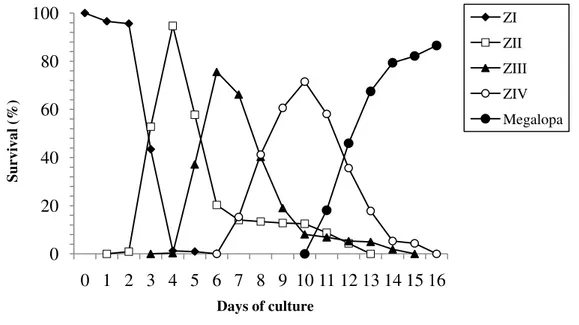
Survival rate and duration of the larval period from hatching to megalopa are shown in the
Fig. 1. Survival (%) and duration of the larval stages zoea to megalopa of Panopeus lacustris
during 16 days of cultivation.
Table 1. Time to the appearance of each larval stage and measurements of Panopeus lacustris.
Stage Time elapsed after hatching (days) Rostrodorsal length (RDL) mean ± SD (mm) Carapace length (CL) mean ± SD (mm) Carapace width (CW) mean ± SD (mm)
Zoea I 0 1.35 ± 0.02 0.33 ± 0.01 —
Zoea II 2 1.7 ± 0.03 0.44 ± 0.01 —
Zoea III 4 2.12 ± 0.04 0.59 ± 0.03 —
Zoea IV 7 2.6 ± 0.06 0.7 ± 0.01 —
Megalopa 11 — 1.01 ± 0.04 0.81 ± 0.03
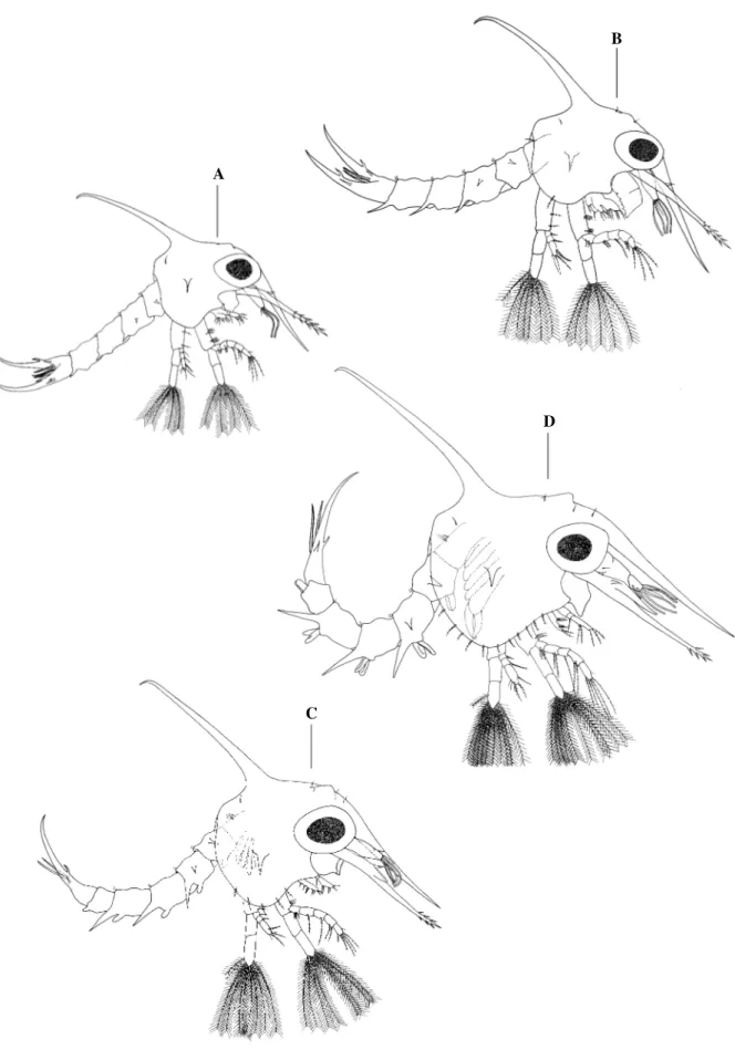
Description
Panopeus lacustris Desbonne, 1867
Zoea I
Carapace (Fig.2A).—Globose, smooth, lacking tubercles and bearing a pair of lateral spine
projected perpendicularly. Dorsal spine present, long and curved distally. Rostral spine as long
as protopod of antenna. Posterodorsal region of carapace with a pair of simple setae. Eyes
sessile. 0 20 40 60 80 100
0 1 2 3 4 5 6 7 8 9 10 11 12 13 14 15 16
Su
rv
iv
al
(%)
Days of culture
Antennule (Fig. 3A).—Uniramous, conical-shaped. Endopod absent. Exopod unsegmented
with 2 aesthetascs and 2 simple unequal setae.
Antenna (Fig. 3F).—Protopod well-developed, armed distally with rows of spines. Endopod
absent. Exopod minute, with 1 terminal simple seta.
Maxillule (Fig. 4A).—Protopod absent. Endopod 2-segmented bearing 1 pappose seta on
proximal segment and 2 subterminal and 4 terminal pappose setae on distal segment. Exopod
lacking setae. Basal endite with 2 cuspidate, 2 plumodenticulate (1 subterminal) and 1
subterminal plumose setae. Coxal endite with 6 plumodenticulate setae (5 terminal and 1
lateral subterminal) and 1 simple lateral subterminal seta.
Maxilla (Fig. 5A).—Scaphognathite with 4 marginal plumose setae and a long posterior
process with michrotrichias. Endopod bilobed with 3 + 5 (2 subterminal and 3 terminal)
sparsely plumose setae. Basal endite bilobed with 5 + 4 sparsely plumose setae. Coxal endite
bilobed, with 4 + 4 sparsely plumose setae.
First Maxilliped (Fig. 6A).—Basis with 2, 2, 3, 3 pappose setae in the inner margin. Endopod
5-segmented, with 3, 2, 1, 2 and 5 (1 subterminal simple seta + 4 terminal) pappose setae from
proximal to distal segment. Exopod 2-segmented with 4 long plumose natatory terminal setae.
Second Maxilliped (Fig. 6F).—Basis with 1, 1, 1, 1 pappose setae on the inner margin.
Endopod 3-segmented with 1 simple, 1 denticulate and 5 (1 subterminal denticulate + 2
pappose terminal + 2 simple terminal) setae from proximal to distal segment. Exopod
2-segmented with 4 long plumose natatory terminal setae.
Abdomen (Fig. 7A).—With 5 somites and telson. Somites 2 and 3 with pair of dorsolateral
spines. Somites 3 - 5 with pair of long posterolateral processes. Somites 2 - 5 with a pair of
minute posterodorsal setae. Telson bifurcated, curved dorsally with 2 lateral unequal spines
and 1 dorsal spine. Inner margin with 2 groups of 3 plumodenticulate setae separated by
Table 2. Morphogical features and setation formulae of the zoeal development of Panopeus lacustris.
Abbreviations: s, seta; a, aesthetasc; seg., segment.
Zoea I Zoea II Zoea III Zoea IV Carapace
Lateral spine present present present present
Anterodorsal s (pairs) 0 2 3 3
Posterodorsal s (pairs) 1 1 1 1
Ventral margin s (pairs) 0 1 5 11
Antennule
Protopod 2a, 2s 4a, 1s 4a, 2s 7a, 2s
Endopod absent absent small bud bud
Antena
Endopod absent absent bud developed
Maxillule
Exopod s absent present present present
Basal endite s 5 8 9 11
Coxal endite s 7 7 8 11
Endopod
Proximal seg s 1 1 1 1
Distal seg s 6 6 6 6
Epipod absent absent absent present
Maxilla
Basal endite s 5 + 4 5 + 4 5 + 5 6 + 6
Coxal endite s 4 + 4 4 + 4 4 + 4 4 + 5
Endopod s 3 + 2 + 3 3 + 2 + 3 3 + 2 + 3 3 + 2 + 3
Scaphognathite 4 + process 11 19 26
First Maxilliped
Coxa s absent absent 1 2
Basis s 2 + 2 + 3 + 3 2 + 2 + 3 + 3 2 + 2 + 3 + 3 2 + 2 + 3 + 3 Endopod
Proximal seg s 3 3 3 3
2nd seg s 2 2 2 2
3rd seg s 1 1 1 1
4th seg s 2 2 2 2
Distal seg s 5 5 6 6
Exopd s 4 6 8 10
Second Maxilliped
Basis s 1 + 1 + 1 + 1 1 + 1 + 1 + 1 1 + 1 + 1 + 1 1 + 1 + 1 + 1 Endopod
Proximal seg s 1 1 1 1
2nd seg s 1 1 1 1
Distal seg s 5 5 5 6
Exopod 4 7 9 11
Third Maxilliped * * * *
Pereiopods * * * *
Abdomen
Proximal so s 0 1 3 3
2nd to 5th so s 2 2 2 2
6th so s absent absent absent absent
Pleopods absent absent buds bilobed
Telson
Furca (outer spine) 3 3 3 3
s (inner) 6 6 8 9
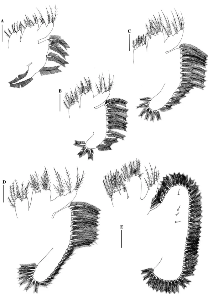
Fig. 2. Panopeus lacustris. Lateral view: A, zoea I; B, zoea II; C, zoea III; D, zoea IV. Scale
bar: A-D= 0.2 mm.
D
C
B
Zoea II
Carapace (Fig. 2B).—With 2 pairs of anterodorsal setae and one pair of posterodorsal setae.
Each ventral margin with 1 sparsely plumose seta. Eyes stalked.
Antennule (Fig. 3B).—Exopod with 4 long aesthetascs and 1 simple seta.
Antenna (Fig. 3G).—Spinous process now less spinose.
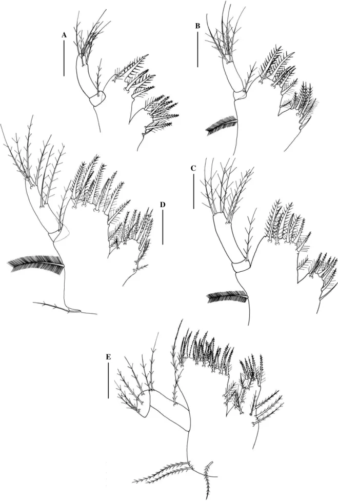
Maxillule (Fig. 4B). —Basal endite with 3 cuspidate, 4 plumodenticulate (2 subterminal) and
1 subterminal lateral plumose setae. Coxal endite with 4 plumodenticulate, 1 plumose and 2
simple (1 subterminal lateral) setae. Exopod present a long plumose marginal seta.
Maxilla (Fig. 5B).—Scaphognathite with 11 marginal plumose seta (last 4 setae showing
reduced in size). Basal endite bilobed with 4 + 5 sparsely plumose setae.
First Maxilliped (Fig. 6B).—Exopod with 6 long natatory plumose setae on distal segment.
Second Maxilliped (Fig. 6G).—Endopod now with 1 pappose seta on distal segment. Exopod
with 1 short subterminal and 6 long terminal natatory plumose setae on distal segment.
Abdomen (Fig. 7B).—First somite with 1 dorsal seta. Posterolateral process on 3 – 5 somites
are more developed. Telson unchanged.
Zoea III
Carapace (Fig. 2C).—With 3 pairs and 1 pair of anterodorsal and posterodorsal setae,
respectively. Each ventral margin with 5 sparsely plumose setae.
Antennule (Fig. 3C).—Exopod with 4 aesthetascs and 1 simple and 1 short subterminal setae.
Endopod bud present.
Antenna (Fig. 3H).—Endopod bud present.
Maxillule (Fig. 4C).—Basal endite with 3 cuspidate, 5 plumodenticulate (3 subterminal) and 1
subterminal lateral plumose setae. Coxal endite with 5 plumodenticulate (1 subterminal
Fig. 3. Panopeus lacustris. Antennule: A, zoea I; B, zoea II; C, zoea III; D, zoea IV; E,
megalopa. Antenna: F, zoea I; G, zoea II; H, zoea III; I, zoea IV; J, megalopa. Scale bar: A, B, E, F= 0.1 mm; C, D, G, H, J= 0.13 mm; I= 0.2 mm.
Maxilla (Fig. 5C).—Scaphognathite with 19 marginal plumose setae (last 11 setae showing
reduced in size). Basal endite bilobed, with 5 + 5 sparsely plumose setae.
G B
A
H
C
D
E I
J
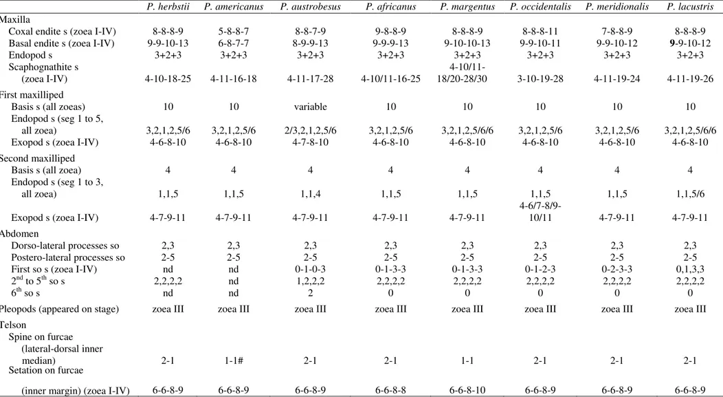
Table 3: Differences and morphologic similarities in the larval stages of Panopeus species: P. herbstii (Costlow and Bookhout 1961); P. americanus (Negreiros-Fransozo 1986); P.
austrobesus (Montú et al. 1988); P. africanus (Rodríguez and Paula 1993); P. margentus (Rodriguez and Spivak 2001); P. occidentalis (Ingle 1985); P. meridionalis (Luppi et al.
2003); and P. lacustris (present study). Abbreviations: s, setation; a, aesthetacs; seg, segment; so, somites; nd, not described.
P. herbstii P. americanus P. austrobesus P. africanus P. margentus P. occidentalis P. meridionalis P. lacustris
Carapace
Dorsal and well well well well well well well well
rostral spaine developed developed developed developed developed developed developed developed Lateral spaine present present present present reduced present present present Anterodorsal s
(pairs, zoea I - IV) nd nd nd 0-2-2-2 0-2-3-3 0-3-3-3 0-2-2-3 0-2-3-3 Posterodorsal s
(pairs, all zoea) nd nd nd 1 1 1 1 1
Ventral margin s
(zoea I-IV) in zoea IV nd 1-2/1-2/0-6/8 0-1-5-9 1-6-9-12 0-1/2-3/5-(?) 0-3-6-12 0-1-5-11 Antennule
Endopod (appeared
on stage) zoea III zoea IV zoea III zoea IV zoea IV zoea IV zoea III zoea III Exopod a, s 3-3-3-11, 3-3-4-6, 3-3-3-5, 3-4-5-8,1 4-5-6-10 3-4-4-9/10, 3-4-5-12,1 2-4-4-7,
(zoea I-IV) 2-2-2-1 1-1-1-1 1-2-2-3 1-1-2-1/2 2-1-2-0
Antena
Protopod spines present present present present* absent present present* present* Endopod (appeared
on stage) zoea II zoea III zoea III zoea II zoea III zoea II zoea III zoea III Exopod apical s (zoea I-V) 1-1-1-1 1-1-1-2 0-0-1-0 1-1-1-1 1-1-1-1 2-1-1-1 1-1-1-1 1-1-1-1
Maxillule
Coxal endite s (I-IV) 7-7-7-9 6-7-5-7 7-7-8-9 7-7-8-9 7-7-8-11 8-8-8-9 7-7-8-11 7-7-8-11 Basal endite s (I-IV) 5-8-9-12 5-8-9-12 5-8-9-12 5-8-9-14 5-8-9-11 5-8-9-11 5-8-9-12 5-8-9-11 Endopod s (proximal,
distal, all zoeas) 1,6 1,6 1,6 1,6 1,6 1,6 1,6 1,6
Exopod s (appeared on stage) zoea III nd zoea II zoea II zoea II zoea II zoea II zoea II Epipod s (appeared
Table 3. Continued.
P. herbstii P. americanus P. austrobesus P. africanus P. margentus P. occidentalis P. meridionalis P. lacustris
Maxilla
Coxal endite s (zoea I-IV) 8-8-8-9 5-8-8-7 8-8-7-9 9-8-8-9 8-8-8-9 8-8-8-11 7-8-8-9 8-8-8-9 Basal endite s (zoea I-IV) 9-9-10-13 6-8-7-7 8-9-9-13 9-9-9-13 9-10-10-13 9-9-10-11 9-9-10-12 9-9-10-12
Endopod s 3+2+3 3+2+3 3+2+3 3+2+3 3+2+3 3+2+3 3+2+3 3+2+3
Scaphognathite s 4-10/11-
(zoea I-IV) 4-10-18-25 4-11-16-18 4-11-17-28 4-10/11-16-25 18/20-28/30 3-10-19-28 4-11-19-24 4-11-19-26 First maxilliped
Basis s (all zoeas) 10 10 variable 10 10 10 10 10
Endopod s (seg 1 to 5,
all zoea) 3,2,1,2,5/6 3,2,1,2,5/6 2/3,2,1,2,5/6 3,2,1,2,5/6 3,2,1,2,5/6/6 3,2,1,2,5/6 3,2,1,2,5/6 3,2,1,2,5/6/6 Exopod s (zoea I-IV) 4-6-8-10 4-6-8-10 4-7-8-10 4-6-8-10 4-6-8-10 4-6-8-10 4-6-8-10 4-6-8-10 Second maxilliped
Basis s (all zoea) 4 4 4 4 4 4 4 4
Endopod s (seg 1 to 3,
all zoea) 1,1,5 1,1,5 1,1,4 1,1,5 1,1,5 1,1,5 1,1,5 1,1,5/6
Exopod s (zoea I-IV) 4-7-9-11 4-7-9-11 4-7-9-11 4-7-9-11 4-7-9-11 4-6/7-8/9-10/11 4-7-9-11 4-7-9-11 Abdomen
Dorso-lateral processes so 2,3 2,3 2,3 2,3 2,3 2,3 2,3 2,3
Postero-lateral processes so 2-5 2-5 2-5 2-5 2-5 2-5 2-5 2-5
First so s (zoea I-IV) nd nd 0-1-0-3 0-1-3-3 0-1-3-3 0-1-2-3 0-2-3-3 0,1,3,3 2nd to 5th so s 2,2,2,2 nd 1,2,2,2 2,2,2,2 2,2,2,2 2,2,2,2 2,2,2,2 2,2,2,2
6th so s nd nd 2 0 0 0 0 0
Pleopods (appeared on stage) zoea III zoea III zoea III zoea III zoea III zoea III zoea III zoea III Telson
Spine on furcae
(lateral-dorsal inner
median) 2-1 1-1# 2-1 2-1 1-1 2-1 2-1 2-1
Setation on furcae
First Maxilliped (Fig. 6C).—Coxal with 1 pappose seta. Basis unchanged. Endopod
5-segmented with 3, 2, 1, 2 and 6 (2 subterminal, 1 simple and 1 pappose + 4 terminal) pappose
setae. Exopod with 8 long natotory plumose setae on distal segment.
Second Maxilliped (Fig. 6H).—Endopod with 2 pappose setae on distal segment. Exopod with
1 short subterminal and 8 long terminal natotory plumose setae on distal segment.
Abdomen (Fig. 7C).—With 6 somites. First somite with 3 dorsal simple setae. Pleopod buds
and present on somites 2 – 5, endopod absent. Telson with 1 additional pair of inner setae on
the mid-posterior margin.
Zoea IV
Carapace (Fig. 2D).—With 11sparsely plumose setae on each ventral margin.
Antennule (Fig. 3D).—Biramous. Basis with 2 simple setae. Exopod with 2 subterminal (1
short) + 5 terminal long aesthetascs.
Antenna (Fig. 3I).—Endopod well-developed.
Maxillule (Fig. 4D).—Endopod with 2 setae in a half of the distal segment and 2 subterminal
and 2 terminal pappose setae. Basal endite with 3 cuspidate, 6 (3 subterminal and 1
subterminal lateral) plumodenticulate and 2 subterminal lateral plumose setae. Coxal endite
with 3 cuspidate, 3 plumodenticulate and 5 subterminal plumose (3 lateral) setae. Basis of the
exopod with 1 sparsely plumose (epipod) setae.
Maxilla (Fig. 5D).—Scaphognathite with 26 marginal plumose setae (last 16 setae showing
reduced in size). Basal endite bilobed with 6 + 6 sparsely plumose setae. Coxal endite bilobed
with 4 + 5 sparsely plumose setae.
First Maxilliped (Fig. 6D).—Coxal with 2 pappose setae. Exopod with 10 long natatory
E
C B A
D
Fig. 4. Panopeus lacustris. Maxillule: A, zoea I; B, zoea II; C, zoea III; D, zoea IV; E,
Fig. 5. Panopeus lacustris. Maxilla: A, zoea I; B, zoea II; C, zoea III; D, zoea IV; E,
megalopa. Scale bar: A-E= 0.05 mm.
A
B
C
D
Second Maxilliped (Fig. 6I).—Endopod with 1 additional simple seta on distal segment.
Exopod with 1 short subterminal plumose and 10 long terminal natatory plumose setae on
distal segment.
Abdomen (Fig. 7D).—First somite with 3 dorsal sparsely plumose setae. Posterolateral
processes of the somites 3-5 more developed. Pleopods with endopods (Fig. 7G), except in the
6th somite (Fig. 7H). Telson with 3 sparsely plumose setae on mid-posterior margin.
Megalopa
Carapace (Fig. 8A).—Subquadrate, longer than broad. Rostrum ventrally deflected, with 2
lateral spines in base. Carapace surface setose with 4 setae on the groove present in the region
posterior dorsal.
Antennule (Fig. 3E).—Peduncle 3-segmented with 4, 2, 2 short simple + 6 long and
denticulate setae, respectively. Endopod unsegmented with 2 subterminal and 4 terminal
simple setae. Exopod 4-segmented with 0, 8, 4, 3 aesthetascs, respectively and 0, 1 serrulate
and 0, 3 simple setae, respectively.
Antenna (Fig. 3J).—Peduncle 3-segmented with 3, 1, 1 setae, respectively. Flagellum
8-segmented, segments 2-5 incompletely segmented (in some individual well- segmented)
bearing 0, 0, 2, 0, 4, 0, 4, 2 terminal setae, respectively.
Maxillule (Fig. 4E).—Endopod unsegmented with 1 proximal sparsely plumose, 2 medial
sparsely plumose, 2 subterminal sparsely plumose and 2 terminal sparsely plumose setae.
Basal endite with 7 cuspidate, 12 subterminal plumodenticulate and 2 subterminal lateral
plumose setae. Coxal endite with 9 subterminal sparsely plumose (3 lateral), 5
plumodenticulate (1 subterminal) setae. Basis of the exopod with 2 + 1 sparsely plumose
E
F A
B C
D
J
I H
G
Fig. 6. Panopeus lacustris. First maxilliped: A, zoea I; B, zoea II; C, zoea III; D, zoea IV; E,
D
G
H
A B C
E F
Fig. 7. Panopeus lacustris. Abdomen, dorsal view: A, zoea I; B, zoea II; C, zoea III; D, zoea
Fig. 8. Panopeu lacustris. Megalopa. A, dorsal view; B, cheliped; C, D, E, F, second to fifth
pereiopod; G, third maxilliped. Abdominal appendages: H, I, J, K, first to fourth pleopod; L,
uropod. Scale bar: A-F= 0.2 mm; G-L= 0.13 mm.
A
G
F
E
D
C
B
L K
J I
Table 4: Differences and morphologic similarities of the megalopa of Panopeus species: P. herbstii (Costlow and Bookhout 1961); P. americanus (Negreiros-Fransozo 1986); P.
austrobesus (Montú et al. 1988); P. africanus (Rodríguez and Paula 1993); P. margentus (Rodriguez and Spivak 2001); P. occidentalis (Ingle 1985); P. meridionalis (Luppi et al.
2003); and P. lacustris (present study). Abbreviations: s, setation; a, aesthetacs; seg, segment; so, somites; nd, not described.
P. herbstii P. americanus P. austrobesus P.africanus P. margentus P. occidentalis P. meridionalis P. lacustris
Carapace
Rostral spines present present present present absent absent present present Antennule
Peduncle s 0,0,6 0,0,3 0,0,2+3 0,1,1+3 3,1,3+6 sparsely 4,2,2+6 4,2,2+6
Endopod s 2+3 0+3 1+3 0,2+4 1+3 0+4 2+3 2+4
Exopod a 8,6,4 4,4,4,0 0,8,6,4,0 6+6+4+0 0,6,4,4 4 seg, 14(15) 0,8,4,4 0,8,4,3 Exopod s 0,0,3 0,0,0,2 0,0,1,0,1 0,1,2,2 0,0,2,2 0,0,1(2),2 0,0,2,2 0,1,0,3 Antena
Peduncle s 2,1,1 0,2,1,0 variable 3,2,1 3,1,1 2,2,2 3,3,1 3,1,1
Flagellum s 0,0,2,0,4,0,4,(?) 0,0,2,0,4,0,4,4 variable,0,4,3 0,0,4,0,4,0,4,3* 0,0,4,0,4,0,4,4 0,0,2,0,4,0,4,3 1,0,3,0,3,0,4,4 0,0,2,0,4,0,4,2 Maxillule
Coxal endite s 14 13 14 12 12 14/15 10+5 14
Basal endite s 22 18 17 2+19 2+18 3+17 11+11 21
Endopod s 3,2+2 1,3 1,4 3+2 1,2+2+2 2+3 1+2+2 1+2+2+2
Maxilla
Coxal endite s 8+6 6+3 8+6 8+7 7+6 9+6 8+6 8+5
Basal endite s 8+10 6+7 8+10 7+10 8+12 6+9 6+1+7+2 5+10
Endopod s 2+2+3 5 8(9) 4+1 2+4 1(2) 3+1+1 3+3+2+2
Scaphognathite
marginal +internal s 49+nd 37+3 44+nd 40+4 45(50)+8 49(50)+nd 44+4 43+4 First Maxilliped
Coxal endite s 10 4(6) 6 8+3 11 11(13) 7+4 5+6
Basal endite s 18 16(18) 20 14(15)+3(4) 20(21) 21(22) 22 4+14
Endopod s 3+2 5(6) 2+5(6) 3 3+2 4(5) 4 3+2
Exopod s 1,6 0,5 1,6 2,4 2,5 2,5 2,6 2,4
Epipod s 7 7 6 3 7(8) 1(2) 7 6
Second Maxilliped
Coxa + Basis s nd nd 1 1 1 2
P. herbstii P. americanus P. austrobesus P.africanus P. margentus P. occidentalis P. meridionalis P. lacustris
Exopod (proximal, distal) s 0,6 1,5+6/7 0,5 1,5 2,5 0,5 2,6 1,5
Epipod s nd absent bud 1+3 3 1+3 0+3 1+2
Third Maxilliped
Coxa + basis s nd 9 16 10 18 3/5 18 14**
Endopod (is,me,ca,pr,da) s 10,11,11,8,8 (?) 7,10,8,8
15-17,11,7/8,8/9,9 20,12,7,9,9
18/22,5/6,4/5,5/6,
7/8 21,11,7,10,10
18,10,7,10, 8 Exopod (proximal, distal) s 0,7 1,7 1,6/7 0,2+5 1,5 0,6 1,7 1,2+5 Epipod (prox. + long distal) s 8+12 4/5+8 0+6 4+13 6+14/15 6/7 5+14 5+13 Pereipods
1st ischium, hook present present present present absent present present present 2nd-4th dactylus, serrate sp nd present present present present present present present
5th dactylus, long s 3 present 3 3 absent 3 3 2
Abdomen
First so s nd nd 6 8 8 10 4 4
Second so s nd nd 2 12 12 12 14 10
Third so s nd nd 2 12 14 10 14 10
Fourth so s nd nd 2 12 12 8 17 13
Fifth so s nd nd 2 10 12 12 13 9
Sixth so s nd nd 2 4 4 4 6 7
Pleopod, exopod nat s 15,15,15,12 14/15,14,13,11 12,15,14,12 14,14/15,12-14,12 14/15,14/15,13/14,12/13 15/16,15,16,13/14 16,15,14,13 15,15,15,13 Pleopod, endopod hooks nd 3,3,2,2 3,3,2,2 3/4,2/3,2/3,2 3/4,3/4,3,3 2(3) 3 3
Uropod, exopod nat s 8 8 8 1+7/8 1+7 10 1+8 1+7
Telson
Maxilla (Fig. 5E).—Scaphognathite with 43 marginal plumose and 4 short lateral setae.
Endopod with 3 basal plumose setae on outer margin and 3 + 2 + 2 sparsely terminal plumose
setae. Basal endite bilobed with 10 (3 subterminal) + 5 (2 subterminal) sparsely plumose setae.
Coxal endite bilobed with 5 (3 subterminal) + 8 (5 subterminal) sparsely plumose setae.
First Maxilliped (Fig. 6E).—Epipod with 6 long setae. Basal endite with 14 terminal (6
plumodenticulate + 4 sparsely plumose + 4 simple) and 4 subterminal (2 sparsely plumose + 1
plumodenticulate + 1 simple) setae. Coxal endite with 6 terminal plumodenticulate and 5
subterminal (4 plumodenticulate + 1 simple) setae. Endopod unsegmented, with 3 lateral and 2
simple terminal setae. Exopod 2-segmented, with 2 distal plumose setae on proximal
segmented and 4 terminal plumose setae on distal segment.
Second Maxilliped (Fig. 6J).—Epipod bilobed with 1 proximal sparsely plumose on the short
lobe and 2 distal simple setae in the elongate lobe. Coxa and basis indistinctively
differentiated with 2 simple setae. Endopod 4-segmented with 5 (2 sparsely plumose + 3
simple), 1 sparsely plumose, 4 plumodenticulate and 10 (7 plumodenticulate + 3 simple) setae.
Exopod 2-segmented, proximal segmented with 1 medial short seta, distal segmented with 5
long terminal plumose setae.
Third Maxlliped (Fig. 8G).—Epipod elongated, with 13 simple setae and 5 proximal plumose
setae. Coxa and basis and 1st segment of endopod fused with 13 pappose setae + 1 simple seta. Endopod 5-segmented with 18 (5 plumodenticulate + 13 simple), 10 (6 plumodeticulate + 4
simple), 7 (3 plumodenticulate + 4 simple), 10 (7 plumodenticulate + 3 simple) and 8 (6
plumodenticulate + 2 simple) setae. Exopod 2-segmented, proximal segment with 1short
medial simple seta, distal segment with 2 subterminal simple and 5 long terminal plumose
setae.
Pereiopods (Fig. 8B-F).—All segments are well differentiated. Chelipeds similar (Fig. 8B)
teeth and setae as illustrated. Pereiopod 2, 3 and 4 (Fig. 8C-E) dactylus bearing 3 serrulate
setae on inner margin and 1 terminal spine. Dactylus of pereiopod 5 (Fig. 8F) with 2
subterminal long simple setae, lacking spines.
Abdomen (Fig. 7E, 7F).—Somites (from proximal to distal) with 4, 10, 10, 13, 9, 7 simple
setae. Telson broader than long with 3 pairs of dorsal simple and 3 terminal (2 sparsely
plumose + 1 plumodenticulate) setae.
Pleopods (Fig. 8H-K).—Endopods unsegmented with 3 coupling hooks on inner margin.
Exopods 1 - 4 with 15, 15, 15, 13 marginal plumose natatory setae. Uropods 2-segment (Fig.
8L) without endopod, exopods with 1 plumose seta on proximal segment and 7 natatory
plumose setae on distal segment.
Discussion
Previous and recent studies on larval development reveal that species of the Panopeus genus,
without exception, are comprised by four zoea stages and one megalopa.
The larval period of the zoeal stages up to metamorphosis into megalopa of P.
lacustris lasted 11 days (Fig. 1), and the larval period was very similar to those reported by
Montú et al. (1988) for P. austrobesus, which reach the post-larval stage after 12 days of
culture. Very much unlike the larval period up to metamorphosis for P. herbstii (senso lato)
and P. americanus, which were recorded as having a longer larval period (over 31 days) when
reared under similar laboratory conditions, (Negreiros-Fransozo 1986; Costlow et al. 1962).
Further larval culture investigations are needed in order to assure that the differences in the
larval period are related to the larval rearing conditions and not to other factors.
Comparisons of the morphological features and setation formulae of the larvae of
Panopeus species (Table 3 and 4) have shown some morphological similarities with P.
lacustris and other species. For P. lacustris, P. herbstii, P. americanus, P. austrobesus, P.
combination of characteristics such as: well developed dorsal and rostral spines with a smooth
rostral; lateral spine development in the zoea stage; protopod antennal spinous as long as the
rostrum, and a reduced exopod with 1 seta (except for P. americanus in stage IV,
Negreiros-Fransozo 1986; P. occidentalis in stage I, Ingle 1985 and P. austrobesus in stage I, II and IV,
Montú et. al. 1988); endopod of maxillule 1, 2+4 setae; endopod of maxilla with eight setae,
arranged 3+2+3; proximal segment of the endopod of first maxilliped with three setae (except
for P. austrobesus in stage I); proximal segment of endopod of second maxilliped with a
single seta; abdominal somites 2nd and 3rd with pairs of dorsolateral spines; the posterodorsal spines on abdominal somites 3-5 of zoea are well developed; telson with 2 lateral and 1 dorsal
spine on the external surface of the furca, but in P. americanus there is only one dorsal spine,
except in zoea I (one small lateral spine).
The zoeae of P. lacustris have showed closed similarities when compared with other
Panopeus species, however, they show some distinct features such as number of aesthetasc
and setal number of the antennule in the stages I and IV; setal number of the scaphognathite
in stage IV; setae number of distal segment of the second endopod maxilliped in stage IV and
the setae number on the ventral margin of carapace in stage IV. The associations of these
referred characteristics are the basis of identification of this species in subsequent plankton
studies.
Felder and Martin (2003) reported that the zoeae of the genus Panopeus, commonly
presented a well developed lateral spine in the carapace (which was sometimes reduced),
antenna with protopodito often armed with a distal spinous process (not dilated at the tip) and
telson with two or three small spines on each side of the furca, located approximately at the
level of the three pairs of setae on its inner margin. However, even though P. margentus
shares many of these characteristics, it is differentiated from the other species of this genus by
the absence of a lateral spine in the carapace, although a small protuberance is observed. This
single spine on the inner margin of the furca. These features are similar to those described for
the former P. bermudensis (now Acantholobulus bermudensis), even though this species does
not present a protuberance in the carapace (Felder and Martin 2003).
According to Rodriguez and Spivak (2001), the larvae of P. margentus are very
similar to those of Hexapanopeus schmitti Rathbun, 1930, now recorded as Acantholobulus
schmitti. Felder and Martin (2003) suggested that P. margentus and Hexapanopeus schmitti
may be the same species and that P. margentus may, in the future, be another species leaving
the genus Panopeus.
The descriptions of megalopa of the panopeid species do not show many similarities
among themselves, but some features among them are noticeably similar, as in: the pair of
anterolateral spines in the base of the rostrum, a spine curved downward in the ischium of the
cheliped, and pereiopod 2-4 with three serrulated setae on the inner margin of the dactylus.
However, even though P. margentus shares fewer similarities when compared with other
panopeids, this species presents a unique similarity in regards to the serrulated setae of the
dactylus.
Evidence from morphological studies shows a close relationship among the different
species of the genus Panopeus. However, phylogenetic analysis clearly suggests the existence
of clusters that were detected through the specific levels of similarity within this genus, as
observed for P. herbstii, P. simpsoni, P. obesus and P. occidentalis., and therefore, P.
lacustris can also be grouped individually (Schubart et al. 2000). The referred author infers
that the morphological similarities among many Panopeus species may occur in response to
similar life forms. These similarities, which are found among the larvae of this group, make it
difficult to describe their specific genus distinction. This is also the case when dealing with
the distinctions that occur between this genus and the genera that belong in the same family,
consequently, this fact remains the main obstacle in phylogenetic studies within the
ACKNOWLEDGEMENTS
We thank Ádila Kelly Rodrigues and Marília Borges Palma for helping in the
laboratory during larval rearing. This study was financed by ‗Fundação de Amparo à Pesquisa
do Estado do Pará‘ (FAPESPA).
LITERATURE CITED
Anger, K., Schreiber, D. and Montú, M. 1995. Abbreviated larval development of Sesarma
curacaoense (Rathbun, 1897) (Decapoda: Grapsidae) reared in the laboratory. Nauplius 3:
127-154.
Brown, K. M. and Haight, E. S. 1992. The foraging ecology of the Gulf of Mexico stone crab
Menippe adina (Williams et Felder). Journal of Experimental Marine Biology and Ecology
160: 67-80.
Clark, P. F., Calazans, D. K. and Pohle, G. W. 1998. Accuracy and standartization of
brachyuran larval descriptions. Invertebrate Reproduction and Development 33: 127-144.
Costlow, J. D. and Bookhout, C. G. 1961. The larval stages of Panopeus herbstii
Milne-Edwards, reared in the laboratory. Journal of the Elisha Mitchell Scientific Society 77: 33–
42.
Costlow, J. D., Bookhout, C. G. and Monroe, R. 1962. Salinity temperature effects on the
larval development of the crab, Panopeus herbstii Milne-Edwards, reared in the laboratory.
Physiological Zoology 35: 79-93.
Desbonne, I. 1867. In: Desbonne and A. Schramm, Crustacés de la Guadeloupe d’aprés un
manuscript du Docteur Isis Desbonne comparé avec les échantillons de Crustacés de sa
collection et les dernières publications de M.M.H. de Saussure et W. Stimpson. I Partie.
Factor, J. R. 1978. Morphology of the mouthparts of larval lobsters, Homarus americanus
(Decapoda: Nephropidae), with special emphasis on their setae. Biological Bulletin 154:
383–408.
Felder, D. L., and Martin, J. W. 2003. Establishment of a new genus for Panopeus
bermudensis Benedict & Rathbun, 1891 and several other xanthoid crabs from the Atlantic
and Pacific oceans (Crustacea: Decapoda: Xanthoidea). Proceedings of the Biological
Society of Washington 116: 438-452.
Garm, A. 2004. Mechanical functions of setae from the mouth apparatus of seven species of
Decapod Crustaceans. Journal Morphology 260: 85-100.
Guida, V. G. 1976. Sponge predation in the oyster reef community as demonstrated with
Cliona celata Grant. Journal Experimental Marine Biology and Ecology 25: 109-122.
Horn, A. C. M. and Buckup, L. 2004. Morfologia setal de Parastacus brasiliensis (von
Martens) (Crustacea, Decapoda, Parastacidae). Revista Brasileira de Zoologia 21: 765-768.
Ingle, R. W. 1985. Larval development of the mud crab Panopeus occidentalis de Saussure,
from Bermuda (Crustacea: Xanthoidea: Panopeidae). Bulletin of the British Museum of
Natural History (Zoology)48: 233–248.
Luppi, T. A., Rodriguez, A. and Spivak, E. D. 2003. Larval morphology of the southwestern
Atlantic mud crab Panopeus meridionalis (Decapoda: Brachyura: Panopeidae) described
from laboratory-reared material. Journal of Crustacean Biology 23: 920-935.
Martin, J. W., Truesdale, F. M. and Felder, D. L. 1985. Larval development of Panopeus
bermudensis Benedict and Rathbun, 1891 (Brachyura, Xanthidae) with notes on zoeal
characters in xanthid crabs. Journal of Crustacean Biology 5: 84–105.
Melo, G. A. 1996. Manual de Identificação do Brachyura (Caranguejos e Sirís) do Litoral
Brasileiro. São Paulo: Ed. Plêiade, FAPESPA (Fundação de Amparo à Pesquisa do Estado
Milne Edwards, H. 1834-1837. Historie naturelle dês Crustacés comprenant l’anatomie, La
physiologie et la classification de ces animaux. Paris, Librairie Encyclopédique de Roret. 1:
i-xxxv, 1-468. 2: 1-531. Atlas, 1837: 1-32, pls. 1-42. 3: 1-638.
Milne-Edwards, A. 1867. Description de quelques espices nouveltes de Crustacés Brachyures.
Annales de la Society Entomologique de France 7: 263-288.
Milne-Edwards, A. 1880. Reports on the results of dredging, under the supervision of
Alexander Agassiz, in the Gulf of Mexico and in the Caribbean sea. 1977, 78, 79. by the
United States Coast Survey Steamer "Blake"... VIII. Études préliminaires sur les
Crustacés. Bulletin of the Museum of Comparative Zoology. Harvard 8: 1-68.
Milke, L. M. and Kennedy, V. S. 2001. Mud crabs (Xanthidae) in Chesapeake Bay: claw
characteristics and predation on epifaunal bivalves. Invertebrate Biology 120: 67-77.
Montú, M., Anger, K., Bakker, C., Anger, V. and Loureiro Fernandes, L. 1988. Larval
development of the Brazilian mud crab Panopeus austrobesus Williams, 1983 (Decapoda:
Xanthidae) reared in the laboratory. Journal of Crustacean Biology 8: 594–613.
Negreiros-Fransozo, M. L. 1986. Desenvolvimento pós-embrionário de Panopeus americanus
Saussure, 1857 (Decapoda, Xanthidae) em laboratório. Revista Brasileira de Biologia 46:
173–188.
Pohle, G. and Telford, M. 1981. Morphology and classification of decapod crustacean larval
setae: a scanning electron microscope study of Dissodactylus crinitichelis Moreira, 1901
(Brachyura: Pinnotheridae). Bulletin of Marine Science 31: 736-752.
Rathbun, M. J. 1898. The Brachyura collected by the U.S. Fish commission steamer Albatross
on the voyage from Norfolk, Virginia, to San Francisco,California, 1887-1888.
Proceedings of the United States National Museum 21: 567-616, pls. 41-44.
Rathbun, M. J. 1930. The cancroid crabs of America of the families Euryalidae, Portunidae,
Atelecyclidae, Cancridae, and Xanthidae. Bulletin of the United States National